RESEARCH ARTICLE
Directional migration of mesenchymal stem cells under an SDF-1α gradient on a microfluidic device Siwan Park1, Hwanseok Jang1, Byung Soo Kim2, Changmo Hwang3, Gi Seok Jeong3*, Yongdoo Park1*
a1111111111 a1111111111 a1111111111 a1111111111 a1111111111
1 Department of Biomedical Engineering, Biomedical Science of Brain Korea 21, College of Medicine, Korea University, Seoul, Korea, 2 Department of Biomedical Science, Graduate School of Medicine, Korea University, Seoul Korea, 3 Biomedical Engineering Research Center, Asan Institute for Life Sciences, Asan Medical Center, Seoul, Korea *
[email protected] (YP);
[email protected] (GSJ)
Abstract OPEN ACCESS Citation: Park S, Jang H, Kim BS, Hwang C, Jeong GS, Park Y (2017) Directional migration of mesenchymal stem cells under an SDF-1α gradient on a microfluidic device. PLoS ONE 12(9): e0184595. https://doi.org/10.1371/journal. pone.0184595 Editor: Nic D. Leipzig, The University of Akron, UNITED STATES Received: April 16, 2017 Accepted: August 25, 2017 Published: September 8, 2017 Copyright: © 2017 Park et al. This is an open access article distributed under the terms of the Creative Commons Attribution License, which permits unrestricted use, distribution, and reproduction in any medium, provided the original author and source are credited. Data Availability Statement: All relevant data are within the paper and its Supporting Information files. Funding: This work was supported by a National Research Foundation of Korea grant funded by the Korean government (NRF2014R1A2A1A11051879), (http://www.nrf.re.kr/ index) and a Korea University Grant. The funder had no role in study design, data collection and analysis, decision to publish, or preparation of the manuscript.
Homing of peripheral stem cells is regulated by one of the most representative homing factors, stromal cell-derived factor 1 alpha (SDF-1α), which specifically binds to the plasma membrane receptor CXCR4 of mesenchymal stem cells (MSCs) in order to initiate the signaling pathways that lead to directional migration and homing of stem cells. This complex homing process and directional migration of stem cells have been mimicked on a microfluidic device that is capable of generating a chemokine gradient within the collagen matrix and embedding endothelial cell (EC) monolayers to mimic blood vessels. On the microfluidic device, stem cells showed directional migration toward the higher concentration of SDF-1α, whereas treatment with the CXCR4 antagonist AMD3100 caused loss of directionality of stem cells. Furthermore, inhibition of stem cell’s main migratory signaling pathways, RhoROCK and Rac pathways, caused blockage of actomyosin and lamellipodia formation, decreasing the migration distance but maintaining directionality. Stem cell homing regulated by SDF-1α caused directional migration of stem cells, while the migratory ability was affected by the activation of migration-related signaling pathways.
Introduction Stem cell homing is a controlled recruitment of stem cells within the vascular endothelium that leads to trans-endothelial and directional migration. Damaged tissues in heart, liver, and other organs can be regenerated by stem cell homing through well-directed migration of stem cells. The directional migration of stem cell is precisely regulated by the homing factors released from the injury sites. The released soluble cytokines, homing factors, contribute to generating the cytokine gradient that determines the direction of stem cell migration. Consequently, the bio-chemical gradient induces stem cells to migrate to the injury site for regeneration.
PLOS ONE | https://doi.org/10.1371/journal.pone.0184595 September 8, 2017
1 / 18
Directional migration of mesenchymal stem cells under an SDF-1α gradient on a microfluidic device
Competing interests: The authors have declared that no competing interests exist.
Although the healing process by stem cells has not been elucidated, it has been shown that homing factors have a pivotal role in tissue regeneration [1]. After tissue damage, homing factors such as SDF-1α also known as the C-X-C motif chemokine 12 (CXCL12) is released from the damaged site. A predominant receptor for the SDF-1α is CXCR4 which is a seven transmembrane G protein-coupled receptor widely expressed in cells and tissues taking a role in vasculogenesis and organogenesis [2, 3]. More importantly, down regulation of CXCR4 and SDF-1α significantly decreased the invasiveness of cancer cells, meaning that expression of CXCR4 is responsible for the cell recruitment [4, 5]. CXCR7 is also a protein known as the receptor of SDF-1α [2, 6]. The released homing factors form a chemical gradient from the injury site to the surrounding area, which initiates the transmigration of stem cells through the endothelium and directional migration into the stromal tissue (Fig 1a) [7]. Dar et al have shown enhanced trans-endothelial migration under a gradient of SDF-1α [8]. Cheng et al showed that stem cells overexpressing CXCR4, contributes to the improvement of cardiac performance in myocardial infarction [9], illustrating that SDF-1α is a key homing factor for stem cells [10]. However, the mechanism behind the directional migration of mesenchymal stem cells (MSC) through the endothelium due to a chemokine gradient has not been clearly elucidated in in vivo or conventional in vitro experimental systems. Directional migration of stem cells during stem cell homing is a key mechanism of homing from the blood vessels to injury sites based on the gradient of homing factors. Peripheral MSCs expressing CXCR4 are trafficked by the gradient of SDF-1α. Binding of SDF-1α leads to activation of signaling pathways related to migratory mechanisms such as Rho-ROCK, Rac, and Cdc42 [11]. Rho-ROCK and Rac pathways are known for their roles in the synthesis of migratory machineries for the cells and are mediated by SDF-1α ligand binding [12, 13]. Although there are limitations in the study of microfluidics [14], the device used in this study is a fascinating system that is able to mimic numerous in vivo microenvironments generating gradients of soluble cytokines. Directional migration was incorporated into a collagen
Fig 1. Mesenchymal stem cell homing mimicked on a microfluidic device. (a) Theoretical schematic of peripheral MSC homing process. (b) Illustration of the microfluidic device and stem cell homing on-chip. https://doi.org/10.1371/journal.pone.0184595.g001
PLOS ONE | https://doi.org/10.1371/journal.pone.0184595 September 8, 2017
2 / 18
Directional migration of mesenchymal stem cells under an SDF-1α gradient on a microfluidic device
matrix-integrated microfluidic device, which could be used for the assessment of stem cell homing [15] (Fig 1b). Chung et al developed the basic three-channel-based microfluidic chips with collagen matrix as a barrier of fluid on a vascular structure [16]. Also, the capability of this device to form endothelial cell (EC) monolayers allowed the observation of EC migration and sprouting through the collagen matrix by Chung et al and Jeong et al [16, 17]. To better understand the homing mechanism, Boyden chambers and transwells have been used as tools to observe the increased migration of MSCs due to the chemokine effect of SDF1α [1, 18]. However, none of these devices showed the chemotaxis of MSCs through the endothelial barrier or the ECM conditions, meaning that the devices were not able to mimic the in vivo spatial environment. The proposed device has a geometric set up for building perfusable vessel structures as well as the ECM environment and has both biological and technical advantages over the Boyden chamber and the transwell systems. Furthermore, the previous studies did not focus on the effect of migratory inhibitors during cellular migration or on the quantification of migration distance or directional migration. In this study, we describe the directional migration of MSCs under a gradient of homing factors using a microfluidic channel. To identify the behaviors of homing factors, directional movement and transmigration of MSCs were observed. To construct an in-vivo-mimicking microenvironment, an EC barrier was constructed by forming an EC monolayer along the center channel of the microfluidic device. Directionality and migratory ability of MSCs were assessed in the presence of different inhibitors. Five conditions including the control environment were created by exposing inhibitors to MSCs undergoing migration; (i) Control group without SDF-1α gradient, (ii) SDF-1α condition, (iii) AMD3100 (CXCR4 antagonist) treatment condition, (iv) Y-27632 (Rho-ROCK inhibitor) treatment condition, and (v) NSC23766 (Rac inhibitor) treatment condition. The inhibitor AMD3100 was used for blocking SDF-1α binding to CXCR4 in order to disrupt the directionality of MSC and Y-27632, and NSC23766 were used to disable the migratory mechanism of the stem cells. Treatment with Y-27632, an inhibitor of the Rho-ROCK signaling pathway, and NSC23766, an inhibitor of the Rac signaling pathway, resulted in decreased migration distance of MSCs without loss of directionality. In contrast, the CXCR4 antagonist AMD3100 disrupted the directionality of the MSCs but did not affect the migration ability of the stem cells resulting in near average migration distances.
Material and methods 2.1 Chip fabrication 2.1.1. Microfluidic device. To visualize the directional migration of MSCs, a polydimethylsiloxane (PDMS; Sylgard 184A, B Dow Chemical, MI, USA) device (Fig 2a) inspired by a microfluidic chip used in a previous study was fabricated [16]. Using conventional soft lithography, PDMS (Sylgard A: Sylgard B = 10:1) was cured on an SU-8 (MicroChem, MA, USA) wafer and placed at 80˚C. An individual chip had a 25mm width and length and a postmolding height of 250μm. The center cell seeding channel was 0.5mm wide, and the two side cell seeding channels were 1mm wide. Each of the collagen gel channels was 1mm wide (S1b Fig). After curing the microfluidic channel, the device was autoclaved and bonded with cover slips under oxygen plasma treatment. Within 10 minutes after bonding, 1mg/ml PolyD-Lysine (PDL; Sigma-Aldrich, St. Louis, MO) was applied to the inside of the chip for enhancement of adhesion of collagen as well as the EC monolayer. After at least two hours of incubation with PDL coating, the channels were washed with triple-distilled water and dried in an oven for 24 hours. 2.1.2 Cell seeding and settings of the microfluidic device. The main set up for this experiment is inhibition of EC migration during induced stem cell migration. Metabolic
PLOS ONE | https://doi.org/10.1371/journal.pone.0184595 September 8, 2017
3 / 18
Directional migration of mesenchymal stem cells under an SDF-1α gradient on a microfluidic device
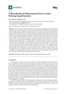
Fig 2. Extravasation of MSCs under a gradient of homing factor. (a) An image of the PDMS-based microfluidic device. (b) The microfluidic device set up for cell seeding. Micropipette tips were used as a media reservoir. (c) A FITC-dextran gradient within the collagen matrix on a microfluidic device with endothelial monolayers (blue squares) embedded in cell channels. Red arrows indicate the direction of MSC migration due to an SDF-1α gradient. The graph indicates the fluorescence intensity of dextran within the chip. (d) A microscopic image of MSC extravasation and migration through the collagen matrix on a microfluidic device. (e) RFP-tagged endothelial cells are used to distinguish migrating MSCs. (f) MSC extravasation. The white arrow indicates MSCs, and red arrow indicates endothelial cells. https://doi.org/10.1371/journal.pone.0184595.g002
balance is controlled by consumption of nutrients and oxygen in the culture media in each channel. Equal numbers of ECs were seeded in all three channels for equal consumption of nutrients and oxygen in culture media, allowing the endothelial cells to remain as a stable monolayer. On the other hand, MSCs are set to migrate toward a higher concentration of metabolic factors from the side channels. In this experiment, we compare the numbers of MSCs migrating both toward and away from the SDF-1α gradient in order to identify the cells migrating under the influence of SDF-1α.
PLOS ONE | https://doi.org/10.1371/journal.pone.0184595 September 8, 2017
4 / 18
Directional migration of mesenchymal stem cells under an SDF-1α gradient on a microfluidic device
2.2 Evaluation of gradients of the microfluidic chip 2.2.1. Collagen gel preparation. In order to prepare the collagen gel, stock type I collagen (BD Bioscience, MA), 10X PBS (Gibco BRL, NY, USA), filtered triple-deionized water, and 0.5N NaOH were kept in an ice bath and mixed to a final pH of 7.4 and concentration of 2.5mg/ml. Just after mixing, collagen solution was carefully introduced into the ECM channels of the microfluidic chip. Then, the chip was placed in the humidified chamber and placed in the incubator at 37˚C for 30 min. Next, 37˚C warmed media was applied in order to prevent dehydration or shrinkage of the collagen matrix. 2.2.2. SDF-1α gradient. Two hours after MSC seeding, cell culture medium containing 250ng/ml of SDF-1α (R&D Systems, MN, USA) was applied only to the conditioning channel to generate an SDF-1α gradient within the collagen gel matrix. The concentration of SDF-1α in vivo is known to be about 0.5 to 0.8 ng/ml in human circulation and 1 to 5 μg/ml in in vivo fluids. However, the optimal concentration range for most chemokines for in vivo cell attraction is 10 to 1000 ng/ml [19]. Here, 250ng/ml of SDF-1α was applied every 24 hours after washing the channels with media in order to reset the gradient. 2.2.3. Dextran gradient test. To ensure that the chemokine gradient is sustained in the collagen matrix, 10 kDa fluorescence isothiocyanate (FITC)-dextran (FITC-dextran, 10kDa, Sigma-Aldrich, St. Louis, MO) was applied to visualize the gradient when an endothelial monolayer was present (Fig 2c). 10 kDa FITC-dextran was used because of its identical molecular weight to SDF-1α. Fluorescence images were taken every 4 hours for up to 44 hours. The dextran gradient was renewed 24 hours after the initial creation. Image J (NIH image, Wayne Rasband) was used for plot profiling and measuring the fluorescence intensity of dextran. The maximum fluorescent intensity (1.0) was shown to slightly decrease over time; plot profiling clearly illustrated the level of the gradient. However, the intensity also seemed to fluctuate while decreasing because the least intense fluorescence is not at the final hours. However, the intensity generally decreased by about 20 to 50% within the collagen matrix throughout the experiment. This evidence suggests that the SDF-1α gradient is sustained within the collagen gel for chemokine-derived migration of MSCs (S3 Fig). The consumption rate of SDF-1α is not well studied; therefore, the results might vary due to the reduction of chemokines in certain areas and deformation of the gradient. However, this graphic data is strong support that the gradient is not saturated within the collagen and sustains the homing process for over 40 hours.
2.3 Directional migration of stem cells under physiological conditions 2.3.1. Cell culture. Red fluorescent protein (RFP) expressing human umbilical vein endothelial cells (RFP-HUVECs) (Olaf Pharmaceuticals, Inc., USA) were cultured in endothelial cell growth medium (EGM TM-2) (Lonza, MD, USA) supplemented with 5% FBS, 0.04% hydrocortisone, 0.4% hFGF-B, 0.1% VEGF, 0.1% R3IFG-1, 0.1% ascorbic acid, 0.1% hEFG, and 0.1% GA-1000. The seeding concentration of RFP-HUVECs in the microfluidic channel was 2 x 10⁶ cells/ml. Human originated mesenchymal stem cells (hMSCs) (Lonza, MD, USA) were cultured in Dulbecco’s modified Eagle’s media (DMEM; Gibco BRL, NY, USA) supplemented with 10% FBS and 1% (v/v) antibiotics. A cell suspension with an MSC concentration of 4.5 x 10⁵ cells/ml was used for microchip injection. MSCs were not used after their ninth passage. Both cells were cultured in regular 75T cell culture plates before they were used in the chip. RFP-HUVECs were seeded twice at a 2-hour interval in order to seed the cells thoroughly in the channels. The collagen gel interface was endothelialized by seeding HUVECs in the
PLOS ONE | https://doi.org/10.1371/journal.pone.0184595 September 8, 2017
5 / 18
Directional migration of mesenchymal stem cells under an SDF-1α gradient on a microfluidic device
main channels. After seeding, the chips were tilted at various angles and placed for 30 minutes to form a uniform EC monolayer (S5 Fig). After 2 to 3 days of culturing EC in the microfluidic device, monolayer formation could be seen under the microscope (Fig 2d and 2e), and MSCs were introduced in the middle channel with different conditioned media. Media was composed of EGM-2 and DMEM in a ratio of 2:1 and was changed every 24 hours. For cell culture, micropipette tips were used as a media reservoir (Fig 2b). 2.3.2. Treatment with cell migration inhibitors. In order to influence the migration of MSCs, AMD3100 (CXCR4 antagonist), Y-27632 (inhibitor of Rho-ROCK pathway), and NSC23766 (inhibitor of Rac pathway) were applied throughout all channels. Each of the inhibitors Y-27632 (25μM, Sigma-Aldrich, St. Louis, MO, USA), NSC23766 (50μM, Sigma Aldrich, St. Louis, MO, USA), and AMD3100 (25μg/ml, Sigma Aldrich, St. Louis, MO, USA) were mixed in 2:1 EGM-2 and DMEM with addition of SDF-1α (250 ng/ml) on the condition channel. A total of five groups including conditions with media only (no SDF-1α, no drugs), SDF1α only group, and three inhibitor-treated groups were tested. Media for each group were replaced every 24 hours, and images were obtained with an EVOS fluorescence microscope (EVOS1 FL Auto, Life Technologies, USA).
2.4 Assessment of stem cell homing in microfluidic chips 2.4.1. Stem cell migration analysis. Images were obtained with EVOS (EVOS1 FL Auto) every 24 hours for 2 days. Migrating cells were counted with Image J software, and after immunohistochemistry, the distance of migration was measured from the starting point of the collagen matrix to the nucleus of the cell with Image J. Confocal images were taken with an LSM710 (Zeiss, Germany) and analyzed with ZEN Black Edition. Stem cells migrated from the endothelial monolayer were counted on EVOS microscopic images. Cells were distinguishable since the cytosol of ECs was expressing RFP and MSCs were not. MSCs in the process of transmigration were also counted for extravasated cells since their branches were within the collagen matrix (Fig 2f). Cell migration displacement was measured by DAPI images obtained also with an EVOS microscope. The distance measurement was performed utilizing both the RFP and DAPI images to distinguish between MSCs and RFP-ECs. The term displacement is used because cells do not migrate in straight lines, although the measurement was in the linear distance. The distance was measured from the start of the collagen matrix or endothelial monolayer to the nucleus of the stem cells with Image J. 2.4.2. Immunohistochemistry. The channels of the microfluidic chip were gently washed with PBS to remove all media. Then, 4% paraformaldehyde was applied and cooled to 4˚C. When the cells were fixed, F-actin was labeled with Alexa Fluor 488 phalloidin (Thermo Fisher Scientific Cat# A12379, RRID:AB_2315147), and nuclei were stained with 4’6-diamidino-2-phenylindole (Thermo Fisher Scientific Cat# D1306, RRID:AB_2629482). Cells were permeabilized with 0.1% Triton X-100 (Sigma-Aldrich, St. Louis, MO, USA) for 30 minutes. After permeabilization, BSA treatment was applied for 30 minutes, after which phalloidin and DAPI dissolved in BSA were applied to all channels. After two hours of staining, the channels were washed with 1X phosphate buffered saline tween-20 (PBST) to remove the fluorescent noise. VE-cadherin was stained with Anti-VE Cadherin antibody—Intercellular Junction Marker (Abcam Cat# ab33168, RRID:AB_870662) and Alexa Fluor 594 goat anti-rabbit lgG (H+L) (Molecular Probes Cat# A-11012, RRID:AB_141359) (Life Technologies, USA) after being fixed with 4% paraformaldehyde.
PLOS ONE | https://doi.org/10.1371/journal.pone.0184595 September 8, 2017
6 / 18
Directional migration of mesenchymal stem cells under an SDF-1α gradient on a microfluidic device
Result and discussion 3.1. Chip fabrication and gradient formation Homing factors are released from an injury site and form a chemokine gradient across the surrounding area to induce MSC migration toward the injury site (Fig 1a). When SDF-1α is bound to CXCR4, the associated MSCs migrate toward the higher end of the gradient. To mimic this event in a microenvironment, the microfluidic device was re-designed and modified from a previously reported version [16]. The differences in device design are 5 times more regions of interest (ROIs) and wider gel channel areas for observation of the migration tendency (Fig 1b). The microfluidic device used in this experiment has three main cell seeding channels separated by two gel channels. (S1a Fig). The three main channels are designed to provide both control and experimental conditions in one chip sample. Also, the separating collagen scaffolds help divide the three main channels to ensure that they are physically independent. Since the device allows cell culture inside the channels, we co-cultured ECs with MSCs for observation of transendothelial migration (Fig 1b). Stem cell migration due to the chemokine effect of SDF-1α has been previously demonstrated on a Boyden chamber. Imitola et al showed increased stem cell migration through a fibronectin-coated membrane depending on the dosage of SDF-1α [18]. The Boyden chamber is a useful tool to examine chemotaxis [20] but has limitations in spatiotemporally mimicking the in vivo environment. The proposed microfluidic device, however, has significance in mimicking the spatial environment with both a chemokine gradient to initiate directional migration and inhibitors to affect the migratory mechanisms of stem cells. Applications of different environmental conditions on the chip produced significantly different results, which provided insights into cellular migration depending on the chemical environment. The main set up for this experiment was to inhibit the migration of ECs while inducing stem cell migration. Differential migration was achieved by controlling the consumption of nutrients in the culture media in each channel [17]. Equal numbers of ECs were seeded in all three channels, which led to equal consumption of the culture media and maintenance of the endothelial cells as a stable monolayer (S2c and S2d Fig). If only one of the channels was seeded, ECs would start sprouting toward the higher concentration of metabolic factors by degrading the collagen gel matrix (S2a and S2b Fig). Conversely, MSCs were seeded only in the center channel in order to easily initiate the migration toward the higher concentration of metabolic factors from the side channels. In this experiment, we compared the numbers of MSCs migrating both toward and away from the SDF-1α knowing that the migration is both caused by the metabolic and chemokine gradient. However, when both sides were compared, more of MSCs were estimated to migrate towards the chemokine gradient. In order to simulate the gradient formation of SDF-1α (10kDa) in microfluidic channels, FITC-dextran of same molecular weight was used for generating a visual gradient. Maximum fluorescent intensity (1.0) was slightly declining over time and with plot profiling, the fluorescent intensity data was analyzed. The intensity was generally decreasing throughout the experiment by about 20 to 50% within the collagen matrix. This evidence suggested that the SDF-1α gradient was sustained within the collagen gel for chemokine derived migrations of MSCs (S3 Fig). A uniform confluence of the endothelial monolayer at the interface with the collagen matrix and the channel surface was a crucial factor in preventing undesirable leakage of the chemokine gradient in the microfluidic devices. VE-cadherin (vascular endothelial-cadherin) is a glycoprotein responsible for cell-cell adhesion, whose expression contributes greatly to endothelial permeability by regulating intercellular junctions [21]. Significant expression of VEcadherin means that the cells are well-adhered to form a strong barrier. VE-cadherin expressed
PLOS ONE | https://doi.org/10.1371/journal.pone.0184595 September 8, 2017
7 / 18
Directional migration of mesenchymal stem cells under an SDF-1α gradient on a microfluidic device
Fig 3. Confocal microscopic images of endothelial monolayer formed within the channel of the microfluidic device. (a) Side view of the endothelial monolayer confluent with collagen matrix. RFP represents the expression of VE-cadherin and blue represents DAPI. (b) Front view of the endothelial monolayer confluent with both the channel and the collagen matrix. GFP represents expression of actin fibers within the cells. (c) Overall view of the endothelial monolayer confluent within the channel of the microfluidic device. (d) Ortho view of the endothelial monolayer. The top image shows homogeneous confluence of cellular monolayer formed throughout the channel. Blue territory represents the PDMS posts. https://doi.org/10.1371/journal.pone.0184595.g003
in seeded endothelial cells in the microfluidic device reflects the formation of a definite EC monolayer which was observed under the confocal microscopy (Fig 3a–3c). The expression level of VE-cadherin confirms that the endothelial monolayer constructed within the microfluidic device is reliable (Fig 3d). To ensure that the endothelial monolayer in the microfluidic device had barrier functions, the plot profile was used to measure the intensity of the FITC-dextran fluorescence over 16 hours with and without the EC monolayer. The result showed stabilized fluorescence intensity in the presence of an EC monolayer. On the other hand, the dextran gradient seemed to randomly flow within the device, showing a significant increase in fluorescence intensity in the ROIs (n = 4, p