(computer search/elongation factor Tu/foot-and-mouth disease virus protein 2C/nucleotide binding). THOMAS E. ... refined the GTP-binding domain consensus sequence and ..... sequence presented in this paper can be used as a tool to.
Proc. Natl. Acad. Sci. USA Vol. 84, pp. 1814-1818, April 1987 Biochemistry
GTP-binding domain: Three consensus sequence elements with distinct spacing (computer search/elongation factor Tu/foot-and-mouth disease virus protein 2C/nucleotide binding)
THOMAS E. DEVER*, MANUEL J. GLYNIASt, AND WILLIAM C. MERRICK* Departments of *Biochemistry and tPharmacology, Case Western Reserve University, School of Medicine, Cleveland, OH 44106
Communicated by Harland G. Wood, November 20, 1986
by using it in a computer search of the Protein Identification Resource (PIR) protein sequence data base. In this paper we report our consensus sequence, a list of known GTP-binding proteins that match the consensus sequence, and the results of our computer search.
A sequence comparison of nine functionally ABSTRACT different GTP-binding protein families has yielded further information on the general characterization ofthe conservation and importance of amino acid sequences in the GTP-binding domain, including (i) a consensus sequence composed of three consensus elements GXXXXGK, DXXG, and NKXD with consensus spacings of either 40-80 or 130-170 amino acid residues between the first and second elements and -40-80 amino acid residues between the second and third sequence elements; (it) the sequence NKXW in place of NKXD in the sequence element responsible for base specificity allows the use of ITP as well as GTP; (fib) dGTP can be used with essentially the same efficiency as GTP; (iv) signal transducing proteins and enzymes have been identified in the nine families; and (v) family conservations allow the identification of the most probable consensus sequence element when more than one is present. Employing these features we have screened the protein sequence data base of the Protein Identification Resource and have identified only known GTP-binding proteins with the exception of protein 2C from foot-and-mouth disease virus as matching the consensus sequence. Based on this finding we predict that foot-and-mouth disease virus protein 2C binds GTP and, by analogy, that protein 2C from several related viruses (polio, rhino, encephalomyocarditis, and cowpea mosaic) will bind a nucleotide as part of its biologic activity.
MATERIALS AND METHODS Search of the PIR Protein Sequence Data Base. We screened the protein sequence data base of the PIR, supported by the Division of Research Resources of the National Institutes of Health, using the Bionet program "Quest" (Intelligenetics), looking for proteins that had the three elements of the consensus sequence (see text) in the proper order. In our initial screen we allowed conservative amino acid replacements: A for G, E for D, Q for N. At the time the data base was screened, August 1986 (release 10.0), =3800 protein sequences were present. The data base was screened for the following sequence: (G/A)XXXX(G/A)K, then (D/E)XX(G/A), then (N/Q)KX(D/E) (where G/A means G or A and X can be any amino acid). The spacing between the three sequence elements was allowed to be any number of amino acids. Calculation of the Probability of the Chance Occurrence of the Consensus Sequence. In the determination of the probability of the occurrence of the consensus sequence strictly by chance, several assumptions were made: (i) all amino acids occur with equal frequency (1/20); (it) the length of the average protein was taken as 1000 amino acid residues; (iii) the spacing between the consensus elements GXXXXGK, DXXG, and NKXD is about 40-80 amino acids. The approximate probability of the three sequence elements appearing in a protein of 1000 amino acids is thus:
The cloning and sequencing of many proteins have stimulated a search for common primary structure motifs that could be used to predict protein function. Nucleotide binding is a property that has been extensively studied and it would be very valuable if it could be predicted based on the primary structure. ATP-binding proteins have been characterized at the structural level by the Rossmann fold (1, 2) and attempts have been made to characterize a common sequence. However, the proposed common sequence elements, typically a glycine-rich region, cannot be characterized by a consensus sequence nor are they unique enough to have predictive value (3, 4). Recently, the x-ray structure of the GTP-binding domain of elongation factor Tu (EF-Tu) was reported by la Cour et al. (5) and a consensus sequence for GTP-binding domains was proposed (6). In contrast to the ATP-binding sequence, the GTP-binding sequence is more extensive and unique. The GTP-binding consensus sequence has been found in a wide variety of proteins performing diverse functions and having a high affinity for GTP. These proteins include the elongation factors, ras p21 protein, phosphoenolpyruvate carboxykinase (GTP) (EC 4.1.1.32) (PEPCK), and guanine nucleotide-binding proteins of adenylate cyclase (G proteins). Based on a further review of the literature we have refined the GTP-binding domain consensus sequence and have tested the predictive value of this consensus sequence
(1/20)3(880)(1/20)2(40)(1/20)3(40)
=
5.5 x 10⁻⁵ or 1/18,182.
Though the assumptions used may not be entirely valid (G and K often exceed 5%, the average molecular size is often taken as 450-600 amino acids), the chance occurrence of the three consensus elements correctly spaced would appear to be between 1/5000 and 1/10,000. Assays. Polyphenylalanine synthesis was monitored by the incorporation of [14C]phenylalanine into hot, trichloroacetic acid-precipitable radioactivity directed by poly(U) using EF-1, EF-2, and ribosomes as described (7). The purification of the protein synthesis factors (EF-1 and EF-2) was as reported (8). PEPCK was provided by T. Nowak (Univ. of Notre Dame, Notre Dame, IN). The PEPCK spectrophotometric coupled assay monitored the conversion of NADH to NAD over time as facilitated indirectly by PEPCK as Abbreviations: EF, elongation factor; PEPCK, phosphoenolpyruvate carboxykinase (GTP); G protein, guanine nucleotide-binding regulatory protein of adenylate cyclase; PIR, Protein Identification Resource; IF, initiation factor; FMDV, foot-and-mouth disease virus.
The publication costs of this article were defrayed in part by page charge payment. This article must therefore be hereby marked "advertisement" in accordance with 18 U.S.C. §1734 solely to indicate this fact. 1814
Biochemistry: Dever et al.
Proc. Natl. Acad. Sci. USA 84 (1987)
described (9), except the assay was performed in the reverse direction as described in ref. 10. The GDP-binding activity was determined as protein-dependent retention of radiolabeled GDP on nitrocellulose filters (7).
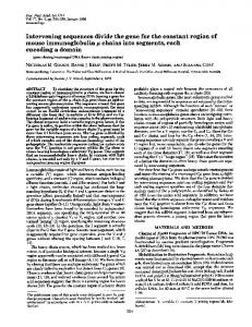
RESULTS GTP-Binding Domain Consensus Sequence. Based on the x-ray data of the GTP-binding domain of EF-Tu (5) and a sequence comparison of other known GTP-binding proteins, a consensus sequence can be found. The consensus sequence contains three consensus elements, GXXXXGK, DXXG, and NKXD, with a consensus spacing requirement of =4080 amino acids between the first and second and between the second and third sequence elements. The first two elements are involved in interactions with the phosphate portion of the GTP molecule and the last element is involved in nucleotide specificity (6). Table 1 shows the alignment of the three elements of the GTP-binding domain consensus sequence for a number of GTP-binding proteins that have been sequenced. Table 1 is spaced to emphasize the different functional families [i.e., proteins from different sources that perform the same function: EF-Tu (EF-1) family, EF-G (EF-2) family, ras family, PEPCK family, G-protein family, plus the three families with one representative each: LepA, initiation factor 2 (IF-2), and GTP:AMP phosphotransferase]. Not all of the known examples for each of the families are listed due to space considerations. It is important to notice that even though there may be extensive amino acid sequence conservation within a family of proteins for the three sequence elements, it would be inappropriate to include these in the consensus sequence since they are not conserved in proteins
1815
from other functional families. Though not emphasized in Table 1, in most of the GTP-binding proteins that have been sequenced the spacing between the parts of the consensus sequence is 40-80 amino acids. The only exceptions to the spacing rule are GTP:AMP phosphotransferase, transducin, and the G proteins, which seem to have a spacing of about 150 amino acids between the first two sequence elements but the conserved spacing between the second and third sequence element. The first consensus element GXXXXGK is similar to the glycine-rich area seen in many ATP-binding proteins (3, 4, 41, 42). However, a significant difference is the variability in the sequence for ATP-binding proteins and the strong conservation in sequence for the GTP-binding proteins (this difference will be further examined in the Discussion). For three of the proteins listed in Table 1 (EF-G, EF-2, and LepA), the first glycine in GXXXXGK is replaced by alanine. This finding of a conservative amino acid replacement was important in defining the rules for our computer search, which will be discussed later. For two of the proteins in Table 1 (rho and GO), only incomplete sequence data are available; however, based on their similarity to the family sequence they seem to match the elements of the consensus sequence. As shown in Table 1, transducin has two matches to the second conserved sequence element, DXXG. We would predict that DVGGQ (196-200) is most likely the proper element and not DSAGY (146-150) as proposed by McCormick et al. (6), since the DVGGQ sequence is also found in the G-protein family. This choice of DXXG sequence also allows for a conservation in the second spacing of 40-80 amino acids for all of the proteins
Table 1. Components of the GTP/GDP-binding site PHOSPHORYL BINDING SEQUENCES
GUANINE SPECIFICITY BINDING SITE
CONSENSUS SEQUENCE
Gly X X X X GlyLys
EF-Tu, E. coli EF-Tu, Euglena chloro. EF-Tu. yeast mito. EF-la, yeast EF-1a, A. salina EF-la, human
Gly His Val Asp His Gly Lys Gly His Val Asp His Gly Lys Gly His Val Asp His Gly Lys Gly His Val Asp Ser Gly Lys Gly His Val Asp Ser Gly Lys Gly His Val Asp Ser Gly Lys
(18-24) (18-24) (55-61) (14-20) (14-20) (14-20)
Asp Cys Pro Gly His Asp Cys Pro Gly His Asp Cys Pro Gly His Asp Ala Pro Gly His Asp Ala Pro Gly His Asp Ala Pro Gly His
(80-84) (80-84) (117-121) (91-95) (91-95) (91-95)
Asn Lys Cys Asp Asn Lys Glu Asp Asn Lys Val Asp Asn Lys Met Asp Asn Lys Met Asp Asn Lys Met Asp
(135-138) (135-138) (172-175) (153-156) (153-156) (153-156)
(11)
EF-G, E. coli EF-2, hamster
Ala His le Asp Ala Gly Lys Ala His Val Asp His Gly Lys
(16-22) (26-32)
Asp Thr Pro Gly His Asp Ser Pro Gly His
(87-91) (104-108)
Asn Lys Met Asp Asn Lys Met Asp
(141-144) (158-161)
(18)
LepA, E. coli
Ala His le Asp His Gly Lys
(11-17)
Asp Thr Pro Gly His
(77-81)
Asn Lys lle Asp
(131-134)
(20)
IF-2, E. coli
Gly His Val Asp His Gly Lys
(398404)
Asp Thr Pro Gly His
(444-448)
Asn Lys lie Asp
(498-501)
(21)
RAS 1. yeast RAS 2, yeast YP2, yeast H-ras, N-ras, K-ras, human p29 ras, rat v-ras, mouse v-ras H, mouse v-ras K, mouse rho, Aplysia rho, human
Gly Gly Gly Gly Val Gly Lys Gly Gly Gly Gly Val Gly Lys Gly Asn Ser Gly Val Gly Lys Gly Ala Gly Gly Val Gly Lys Gly Ala Arg Gly Val Gly Lys Gly Ala Lys Gly Val Gly Lys Gly Ala Arg Gly Val Gly Lys Gly Ala Ser Gly Val Gly Lys Gly Asp Gly Ala Cys Gly Lys
(17-23) (17-23) (15-21) (10-16) (69-75) (10-16) (10-16) (10-16) (12-18)
Asp Thr Ala Gly Gin Asp Thr Ala Gly GIn Asp Thr Ala Gly Gin Asp Thr Ala Gly Gin Asp Thr Ala Gly Gin Asp Thr Ala Gly GIn Asp Thr Thr Gly GIn Asp Thr Thr Gly GIn Asp Thr Ala Gly GIn Asp Thr Ala Gly GIn
(64-68) (64-68) (63-67) (57-61) (116-120) (57-61) (57-61) (57-61) (59-63) (same)
Asn Lys Leu Asp Asn Lys Ser Asp Asn Lys Cys Asp Asn Lys Cys Asp Asn Lys Cys Asp Asn Lys Cys Asp Asn Lys Cys Asp Asn Lys Cys Asp Asn Lys Lys Asp Asn Lys Lys Asp
(123-126) (123-126) (121-124)
(22, 23) (23, 24) (25) (26-28) (29) (30)
PEPCK, chicken PEPCK, rat liver
Gly Asn Ser Leu Leu Gly Lys Gly Asn Ser Leu Leu Gly Lys
(237-243) (237-243)
Asp Glu Leu Gly Asn Asp Ala Gln Gly Asn
(318-321) (318-321)
Asn Lys Asp Trp Asn Lys Glu Trp
(388-391) (388-391)
(33) (33) (34) (35)
GTP:AMP phosphotransferase bovine
Gly Ala Pro Gly Ser Gly Lys
(12-18)
Asp Leu Thr Gly Glu
(150-154)
Asn Lys Ile Trp
(200-203)
(36)
Transducin a, bovine
Gly Ala Gly Glu Ser Gly Lys
(36-42)
Asp Ser Ala Gly Tyr Asp Val Gly Gly Gln
(146-150) (196-200)
Asn Lys Lys Asp
(265-268)
(37, 38)
Gs protein, bovine adrenal Gs protein, rat brain Go protein, rat brain
Gly Ala Gly Glu Ser Gly Lys Gly Ala Gly Glu Ser Gly Lys
(47-53) (47-53)
(223-227) (223-227)
Asn Lys Gln Asp Asn Lys Gln Asp Asn Lys Lys Asp
(292-295) (292-295)
(39) (40)
G, protein, rat brain
Gly Ala Gly Glu Ser Gly Lys
Asp Val Gly Gly GIn Asp Val Gly Gly GIn Asp Thr Leu Gly Val Asp Val Gly Gly GIn Asp Leu Ser Gly Val Asp Val Gly Gty Gin
(123-127) (201-205)
Asn Lys Lys Asp
(270-273)
Asp X X Gly
not determined
Gly Lys (40-46)
Asn Lys X Asp
REFERENCES
(116-119)
(175-178) (116-119)
(116-119)
(116-119) (117-120) (same)
(12) (13) (14, 15) (16) (17) (19)
(31)
(32)
(40)
The numbers in parentheses represent the amino acid residue number determined by protein sequencing or inferred protein from recombinant DNAs. E. coli, Escherichia coli; A. salina, Artemia salina; chloro., chloroplast; mito, mitochondrion.
(40) sequence
1816
Biochemistry: Dever et al.
Proc. Natl. Acad. Sci. USA 84 (1987)
in Table 1, whereas the DSAGY sequence requires a second spacing of 115 amino acids. A careful examination of Table 1 will show that PEPCK and GTP:AMP phosphotransferase do not adhere to the final (nucleotide specificity) consensus sequence element with a tryptophan in place of the consensus aspartic acid, NKXW (not D). From x-ray studies the asparagine residue in this sequence is proposed to interact with the keto group of the guanine ring, the lysine forms part of the hydrophobic pocket, and the aspartic acid interacts with the amino group of the guanine ring (5, 6, 43). The deviation in consensus sequence is consistent with the ability ofboth enzymes to use either guanine or inosine nucleotides (44, 45), whereas most of the other proteins cited will use only guanine nucleotides (46, 47). Further support for the importance of this sequence is the finding by Feig et al. (48) that a ras p21 mutant protein with the aspartic acid of NKXD replaced by asparagine (NKXN) has a lower affinity for GTP by a factor of 100. Additionally, Clanton et at. (49) have recently shown that in a ras p21 mutant protein a lysine or tyrosine in place of the asparagine in NKXD abolishes GTP-binding activity. Therefore, the altered sequence of the ITP-utilizing proteins and the GTP-binding properties of the ras p21 mutants verify the importance of the sequence NKXD for GTP binding. Search of the PIR Protein Sequence Data Base. To examine the validity of the proposed GTP-binding domain consensus sequence, we have screened the PIR protein sequence data base. Since in EF-G, EF-2, and LepA the first glycine was conservatively replaced by alanine, we allowed for conservative amino acid substitutions, as defined in Materials and Methods, during the screening. In the initial screening of the data base we did not include our spacing restriction, and therefore we visually checked the spacing of the positive matches. Table 2 is a list of those proteins, not listed in Table 1, that were identified as potential GTP-binding proteins by
means of the computer search. Based on our spacing rules, only consensus sequences 80-160 amino acids in length are acceptable (except G protein family members, which are
:'190-225 amino acids in length), and this restriction will eliminate many of the candidates in Table 2. Two proteins, which had not previously been determined to be GTP-binding proteins, were found that exactly match our three consensus elements: foot-and-mouth disease virus (FMDV) protein 2C and a2-macroglobulin. In FMDV the consensus elements are GKSGQGK (110-116), DDLG (160163), and NKLD (243-246), whereas in a2-macroglobulin the consensus elements are GLYTYGK (229-235), DCHG (254-257) or DGHG (350-353) or DEHG (377-380) or DMKG (496-499) or DVIG (527-530), and NKVD (543-546). The numbers in parentheses represent the amino acid residue number taken from the published sequences of FMDV protein 2C (50) and a2-macroglobulin (51). Even though a2-macroglobulin has multiple DXXG sequences and two meet the second spacing requirement of 40-80 amino acids, based on the overall spacing requirement we would predict that a2-macroglobulin does not bind GTP since the consensus length of 317 amino acids is at least 100 amino acids too long. When tested, a2-macroglobulin (and bovine serum albumin) lacked GDP-binding activity (although no activity was detected, the maximal possible level of activity as statistical error was