Sep 19, 2013 - John r. dimar, ii, md, Chair ... John C. France, md ... of patients. medical students, residents, fellows and researchers with an interest in spinal.
Half-Day Courses Non-Operative Spinal Deformity Treatment Techniques Sagittal Plane Deformity Corrective Techniques Spinal Deformity in Myelomeningocele www.srs.org Sponsored by the Scoliosis Research Society
Scoliosis Research Society
th
Annual Meeting & Course September 18–21, 2013 • Cité Centre de Congrès
Lyon, France
Educational Program Scoliosis Research Society • Half-Day Courses
Non-Operative Spinal Deformity Treatment Techniques Room: Forum 4, Forum Level Sagittal Plane Deformity Corrective Techniques Room: Forum 5/6, Forum Level Spinal Deformity in Myelomeningocele Room: Forum 1, Forum Level Thursday, September 19th, 2013 1:30 – 4:30pm Cité Centre De Congrès • Lyon, France
2012-2013 Education Committee
John R. Dimar, II, MD, Chair Mark B. Dekutoski, MD, Past-Chair Lori A. Karol, MD, Chair-Elect Terry D. Amaral, MD Christopher P. Ames, MD Sigurd H. Berven, MD Michael S. Chang, MD Robert H. Cho, MD
John C. France, MD Daniel W. Green, MS, MD, FACS Brian Hsu, MD Andrew H. Jea, MD Elias C. Papadopoulos, MD S. Rajasekaran, MD, FRCS, MCh, PhD Scott S. Russo, MD Frank J. Schwab, MD
Suken A. Shah, MD Kit M. Song, MD, MHA Paul D. Sponseller, MD Mark Weidenbaum, MD Adam L. Wollowick, MD Lukas P. Zebala, MD
Course Overview
These interactive courses, presented by internationally renowned faculty, by instructional lecture and case examples, will address principles of improvement and techniques for optimizing outcomes and safety for deformity patients.
Target Audience
Presentations at the SRS Annual Meeting & Course will have value for physicians and allied health personnel who treat spinal deformities at all levels and in all ages of patients. Medical students, residents, fellows and researchers with an interest in spinal deformities will also benefit from the materials presented.
Disclosure of Conflict of Interest
It is the policy of SRS to insure balance, independence, objectivity and scientific rigor in all of their educational activities. In accordance with this policy, SRS identifies conflicts of interest with instructors, content managers and other individuals who are in a position to control the content of an activity. Conflicts are resolved by SRS to ensure that all scientific research referred to, reported, or used in a CME activity conforms to the generally accepted standards of experimental design, data collection and analysis. Complete faculty disclosures are included in front section of this book.
FDA Statement
All drugs and medical devices used in the United States are administered in accordance with Food and Drug Administration (FDA) regulations. These regulations vary depending on the risks associated with the drug or medical device, the similarity of the drug or medical device to products already on the market, and the quality and scope of clinical data available. Some drugs and medical devices demonstrated in Scoliosis Research Society meetings or described in Scoliosis Research Society print publications have FDA clearance for use for specific purposes or for use only in restricted research settings. The FDA has stated that it is the responsibility of the physician to determine the FDA status of each drug or device he or she wishes to use in clinical practice, and to use the products with appropriate patient consent and in compliance with applicable law.
Disclaimer
The material presented at the SRS Annual Meeting & Course has been made available by the Scoliosis Research Society for educational purposes only. This material is not intended to represent the only, nor necessarily best, method or procedure appropriate for the medical situations discussed, but rather is intended to present an approach, view, statement or opinion of the presenter which may be helpful to others who face similar situations. SRS disclaims any and all liability for injury or other damages resulting to any individuals attending a session for all claims which may arise out of the use of the techniques demonstrated there in by such individuals, whether these claims shall be asserted by a physician or other party
Half-Day Courses • September 19, 2013 •
Lyon, France
1
Non-Operative Spinal Deformity Treatment Techniques
Course Co- Chairs: Theodoros B. Grivas, MD, PhD & Nigel Price, MD Faculty: Josette A. Bettany-Saltikov, MD; Jean-Claude deMauroy, MD; Patrick T. Knott, PhD, PA-C; Tomasz Kotwicki, MD; Hubert Labelle, MD; Stefano Negrini, MD; Eric C. Parent, PhD; Nigel J. Price, MD; Manuel Rigo, MD; Michele Romano, PT; James O. Sanders, MD; Luke Stikeleather, CO; Mónica Villagrasa-Escudero, PT, MSc, DO; Stuart L. Weinstein, MD; James H. Wynne, CPO; Fabio Zania, MD
Half-Day Courses • September 19, 2013 •
Lyon, France
5
Half-Day Course: Non-Operative Spinal Deformity Treatment Techniques Panel case presentation: Radiological appraisal of thoracic deformity - improvement or deteriorationusing the convex/concave rib-hump index (‘double rib contour sign’) in curves Lenke Type 1, 3, 5 and 6 Dr Theodoros B. Grivas, MD, PhD Orthopaedic and Spinal Surgeon Director of the Trauma and Orthopaedic Department “Tzanio” General Hospital of Piraeus, Greece IRSSD (2009-2010) and SOSORT (2008) Past President Chief Editor of Scoliosis http://www.scoliosisjournal.com/
Case presentation
curves Lenke Type 1, 3, 5 and 6. Imaging studies assessing thoracic asymmetries include: (1) the segmental rib-vertebra angles (RVA) [1,2,3], (2) the thoracic ratios [4], (3) the convex/concave rib-hump index (‘double rib contour sign ) [5,6], (4) the ultrasound axial vertebral and rib rotations [7], (5) the ultrasound spine-rib rotation difference (SRRD) [8,9]. The vertebra rotation and the Rib Vertebra Angles (RVAs) are seldom measured on the plane anteroposterior radiographs in the setting of the outpatient clinics. The ultrasound examination is also unusual for the every day clinical praxis. More recently a 3D RVADs study was implemented [10]. A limited number of articles on the RVAs utility have been published. RVAs were used for prognosis of infantile idiopathic scoliosis, [apical RVAD [11], apical RVA [12], for the assessment of brace treatment of juvenile IS [apical RVAD [13] and for the study of the thoracic cage deformity in pre-op & post-op AIS, (T4 to T12) [14, 15, 16, 17, 18]. The analysis of segmental RVAs in a cross-sectional study in chest radiographs of nonscoliotics also provided information on the development of the ribcage morphology during growth [19,1]. The reason of the reluctance to do all the above mentioned measurements is that, till now, the aetiological theories for IS were mainly oriented to the central axis, the spine, as this was believed to be the “heart of the problem”. Furthermore, in a busy clinical setting it is not very convenient to do all these measurements. However, the contribution of the thoracic cage deformity is increasingly taken in consideration as a major casual factor for Scoliogeny, based on recent research. Therefore the assessment of the brace treatment impact on the thoracic cage deformity becomes increasingly significant.
Surface measurements of both girls were documented using the Prujis scoliometer.
Discussion
Traditionally in the setting of the outpatient clinics the assessment of early or late outcomes of brace treatment includes the Cobb angle readings and in a best scenario the assessment of the trunk surface asymmetry, using either a scoliometer or the currently existing surface topography technology facilities.
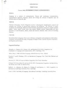
One way to assess the thoracic deformity and especially the RH is the use of the convex/concave rib-hump index - the double rib contour sign (DRCS) in lateral spinal radiographs. The RI is calculated by the ratio of distances d1/d2, where d1 is the distance between the most extended point of the most extending rib contour and the posterior margin of the corresponding vertebra on the lateral scoliosis films, while d2 is the distance from the least projection rib contour and the posterior margin of the same vertebra. The rib-index is the ratio d1/d2 [5]. Figure 1. The DRCS and the RI were introduced for AIS aetiological reasons. Figure 1. The DRCS and the rib index
However the scoliotic children & their parents are very much concerned about the trunk deformity (TD). One of the TD components is the rib hump (RH), which mainly expresses the rib (thoracic) abnormality. The brace treatment aims not only to stop the progression or correct the scoliotic curve of the central axis - the spine - but also the TD in the thorax - the RH. The RI can be used in every curve with a thoracic component, as in
28
th Annual Meeting & Course • Scoliosis Research Society
Half-Day Course: Non-Operative Spinal Deformity Treatment Techniques The DRCS and the RI were initially used for clinical reasons, for the assessment of rib hump deformity correction in the operative management of AIS with or without costoplasty [20]. In this publication it was noted that the RI is used due to its simplicity and to the ability to be calculated on the lateral spinal films with no need for special imaging or additional exposure to radiation. The application of RI method for the assessment of thoracic deformity - RH - in scoliotics using brace treatment was introduced in Chicago 2013 [21]. It was concluded that the RI based on DRCS could easily be used to assess any brace effectiveness on the rib hump deformity correction. Traditionally the early in-brace Cobb angle correction was used to predict the brace treatment outcome. The amount of reduction of the Cobb angle during the early treatment in-brace radiographs was used to predict the brace treatment result. Cobb angle reduction by 25 - 50 percent is reported necessary for a satisfactory outcome [22, 23, 24, 25, 26, 27, 28,29,30,31,32]. Considering that the improvement of the thoracic deformity is as important as that of the spine, in parallel to the study of early in-brace Cobb angle reduction, it is suggested that it is worth measuring the initial in-brace RI correction as well. Further research may confirm that this RI study will be able to predict the brace treatment outcomes on the thoracic cage deformity correction.
Literature
1. Grivas T, Burwell R, Purdue M, Webb J, Moulton A: Segmental patterns of rib-vertebra angles in chest radiographs of children: changes related to rib level, age, sex, side and significance for idiopathic scoliosis. Clin Anat 1992, 5(4):272-288. 2. Grivas TB, Samelis P, Chadziargiropoulos T, Polyzois B: Study of the rib cage deformity in children with 10 degrees-20 degrees of Cobb angle late onset idiopathic scoliosis, using rib-vertebra angles – aetiologic implications. Stud Health Technol Inform 2002, 91:20-24 3. Burwell RG, Aujla RK, Freeman BJ, Dangerfield PH, Cole AA, Kirby AS, Polak FJ, Pratt RK, Moulton A: The posterior skeletal thorax: rib-vertebral angle and axial vertebral rotation asymmetries in adolescent idiopathic scoliosis. Stud Health Technol Inform 2008, 140:263-268. 4. Grivas TB, Burwell RG, Purdue M, Webb JK, Moulton A: A segmental analysis of chest radiographs of children. Changes related to spinal level, age, sex, side and significance for lung growth and scoliosis. J Anat 1991, 178:21-38. 5. Grivas TB, Dangas S, Polyzois BD, Samelis P: The double rib contour sign (DRCS) in lateral spinal radiographs: aetiologic implications for scoliosis. Stud Health Technol Inform 2002, 88:38-43.
6. Grivas TB, Vasiliadis ES, Mihas C, Savvidou O: The effect of growth on the correlation between the spinal and rib cage deformity: implications on idiopathic scoliosis pathogenesis. Scoliosis 2007, 14(2):11. 7. Kirby AS, Burwell RG, Cole AA, Pratt RK, Webb JK, Moulton A: Evaluation of a new real-time ultrasound method for measuring segmental rotation of vertebrae and ribs in scoliosis. Stud Health Technol Inform 1999, 59:316-320. 8. Burwell RG, Aujla RK, Cole AA, Kirby AS, Pratt RK, Webb JK, Moulton A: Relation of rib deformity to vertebral deformity in the transverse plane at the curve apex in preoperative adolescent idiopathic scoliosis (AIS): an ultrasound, radiological and surface study of pathomechanisms. In International Research Society of Spinal Deformities Symposium 2004. Edited by Sawatzky BJ. University of British Columbia; 2004::302-306. 9. Grivas TB, Burwell RG and Dangerfield PH. Body mass index in relation to truncal asymmetry of healthy adolescents, a physiopathogenetic concept in common with idiopathic scoliosis: summary of an electronic focus group debate of the IBSE. Scoliosis 2013, 8:10 doi:10.1186/1748-7161-8-10 10. Foley G, Aubin CE, Parent S, Labelle H, d’Astous J, Johnston C, Sanders J. Physical Significance of the Rib Vertebra Angle Difference and Its 3-Dimensional Counterpart in Early-Onset Scoliosis. Spine Deformity 1 (2013) 259 - 265 11. Mehta MH: the rib –vertebra angle in the diagnosis between resolving and progressing infantile scoliosis. JBJS, 1972, 54B, 230-243. 12. Kristmundsdottir F, Burwell R G, James J I: The rib-vertebra angles on the convexity and concavity of the spinal curve in infantile idiopathic scoliosis. CORR 1985, (201):205-209. 13. Tolo VT, Gilespie R: the characteristics of juvenile idiopathic scoliosis and results of its treatment. J Bone Joint Surg, 1978, 60-B:181. 14. Wojcic A S, Webb J K, Burwell R G: An analysis of the effect of the Zielke Operation on the rib cage of S-shaped curves in idiopathic scoliosis. Spine, 1990, 15(2):81-86. 15. Wythers DJ, Burwell RG, Webb JK, and Wojcik AS: 1992 . The segmental surface and rib deformity of progressive adolescent idiopathic scoliosis: a preoperative segmental appraisal suggesting aetiological factors in the lumbar spine. In 6th International Symposium on Surface Topography and Spinal Deformity, Hotel Estoril Eden, 19- 20 September 1990. ed. A. Alberti, B. Drerup, and E. Hierholzer (eds. ). Stuttgart: Gustav Fischer Verlag, pp. 119-135. 16. Thirlwall, A.S. 1991 The relation of King curve type to the surface and radiological deformity of preoperative scoliosis. Thesis: University of Nottingham
Half-Day Courses • September 19, 2013 •
Lyon, France
29
Half-Day Course: Non-Operative Spinal Deformity Treatment Techniques 17. Wemyss-Holden SA, Jacobs KJ, Burwell RG, McNeill, Polak FJ, Webb JK, and Moulton A, 1991 The rib cage in adolescent idiopathic scoliosis (AIS): a segmental anal ysis of rib-vertebra angles (RVAs) revealing crossed RVA asymmetry with aetiological implications. Proceedings of a Joint Meeting of the British Association of Clinical Anatomists and the American Association of Clinical Anatomists, Norwich, 8-1 1 July 1991. Clin. Anat. 18. Wemyss-Holden SA, Burwell RG, Polak FJ, Jacobs KJ, McNeill AS, Webb JK, Moulton A, Wojcik AS. Segmental evaluation of the surface and radiological deformity after CotrelDubousset (CD) instrumentation for King type II and III. adolescent idiopathic scoliosis (AIS): surgical and etiological implications. Acta Orthop Belg. 1992;58 Suppl 1:135-8. 19. Grivas TB, Burwell RG Purdue M, Webb JK, Moulton A: The rib cage deformity in idiopathic scoliosis – the funnel shaped upper chest I relation to specific rotation as a prognostic factor. An evaluation of chest shape in progressive scoliosis and control children during growth. Surface Topography and Spinal deformity VI, Alberty, Drerup, Hierholzer (ed). Gustav Fischer Verlag, Stuttgart, Jena, New York, 1992, pp.93-109. 20. Lykissas MG, Sharma V, Crawford AH. Assessment of Rib Hump Deformity Correction in Adolescent Idiopathic Scoliosis With or Without Costoplasty Using the Double Rib Contour Sign. J Spinal Disord Tech. 2012 Sep 28. [Epub ahead of print]
26. Katz DE, Durrani AA. Factors that influence outcome in bracing large curves in patients with adolescent idiopathic scoliosis. Spine 2001;26:2354-2361. 27. Katz DE, Richards S, Browne RH, et al. A comparison between the Boston brace and the Charleston bending brace in adolescent idiopathic scoliosis. Spine 1997;22:1302-1312. 28. Katz «Experts debate role of Cobb angle in scoliosis bracing”, Biomechanics Magazine May, 2006 29. Landauer F, Wimmer C, Behensky H: Estimating the final outcome of brace treatment for idiopathic thoracic scoliosis at 6-month follow-up. Pediatr Rehabil 2003, 6(3-4):201-7. 30. Clin J, Aubin CE, Sangole A, Labelle H, Parent S: Correlation between immediate in-brace correction and biomechanical effectiveness of brace treatment in adolescent idiopathic scoliosis. Spine 2010, 35(18):1706-13. 31. Weiss HR, Rigo M: Expert-driven Chκneau applications: description and in-brace corrections. Physiother Therory Pract 2011, 27(1):61-67. 32. Zaina F, Donzelli S, Lusini M, Negrini N Correlation between in-brace radiographic correction and short time brace results. Zaina et al. Scoliosis 2012, 7(Suppl 1):O27, http://www.scoliosisjournal.com/content/7/S1/O27
Notes
21. Grivas TB, Triantafyllopoulos G, Mazioti C (2013): Assessment of early rib hump deformity correction in adolescent idiopathic scoliosis treated with a dynamic derotation brace using the double rib contour sign. 10th International Conference on Conservative Management of Spinal Deformities, 8th SOSORT Annual Meeting, Chicago, Illinois, USA Wednesday, May 8 through Saturday, May 11, Meeting Proceedings Book, page 37. 22. Rigo E, Negrini S, Weiss HR, Grivas TB, Maruyama T, KotwickiT, SOSORT consensus paper on brace action: TLSO biomechanics of correction (investigating the rationale for force vector selection. Scoliosis 2006, 1:11 doi:10.1186/1748-7161-1-11 23. Emans JB, Kaelin A, Bancel P, Hall JE, Miller ME. The Boston bracing system for idiopathic scoliosis: follow-up results in 295 patients. Spine 1986;11:792–801. 24. Emans, Brace Treatment of Idiopathic Scoliosis: What makes sense in the New Millennium Spine: State of the Art Review, Vol. 14, No.1, January 2000. 25. Rowe, et al SRS Natural History Committee .A MetaAnalysis of the Efficacy of Non-Operative Treatment of AIS JBJS 79:664-74, 1997
30
th Annual Meeting & Course • Scoliosis Research Society