PROCEEDINGS OF WORLD ACADEMY OF SCIENCE, ENGINEERING AND TECHNOLOGY VOLUME 6 JUNE 2005 ISSN 1307-6884
HERMES System: a Virtual Reality Simulator for the Angioplasty Intervention Training Giovanni Aloisio, Lucio T. De Paolis, Luciana Provenzano, Lucio Colizzi, and Gianluca Pantile addition it is possible to have objective measures of surgical skill. Many minimally invasive procedures need to be learned by repetition; using a real cadaver, in case of a mistake, a given procedure cannot be repeated because the body organs are altered. Realism and real-time interactions are the essential features for surgery simulators in order to be used as training systems. The realism of the simulation strictly depends on the accuracy of the human tissue modelling and the use of force feedback devices. Therefore, the most critical issues in designing surgical simulators are accuracy - the simulator should generate visual and haptic sensations very close to the reality and efficiency - deformations must be rendered in real-time on the graphic display and the haptic device. Accuracy and efficiency are two opposite requirements; in fact, increased accuracy implies higher computational time and vice versa. So, it is necessary to find a trade-off according to the application. For surgery training, real-time visual and haptic feedback is more important than deformation accuracy. However, substantial differences between the real and the virtual deformations may lead to a wrong learning of the procedure [3], [4]. Our research objective is to build a computer-based training system for the simulation of the coronary stent implant procedure. In this work, we have mainly focused on the realtime constraint and on the accuracy of the interactions in the virtual environment rather than on the visual accuracy. The system we developed is provided with a haptic interface and a collision detection and response algorithm, designed exploiting the coronary stent implant features, it allows the user to interactively navigate in the artery and to produce a force feedback when a contact occurs. This work takes into account the results of the HERMES (HEmatology Research virtual MEdical System) Project and it is the first step in the progress of building a computer-based training system for the coronary stent implant simulation.
Abstract—One of the essential requirements in order to have a
realistic surgical simulator is real-time interaction by means of a haptic interface is. In fact, reproducing haptic sensations increases the realism of the simulation. However, the interaction need to be performed in real-time, since a delay between the user action and the system reaction reduces the user immersion. In this paper, we present a prototype of the coronary stent implant simulator developed in the HERMES Project; this system allows real-time interactions with a artery by means of a specific haptic device; thus the user can interactively navigate in a reconstructed artery and force feedback is produced when contact occurs between the artery walls and the medical instruments
Keywords—Collision Detection, Haptic Interface, Real-Time Interaction, Surgical Simulator.
V
I. INTRODUCTION
IRTUAL reality technology brings numerous advantages to the medical community including improved surgical training. Training on patients can to some extent be avoided by using live animals and human cadavers. However, with the continuously increasing speed of computers, surgical simulators are now being offered to hospitals as a means of improving training and reducing the costs of education. Some simulators are based on phantoms (e.g. plastic structures) and others are virtual reality computer based simulators [1], [2]. Although phantoms may provide realism with regard to tissue behaviour, computer based simulators will increasingly become more eligible as a training aid, especially due to their extensive range of educational features. By means of kind of simulator it is possible to model unusual and rare cases and to practise new procedures avoiding risk for real patients; in
Manuscript received May 18, 2005. Giovanni Aloisio is with the Center for Advanced Computational Technologies (CACT/ISUFI) - University of Lecce & SPACI Consortium (Southern Partnership for Advanced Computational Infrastructure), 73100 Lecce, Italy, (e-mail:
[email protected]). Lucio T. De Paolis is with the Center for Advanced Computational Technologies (CACT/ISUFI) - University of Lecce & SPACI Consortium (Southern Partnership for Advanced Computational Infrastructure), 73100 Lecce, Italy, (e-mail:
[email protected]). Luciana Provenzano is with the Center for Advanced Computational Technologies (CACT/ISUFI) - University of Lecce & SPACI Consortium (Southern Partnership for Advanced Computational Infrastructure), 73100 Lecce, Italy, (e-mail:
[email protected]). Lucio Colizzi is with the Consorzio CETMA, 72100 Brindisi, Italy, (email:
[email protected]). Gianluca Pantile is with the Consorzio CETMA, 72100 Brindisi, Italy, (email:
[email protected]).
PWASET VOLUME 6 JUNE 2005 ISSN 1307-6884
II. RELATED WORKS Building a surgical simulator includes activities such as real-time rendering of dynamic models, real-time collision detection among deformable objects, real-time physical modelling and force computation, real-time interactions using haptic interfaces. Several simulators have been developed for training on a specific procedure, e.g. endoscopy, angiography, cardiology [5], [6].
221
© 2005 WASET.ORG
PROCEEDINGS OF WORLD ACADEMY OF SCIENCE, ENGINEERING AND TECHNOLOGY VOLUME 6 JUNE 2005 ISSN 1307-6884
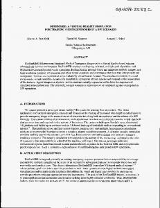
for coronary bypass surgery. The stent catheter is threaded into the artery and the stent is placed around a deflated balloon. When this is correctly positioned in the coronary artery, the balloon is inflated, expanding the stent against the walls of the coronary artery. The balloon catheter is then removed, leaving the stent in place to hold the coronary artery open. Fig. 1 shows the steps of this procedure.
Reference [7] presents an interventional cardiology training system conducted at Mitsubishi Electric Research Lab, in collaboration with CIMIT and the Massachusetts General Hospital. The simulator incorporates synthetic fluoroscopy, real-time three-dimensional interactive anatomy display, and coronary catheterization and angiography. The system simulates the physics and physiology of the human cardiovascular system; it is interfaced with a haptic interface that measures catheter translation and rotation and independently controlled servomotors that produce force and torque resistance. References [8], [9] present an experimental catheter insertion system supporting head-tracked stereoscopic viewing of volumetric reconstruction registered with direct haptic 3D interaction. The system takes as input patient data acquired with standard medical imaging modalities and regards it as a visual and haptic environment whose parameters are defined using look-up tables. By means of a mirror, the screen seems to be positioned like a surgical table giving the impression of looking down at the patient in a natural way. Co-registering physical and virtual spaces beforehand means that, the patient appears at a fixed physical position on the surgical table and inside the workspace of the PHANToM device which controls catheter insertion. During the insertion procedure the system provides perception of the force of penetration and positional deviation of the inserted catheter. Immersion Medical Corporation developed the CathSim intravascular catheterization simulator with a special-purpose haptic interface called AccuTouch. The CathSim system combines cognitive and motor skills training into an integrated learning experience. This simulator provides a variety of patient types encountered in real life and even creates complications. With the AccuTouch tactile feedback device, the student is able to feel the needle and catheter insertion into the skin and vein lumen [10]. The Swedish Company Mentice Corporation [11] is a supplier of several virtual reality based applications regarding minimally invasive surgery. An example of their products in the cardiology field is the VIST system. It allows a high realistic simulation for training in angiography and angioplasty. The system consists of an interface device, a computer and two displays – one for the fluroscopic image and one for the instructional system. The interface device is the virtual patient, with an introducer in place. Through this, the different real life tools and devices can be introduced.
Fig. 1 Steps of the coronary stent implant procedure
Therefore, a realistic stent implant simulator has to accurately model the anatomy of the human coronary artery and provide a real-time tissue-tools interaction and a fair force feedback by means of the haptic device. Fig. 2 shows the proposed architecture for the HERMES simulator. The user interacts with the simulator using the haptic interface. Data acquired from the haptic device sensors are used both to graphically represent the instruments and their positions in the virtual environment and to determine possible collisions between the virtual objects. Movements of the haptic device lead to changes in the virtual scene. force feedback
Haptic Control
Data from Sensors
Geometric Model
Haptic Interface Collision Detection forces
III. HERMES SIMULATOR ARCHITECTURE The coronary stent is a relatively new tool used to keep coronary arteries expanded. Balloon angioplasty is used in patients with coronary artery disease. The arteries become narrow and this disease reduces blood flow to the heart muscle. The primary cause of coronary artery disease is fat deposits blocking the arteries. By forming a rigid support, the stent can prevent restenosis and reduce the need
PWASET VOLUME 6 JUNE 2005 ISSN 1307-6884
USER
visual feedback
Collision Response
Rendering of Virtual Scene
Graphical Interface
Fig. 2 Architecture for the HERMES simulator
Collisions between virtual objects produce both forces, which are to be replicated on the user’s hand by the haptic interface, and virtual organs deformations, which are to be rendered by the graphical interface. In particular, the force computation and organ deformation strictly depends on the
222
© 2005 WASET.ORG
PROCEEDINGS OF WORLD ACADEMY OF SCIENCE, ENGINEERING AND TECHNOLOGY VOLUME 6 JUNE 2005 ISSN 1307-6884
collision detection algorithm. In particular, the collisions we are interested in are those occurring on the tip of the surgical instruments. As regards to collision detection the tip is a geometric point and the artery wall is a triangulated mesh. The artery is fixed in the virtual scene, whereas the tools move inside it. The tool motion is not pre-determined, that is the user can move the instruments in an unknown sequence of displacements. The virtual objects are rigid. Given the new position of the instrument tip and the set of triangles which form the artery, the aim of our collision detection algorithm is to calculate, for each simulation step, the exact contact point, determining if there is a not empty intersection between a segment and a triangle [13], [14], [15].
physical model which describes the mechanical properties of the virtual bodies. IV. HAPTIC RENDERING ALGORITHM The haptic rendering algorithm is made of two parts: collision detection and collision response. As a user manipulates the probe of the haptic device, the new position and the orientation of the haptic probe are acquired and collisions with the virtual artery walls are detected. If a collision is detects, interaction forces are computed using preprogrammed rules for the collision response and conveyed to the user through the haptic device. The collision detection and the collision response algorithms have to guarantee a stable interaction with the force feedback device [12].
A. Pre-Processing Broad Phase Since the virtual artery is considered a rigid body, we can exploit the pre-computed axis-aligned bounding box (AABB) subdivision to reduce the number of triangles checked for intersection at each simulation step, thus optimizing the collision detection algorithm [16]. In a pre-computation step, the AABBs for the artery are computed; each AABB encloses a strip of triangles, that is a subset of the triangles which forms the whole artery. The AABBs are not updated during the simulation, since the artery is fixed in the virtual space and does not deform over time. The output of this phase is a list of AABBs in which the triangulated artery has been divided up.
triangulated artery
Pre-processing Broad Phase list of AABBs
Broad Phase Test
list of triangles Narrow Phase Collision Detection
current position
Haptic Interface
B. Broad Phase Test At discrete time instants the information about the position of the tip, provided by the haptic interface, is used to determine the bounding box which contains the triangles which are most likely to collide with the tip. Only the triangles in the selected AABB are checked for exact collision at each simulation step.
current and previous positions
yes/no collision
C. Narrow Phase Collision Detection In the Narrow Phase Collision Detection step, the triangles in the selected bounding box are checked for exact intersection. It is worth noting that, at each simulation step, the narrow phase collision detection algorithm is provided with the current position of the tip and its position at the previous step, guaranteeing continuous collision detection.
Collision Response force feedback
Fig. 3 Force feedback in a real-time interaction
In the first version of the coronary stent implant simulator we addressed the real-time constraint, which imposes a very high frequency in contact determination and force computation. To achieve this aim, the virtual objects involved in the simulation (artery and surgical instruments) were considered as rigid bodies. The system provides the user with a haptic interface which realistically reproduces the real surgical tools, in order to feel the forces generated during the interaction. In fact, force feedback is the main requirement for the cardiologist who executes this surgical procedure. Fig. 3 shows the steps followed by our simulator to provide force sensations generated during a real-time interaction with an artery. We exploited stent implant features in order to speed up the
PWASET VOLUME 6 JUNE 2005 ISSN 1307-6884
q
q q
p p
p
r
r
r
a) p in a vertex
b) p on an edge
c) p in a face
Fig. 4 Segment-triangle classification
The collision detection algorithm solves a segment in the triangle problem. In particular, the intersection test between the segment and the triangle is performed as follows. Let T be
223
© 2005 WASET.ORG
PROCEEDINGS OF WORLD ACADEMY OF SCIENCE, ENGINEERING AND TECHNOLOGY VOLUME 6 JUNE 2005 ISSN 1307-6884
To estimate the computational time of the “segmenttriangle” procedure in the worst case (that is no collision) the tests have been performed varying the number of triangles in input. Tests have been executed on a PC Pentium IV 2,4 GHz, 512 MB RAM. The results of these tests are shown in Fig. 6. Furthermore, the computational time to determine the contact between the segment and the triangle when an endpoint of the segment coincides with a vertex of the triangle is 4 µs. When the intersection point is in the relative interior of a face of the triangle, the computational time is 6 µs. Considering AABBs which include 300 triangles, the performance of the segment-triangle procedure is about 1 KHz. The exact collision detection procedure guarantees the real time interactive rate required by the haptic device.
the triangle and qr the segment, where q is the endpoint corresponding to the current position and r the endpoint relating to the previous one. The first step is to determine whether the segment qr intersects the plane containing T. If so, the collision detection algorithm uses the intersection point p between the segment qr and the plane containing the triangle T, to classify the relationship between p and T, as shown in Fig. 4. If a contact is found, the algorithm also computes the angle of incidence (between the segment and the triangle plane) and the penetration distance. These data are given to the collision response algorithm to compute the force to be replicated by the haptic interface on the user’s hand.
D. Collision Response
V. HERMES HAPTIC DEVICE
Since the virtual objects involved in the simulation are supposed rigid, the computation of the force to be replicated by the haptic interface is based on the penetration distance. In particular, this force is proportional to the penetration distance and the angle of incidence, as shown in Fig. 5.
q
An interactive environment should be able to get the result of the manipulation like a visual-kinaesthetic interaction, very similar to the eye-hand coordination required in the real situation. Moreover, the haptic device has to be able to reproduce, without distortion, the sensations associated with the interaction in the virtual environment; the workspace has not been reduced by mechanical constraints [17], [18] . In order to achieve realism in the simulation, no commercially available haptic device has been used, but the interface has been planed ad hoc for the coronary stent implant simulation. The HERMES haptic interface has been designed and built at the PERCRO Laboratory of Scuola Superiore S. Anna of Pisa, Italy; this device reproduces the real shape and dimension of the surgical tools used in the stent implant, allowing the user to interact in a more natural way. This device is provided with two degrees of freedom controlled by means of motors that produce force and torque resistance. Fig. 8 shows the HERMES haptic interface and in Fig. 7 is reproduced the scheme of this device.
q
q r
r
r
a) no penetration
b) penetration
c) maximum
Fig. 5 Examples of penetration distance
Com putational tim e (us)
In order to have real-time interactions with the reconstructed artery using the haptic interface, the narrow phase collision detection algorithm has to be executed at least 300 Hz (that is the frequency required to assure a stable interaction with the haptic device). We focused solely on the performances of this algorithm because it is executed at each simulation step.
100 80 60 40 20 0 50
100
150
200
300
Number Triangles Fig. 6 Computational time for the “segment-triangle” procedure (no collision)
PWASET VOLUME 6 JUNE 2005 ISSN 1307-6884
Fig. 7 HERMES haptic device
224
© 2005 WASET.ORG
PROCEEDINGS OF WORLD ACADEMY OF SCIENCE, ENGINEERING AND TECHNOLOGY VOLUME 6 JUNE 2005 ISSN 1307-6884
the virtual scene [21]. Before beginning the simulation, the user has to carry out the calibration of the system and to this end the software interface is provided with a control panel that allows the user to modify some parameters in order to configure the haptic device in the correct way and to modulate its sensibility. As far as the 3D reconstruction of the virtual scene, the software allows the on-line and the off-line operating modalities. The on-line modality is the operating one, where the haptic device is active and connected to the workstation; the off-line is the non-operating one, where the haptic device is not connected to the system. The user can select two types of sight that correspond to different positions of the virtual camera; it is possible to choose between an inner sight of the artery, similar to a classic endoscopy, and an external sight which permits viewing of the arterial map near to the point of interest. All the phases of the coronary stent implant are simulated. Fig. 9 shows two different details of the graphical interface.
1st dof E1
E2
M1
M2
2nd dof Fig. 8 Scheme of the HERMES haptic interface
The system responds to the following user applied forces: the longitudinal forces in the form of push and pull movements; the torque forces in the form of twisting around the longitudinal axis. The device can generate any combination of the two following components of force: an axial force directed along the cylinder axis; a torsional moment directed along the cylinder axis. The implemented mechanical system is a hybrid solution. The translational degree of freedom is actuated by an electrical motor (M2) fixed to the chassis and a tendon transmission with both a motor and an idle pulley; the rotational degree of freedom is directly actuated by an electrical motor (M1) fixed to a slide flowing on a linear guide. Each motor has an optical encoder (E1 and E2) and the encoder’s position signals are used to realize the position and speed controls of the two joints [19]. Data acquired from haptic device sensors are used to represent the surgical instrument position in the virtual environment and to determine possible collisions between the virtual objects. VI. VIRTUAL ENVIRONMENT Computed tomography and magnetic resonance cannot be used to build the anatomic representation of the coronary arteries because of the heartbeat. For this reason, the virtual arteries model is constructed using anatomical models described in medical literature and refined in collaboration with cardiologist in order to ensure accuracy. The system allows students to practice the stent implant using different case studies, corresponding to virtual patients, each of them exhibits a particular difficulty depending on the different artery geometry and the number and type of stenosis. The rendering software has been realized on a LINUX platform, using MOTIF for the building of the graphic user interface and OpenGL Performer as a rendering engine for the virtual reconstruction of the 3D scene [20]. The software architecture is based on a classic multiprocess application. In particular, it consists of three processes: the first one is dedicated to the management of the events generated from the interaction of the user with the GUI, the second one concerns the management and the data exchange between the haptic device and the virtual environment and the third process concerns the rendering of
PWASET VOLUME 6 JUNE 2005 ISSN 1307-6884
Fig. 9 Details of the HERMES graphical interface
225
© 2005 WASET.ORG
PROCEEDINGS OF WORLD ACADEMY OF SCIENCE, ENGINEERING AND TECHNOLOGY VOLUME 6 JUNE 2005 ISSN 1307-6884
VII. CONCLUSIONS AND FUTURE WORK
[9]
In this paper the results of the HERMES Project are presented and a first attempt to interact with a reconstructed artery is described. We mainly focused on the real-time constraint and on the accuracy of the interactions in the virtual environment rather than on visual accuracy. In order to perform a realistic training on the coronary stent implant procedure a haptic interface is used, but not by means of a commercially available haptic device; an ad hoc interface has been designed and built for this kind of surgical operation. The HERMES haptic interface is provided with two degrees of freedom and the simulation guarantees real-time interactions. Collisions between virtual objects produce both forces, which are to be replicated on the user’s hand by the haptic interface, and virtual organs deformations, which are to be rendered by the graphical interface. A test phase in collaboration with cardiologists will be necessary to validate the system and to improve, by tuning some parameters of the system, the accuracy of the simulation. An improvement could be obtained by building a virtual environment which is more similar to that which is seen by the cardiologist during the real surgical procedure. Furthermore, the realism of the virtual scene needs improving; the characteristics of the real heart - contraction and beating should be represented. The simulator developed within the HERMES Project considers the human organs and the surgical instruments involved in the stent implant procedure rigid; since the artery is a visco-elastic body and the surgical instruments are flexible devices they should be modelled as deformable bodies, in order to have a more realistic haptic and visual feedback. A study using the FEM to model the artery as a deformable organ is in progress.
[10] [11] [12]
[13] [14] [15] [16] [17] [18] [19] [20] [21]
A. Zorcolo, E. Gobbetti, P. Pili, M. Tuveri, “Catheter Insertion Simulation with Combined Visual and Haptic Feedback”, Proc. First PHANToM Users Research Symposium, Heidelberg, Germany, 1999. M. Urbino, J. Tasto, B. Nguyen, Cunningham R. Merrill G., “CathSim: an Intravascular Catheterization Simulator on a PC”, Proc. Medicine Meets Virtual Reality Conf., IOS Press, Amsterdam, pp. 360-366, 1999. Mentice Corporation, Sweden, http://www.mentice.com A. Gregory, M. C. Lin, S. Gottschalk, R. Taylor, “Fast and Accurate Collision Detection for Haptic Interaction Using a Three Degree-ofFreedom Force-Feedback Device”, Computational Geometry: Theory and Applications, vol. 15, pp. 1-3, 2000. R. Bridson, R. Fedkiw, J. Anderson, “Robust Treatment of Collisions, Contact and Friction for Cloth Animation”, ACM Trans. Graphics, vol. 21, no. 3, pp. 594-603, 2002. J. Lombardo, M. P. Cani, F. Neyret, “Real-time Collision Detection for Virtual Surgery”, Proc. Computer Animation ’99, pp. 33-39, 1999. G. Van den Bergen, “Collision Detection in Interactive 3D Environments”, Elsevier Morgan Kaufmann Publishers, San Francisco, 2004. A. Watt, F. Policarpo, “3D Games: Real-time Rendering and Software Technology”, Addison Wesley Publishing Company, 2001. B. Jackson, L. Rosenberg, “Force Feedback and Medical Simulation”, Interactive Technology and the New Paradigm for Health-Care, chapter 24, IOS Press, pp.147-151, 1995. J. Davanne, P. Meseure, C. Chaillou, “Stable haptic interaction in a dynamic virtual environment”, Proc. Intelligent Robots and Systems Conf, Lusanne, Switzerland., 2002. Aloisio G., Bergamasco M et al., “Computer-Based Simulator for Catheter Insertion Training”, Medicine Meets Virtual Reality 12 , J.D. Westwood et al. (Eds.), IOS Press, vol. 98, pp. 4-6, 2004. D. Shreiner, M. Woo, J. Neider, T. Davis, “OpenGL Programming Guide: The Official Guide to Learning OpenGL”, 4th Edition, AddisonWesley Publishing Company, 1994. L.Y. Pao, D.A. Lawrence, "Synergistic Visual/Haptic Computer Interfaces" Proc. Japan/USA/Vietnam Workshop on Research and Education in Systems, Computation, and Control Engineering, Hanoi, Vietnam, pp. 155-162, 1998.
REFERENCES [1] [2] [3] [4] [5] [6] [7]
[8]
P. J. Gorman, A. H. Meier, T. M. Krummel, “Simulation and Virtual Reality in Surgical Education”, Archives of Surgery , vol. 134, pp. 12031208, 1999. S. L. Dawson, J. A. Kaufman, “The Imperative for Medical Simulation”, Proc. of the IEEE: Special Issue on Virtual & Augmented Reality in Medicine, pp. 479-483, 1998. N. Ayache, S. Cotin, H. Delingette, “Surgery Simulation with Visual and Haptic Feedback”, Robotics Research, Shirai and Hirose (eds.), Springer, Santa Clara, pp. 311-316, 1998. H. Delingette, “Towards Realistic Soft Tissue Modelling in Medical Simulation”, IEEE Special Issue on Surgery Simulation, pp. 512-523, 1998. B. Geiger, R. Kikinis, “Simulation of endoscopy”, Computer Vision, Virtual Reality and Robotics in Medicine, Lecture Notes in Computer Science, vol. 905, 1995. G. Abdoulaev, S. Cadeddu, G. Delussu et al., “ViVa: The Virtual Vascular Project”, IEEE Transactions on Information Technology in Biomedicine, vol. 2, No. 4, pp. 268-274, 1998. S. L. Dawson, S. Cotin, D. Meglan, D. W. Shaffer, M. A. Ferrell, “Designing a Computer-Based Simulator for Interventional Cardiology Training”, Catheterization and Cardiovascular Interventions, Wiley Press, vol. 51, pp. 522-527, 2000. E. Gobbetti, P. Pili, A. Zorcolo, M. Tuveri, “Interactive Virtual Angiography”, IEEE Computer Society Press, pp. 435-438, 1998.
PWASET VOLUME 6 JUNE 2005 ISSN 1307-6884
226
© 2005 WASET.ORG