International Journal of Computer Theory and Engineering, Vol. 7, No. 2, April 2015
Hierarchical Region Based Template Matching Technique for Global Motion Reduction of Coronary Cineangiograms K. A. S. H. Kulathilake, L. Ranathunga, G. R. Constantine, and N. A. Abdullah
within the sequence of frames in CCA due to the motion artifacts [5] (see Fig. 1). Therefore, we aim to develop a mechanism that can reduce the motion artifact in the CCA to obtain visual alignment of the vessel structures among the CCA frames. Our proposed method introduces a Hierarchical Region based Template Matching (HRTM) technique to reduce the global motion from the CCA to maintain the visual alignment among the Coronary Arteries shown in the frames. The rest of the paper is organized as follows: The recent research findings related to the motion compensation of Angiography are explained in Section II. The methodology of the proposed technique is described in Section III. Experimental results of the method are reported in Section IV, followed by discussion on the results. Finally, the conclusion section briefly explains the future work that can be extended using the results of this proposed technique.
Abstract—The Coronary Cineangiogram (CCA) is an invasive medical image modality which is used to determine the stenosis in the Coronary Arteries. The global motion occurring due to the heart beat makes great disturbance to obtain the visual alignment among the vessel structure shown in the CCA frames. Therefore, the recorded vessel structure’s position in CCA varies within the frame sequence. This paper describes a hierarchical region based template matching technique to reconstruct the CCA by reducing the global motion artifacts. This proposed motion reduction technique is efficient and it reconstructs the CCA by reducing the background motion as desired. Experimental results of this method have shown its’ ability to maintain the visual alignment of the internal blood flow among the frames. Index Terms—Angiography, computer aided diagnosis, image motion analysis, medical diagnostic imaging component.
I. INTRODUCTION II. BACKGROUND
Coronary Angiogram is one of the invasive medical image modalities used by the clinical practitioners as a preliminary diagnostic technique to detect luminal obstruction or the degree of stenosis. Coronary Angiograms are produced either as still images or as videos (Cineangiogram) [1]. The diagnosis based on Coronary Angiogram is subjective because, the results are produced through the visual judgments by considering the contrast agent flow. Hence, in most cases, these assessments fail due to the visual hindrance and lack of quantification capabilities not only in image based Coronary Angiograms but also Coronary Cineangiograms (CCA) [2], [3]. It is desirable to do an objective assessment of detected stenosis based on the functional significance of Coronary Arteries such as flow rate and flow velocity recorded in the CCA [1], [4]. However, the motion artifact occurs due to the heart beat (global motion) and patient movement makes some visual disturbances to do the objective assessments using CCA. Further, it is difficult to envisage the visual alignment among the vessel structure recorded in the frame sequence of CCA because, the position of the vessel structure varies
This section briefly reviews some selected recent publications which explain some research attempts to estimate and eliminate the motion artifacts from Coronary Angiography. Scale Invariant Feature Transform (SIFT) based motion estimation and video stabilization technique was explained in [5]. In this study, the SIFT was used to estimate the translational distance and direction between two frames and the motion compensated video was created using those estimations. Image registration based motion compensation in Angiography has been reported in many research publications. Kumar and team has explained the application of image registration techniques for motion correction in Digital Subtraction Angiograms [6]. As the image registration techniques; a light weight modified demons method and a constraint-based inverse consistence image registration method has been applied. Meijering et al. have also applied the image registration technique for Coronary Angiograms to produce motion compensated Angiograms effectively and efficiently [7]. Ko, Mao and Sun have introduced a multiresolution based image registration approach and their method automatically registers the arterial structures in the areas of interest selected from a pair of sequential images [8]. Further, it provides a sub pixel precision in registration [8]. Reference [9] discusses about an intensity based medical image registration tool box which consists of a collection of algorithms that are commonly used to solve medical image registration problems. Meunier and team proposed optical flow based method to compute the regional epicardial deformation from CCAs [10]. The proposed algorithm can track the motion of the Coronary
Manuscript received March 18, 2014; revised May 14, 2014. This investigation received financial support from the National Science Foundation, Sri Lanka under Grant No. NSF/SCH/2013/06. K. A. S. H. Kulathilake and L. Ranathunga are with the Department of Information Technology, University of Moratuwa, Moratuwa, Sri Lanka (e-mail:
[email protected],
[email protected]). G. R. Constantine is with the Department of Clinical Medicine, Faculty of Medicine, P.O. Box 271, No 25, Kynsey Road, Colombo 08, Sri Lanka (e-mail:
[email protected]). N. A. Abdullah is with the Department of Computer System and Technology, Faculty of Computer Science & Information Technology Building, University of Malaya, 50603 Kuala Lumpur, Malaysia (e-mail:
[email protected]).
DOI: 10.7763/IJCTE.2015.V7.948
156
International Journal of Computer Theory and Engineering, Vol. 7, No. 2, April 2015
Arteries as a whole and quantify the two dimensional deformation of the epicardial surface locally. Zheng and
Weirong also analyzed the Arterial dynamics using an optical flow based technique and elastic registration [11].
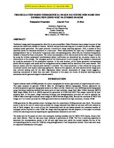
Fig. 1. Motion artifacts in CCA; Catheter engaged area is marked using whit colored square and the placement of it varies in the consecutive frames shown in a,b,c and d, as a result of the global motion. The distribution of contrast agent is gradually increasing in all images and it is denoted as local motion.
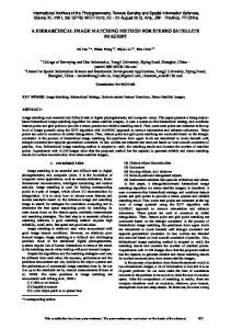
origin to obtain the cutoff frequency, D(u,v) is the radial distance from the origin and n is the order. After the filtering, inverse DFT has been applied to transform the image from frequency domain to the spatial domain. Then the illuminated component is extracted from the processed image to obtain the uniformly illuminated image. The processed image is normalized to obtain the best quality contrast among the vessel structures shown in each frame. Fig. 2 depicts the original CCA frame and the respective preprocessed CCA frame for further clarification.
III. METHODOLOGY In order to achieve the vessel alignment within the frame sequence of CCA, the global motion must be calculated and reduced from each frame of the CCA. There are mainly four steps available in our proposed HRTM motion reduction technique: preprocessing, motion estimation, frame alignment and motion eliminated video creation. An original CCA of frame rate 15 fps is input to the proposed method and it produces a global motion compensated CCA as the output. A. Preprocessing It was revealed that the CCA frames consist of noise and nonuniform illumination problem [3], [5]. As a result of that, the visual quality of the recorded CCA is degraded. Further, the illumination variations provide some incorrect results in template matching procedures. Therefore, the objective of the preprocessing stage is to apply possible image enhancement techniques to obtain the required visual quality of the vessel structures recorded in CCAs. A median filter with kernel size 3×3 was applied to the CCA frames as a noise removal technique. Afterwards a homomorphic filter was applied to extract the nonuniform illumination component of the CCA [12]. An image (P) with spatial coordinates (x,y) consists of illumination (i) and reflectance (r) components and it can be expressed by taking the natural logarithmic as follows;
P x, y ln i x, y ln r x, y
Fig. 2. Preprocessing (a) Original CCA frame with nonuniform illumination (b) Uniformly illuminated CCA frame.
B. Motion Estimation Motion estimation process of the proposed method is based on template matching image processing technique and mainly consists of two phases namely; (i) Template selection and (ii) applying HRTM. Following section explains those two phases briefly. Template selection is done as an interactive activity using the first frame of the input CCA to be processed. The user can arbitrarily select a best featured area as a template and this selected template is located at Fx,y coordinate point as shown in Fig. 3. It has w pixel width and h pixel height. As the next step, the selected template is saved as a sub image and this template image is used in subsequent template matching steps of the proposed method. All the template matching steps contain in the proposed method totally depend on selected template and its visual contents. Therefore, it is possible to select a template from the initial CCA frame which contains a good feature to match in subsequent frames. Based on the empirical results and strong observations of CCA, it is recommended to select a template around the catheter
(1)
We applied the Discrete Fourier Transform (DFT) to the logarithmic image and as a result of that, the illumination components of the image can be easily identified through the low frequency content in the frequency domain because the illumination is considered as a slowly varying pattern in an image. Afterwards, the butterworth low pass filter was applied to extract the low frequency components from the frame to be processed and the transfer function of the butterworth low pass filter is given in (2);
S (u, v)
1 1 D u, v / D0
2n
(2)
where, S(u,v) is the result image, D0 is the distance from 157
International Journal of Computer Theory and Engineering, Vol. 7, No. 2, April 2015
engaged area visualized in the first frame of selected CCA, as the best featured area to match during the matching step.
following threshold function is applied on R(x,y) image to obtain a binary image of it; if (0.94