RESEARCH ARTICLE
High Prevalence of Schistosoma japonicum and Fasciola gigantica in Bovines from Northern Samar, the Philippines Catherine A. Gordon1,2*, Luz P. Acosta3, Geoffrey N. Gobert1, Mario Jiz3, Remigio M. Olveda3, Allen G. Ross4, Darren J. Gray1,2,5, Gail M. Williams6, Donald Harn7, Yuesheng Li1, Donald P. McManus1* 1 Molecular Parasitology Laboratory, Infectious Diseases Division, QIMR Berghofer Institute of Medical Research, Brisbane, Australia, 2 Infectious Disease Epidemiology Unit, School of Population Health, University of Queensland, Brisbane, Australia, 3 Department of Immunology, Research Institute of Tropical Medicine, Manila, Philippines, 4 Griffith Health Institute, Griffith University, Brisbane, Australia, 5 Research School of Population Health, the Australian National University, Canberra, Australia, 6 Discipline of Epidemiology and Biostatistics, School of Population Health, University of Queensland, Brisbane, Australia, 7 University of Georgia, College of Veterinary Medicine, Athens, Georgia, United States of America *
[email protected] (CAG);
[email protected] (DPM)
OPEN ACCESS Citation: Gordon CA, Acosta LP, Gobert GN, Jiz M, Olveda RM, Ross AG, et al. (2015) High Prevalence of Schistosoma japonicum and Fasciola gigantica in Bovines from Northern Samar, the Philippines. PLoS Negl Trop Dis 9(2): e0003108. doi:10.1371/journal. pntd.0003108 Editor: Akira Ito, Asahikawa Medical College, JAPAN Received: March 19, 2014 Accepted: July 10, 2014 Published: February 2, 2015 Copyright: © 2015 Gordon et al. This is an open access article distributed under the terms of the Creative Commons Attribution License, which permits unrestricted use, distribution, and reproduction in any medium, provided the original author and source are credited. Data Availability Statement: All relevant data are within the paper and its Supporting Information files. Funding: Funding was provided by the National Health and Medical Research Council (NHMRC) of Australia (Program grant ID 496600 and ID 1037304, and project grant ID 613671) and UBS Optimus. YL is an Australian Research Council (ARC) Future Fellow; DPM is an NHMRC Senior Principal Research Fellow; and DJG is an ARC Fellow (DECRA). The funders had no role in study design, data collection and analysis, decision to publish, or preparation of the manuscript.
Abstract The cause of zoonotic schistosomiasis in the Philippines is Schistosoma japonicum, which infects up to 46 mammalian hosts, including humans and bovines. In China, water buffaloes have been identified as major reservoir hosts for schistosomiasis japonica, contributing up to 75% of human transmission. In the Philippines, water buffaloes (carabao; Bubalus bubalis carabanesis) have, historically, been considered unimportant reservoirs. We therefore revisited the possible role of bovines in schistosome transmission in the Philippines, using the recently described formalin-ethyl acetate sedimentation (FEA-SD) technique and a qPCR assay to examine fecal samples from 153 bovines (both carabao and cattle) from six barangays in Northern Samar. A high prevalence of S. japonicum was found using qPCR and FEA-SD in both cattle (87.50% and 77.08%, respectively) and carabao (80.00% and 55.24%, respectively). The average daily egg output for each bovine was calculated at 195,000. High prevalence and infection intensity of F. gigantica was also found in the bovines by qPCR and FEA-SD (95.33% and 96.00%, respectively). The identification of bovines as major reservoir hosts for S. japonicum transmission suggests that bovine treatment and/or vaccination, as one becomes available, should be included in any future control program that aims to reduce the disease burden due to schistosomiasis in the Philippines.
Author Summary Schistosomiasis japonica, a zoonosis of over 40 different mammalian species, is endemic to China, the Philippines and Indonesia. In China, water buffaloes have been shown to be major reservoir hosts, while in the Philippines, the smaller sub species (carabao) has been
PLOS Neglected Tropical Diseases | DOI:10.1371/journal.pntd.0003108
February 2, 2015
1 / 13
High Prevalence of S. japonicum and F. gigantica in the Philippines
Competing Interests: The authors have declared that no competing interests exist.
been considered unimportant in transmission, possibly due to the lack of sensitive coproparasitological techniques employed. We used an exhaustive microscopic technique, the FEA-SD, and a sensitive qPCR assay on a cohort of bovines to assess their potential role in transmission in the Philippines. Both cattle and carabao were highly infected with Schistosoma japonicum and Fasciola gigantic and co-infection was common. The high prevalence and intensity of bovine infection with S. japonicum suggest their heavy involvement in human transmission and that future control programs should target these reservoirs to reduce human infection.
Introduction Schistosoma japonicum, the cause of zoonotic schistosomiasis, infects more than 40 species of wild and domestic animals (including bovines, pigs, horses and goats) [1], complicating control efforts. Mathematical modelling predicts that up to 75% of S. japonicum transmission to humans is attributable to bovines in the lake and marshland areas of China [2]. This is due to the fact that infected water buffaloes and cattle excrete daily up to 60 kg of stool per individual [3–5]. With such a large volume of feces excreted daily, the potential number of eggs excreted is similarly high. This contrasts with rodents which excrete approximately 1 g of feces per day and humans which produce around 250 g daily [6]. Water buffaloes habitually spend much of their time immersed in water bodies, such as rivers, lakes and water holes, into which they tend to defecate directly, so that if Oncomelania hupensis are present, the likelihood of transmission is high. While extensive studies have been undertaken on reservoir hosts in China [4,5,7–10], there are limited reports on the zoonotic potential of schistosomiasis japonica in the Philippines. This is despite the 2.88 million carabao present in the Philippines [11] (Bubalis bubalis carabenensis), a smaller sub-species of the Chinese water buffalo. Previous reports from the Philippines had recorded only low S. japonicum prevalence in carabao, suggesting that these bovines play a limited role in transmission [12–15]. However, a study in Leyte, a province located in the Eastern Visayas region, reported a S. japonicum prevalence of 52% in carabao using a quantitative real time polymerase chain reaction (qPCR) method [16]. Much lower prevalence values were obtained using the Kato Katz (KK) method (4%), miracidial hatching test (MHT) (0%) and the Danish Bilharziasis Laboratory (DBL) technique (4%) [16], suggesting caution regarding the involvement of carabao in transmission of schistosomiasis japonica. The Leyte study also highlighted the need for a more sensitive copro-parasitological technique for comparison with the qPCR. Accordingly, in a pilot study conducted in Western Samar, located in the Eastern Visayas region of the Philippines, we recorded a high prevalence of S. japonicum in carabao using a validated real-time PCR (qPCR) and a new copro-parasitological tool, the formalin-ethyl acetate sedimentation (FEA-SD) technique [17,18]. A much lower prevalence of S. japonicum was recorded for the same fecal samples using conventional PCR, the Kato-Katz technique and MHT [17,18]. Here we report on a larger study in Palapag, a municipality in the province of Northern Samar where we determined the prevalence of S. japonicum in cattle and carabao. We also determined the prevalence of Fasciola gigantica in these bovines and investigated whether there is any cross-protective effect between this trematode species and S. japonicum. Fascioliasis in animals is a chronic disease and causes anaemia, lethargy, weight loss and lower fertility [19,20]. F. gigantica is the main causative agent of fascioliasis in the Philippines where it is the leading cause of bovine morbidity and mortality [20].
PLOS Neglected Tropical Diseases | DOI:10.1371/journal.pntd.0003108
February 2, 2015
2 / 13
High Prevalence of S. japonicum and F. gigantica in the Philippines
Materials and Methods Ethics Informed written consent was received from all animal owners in the study area and ethical approval for the animal work was provided by the Ethics Committee of the Research Institute of Tropical Medicine and the QIMR Berghofer Medical Research Institute Animal Research Ethics Committee (P288). This study was performed in accordance with the recommendations of the Australian code of practice for the care and use of animals for scientific purposes, 2004.
Study design We carried out a cross-sectional survey (July-September 2011) in the municipality of Palapag, Northern Samar Province, the Philippines, to determine the level of S. japonicum and F. gigantica infection in animals using the FEA-SD and qPCR methods. Primary endpoints were bovine prevalence and intensity of infection; secondary end points were sensitivity and specificity of the FEA-SD and qPCR techniques.
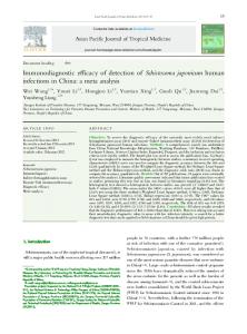
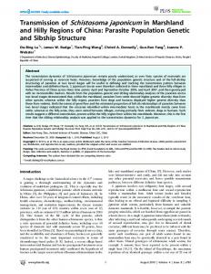
Study area The study was undertaken in six barangays; Napo, Capacujan, Matambag, Mabaras, Magsaysay and Manajao, all in the municipality of Palapag in Northern Samar Province (Fig. 1). Palapag
Figure 1. Map of the Philippines showing Northern Samar province highlighted red (Left). Map of the municipality of Palapag, showing barangay locations and rivers (Right). doi:10.1371/journal.pntd.0003108.g001
PLOS Neglected Tropical Diseases | DOI:10.1371/journal.pntd.0003108
February 2, 2015
3 / 13
High Prevalence of S. japonicum and F. gigantica in the Philippines
was chosen due to the known endemicity of the municipality from government control records. No praziquantel treatment of bovines for schistosomiasis had occurred in the area prior to the study. A total of 153 bovine samples (48 cattle, 105carabao) were collected for analysis for S. japonicum; 150 bovine samples (45 cattle, 105 carabao) were analysed for the presence of F. gigantica infection. Age and gender of bovines was ascertained by use of a questionnaire given to the animal owners prior to fecal collection. Bovines surveyed in this study came from 112 different households, as determined by a household questionnaire, however there are many communal areas (rice fields and rivers) where bovines from different households are co-held.
Study procedures Bovine owners were requested to bring their animals to a central area on a stated day for faecal collection. Fecal samples from these animals were collected intra-rectally by a team of local veterinarians in plastic, re-sealable bags, and brought back to the local Palapag medical center. Once there, samples were stored for later qPCR analysis and for processing of the FEA-SD technique. For molecular analysis approximately 3 g of feces was placed into 5 ml tubes with sufficient 80% (v/v) ethanol to completely cover the sample. Tubes were stored at 4°C transported at room temperature to the QIMR Berghofer laboratory in Brisbane where DNA was extracted from the samples and qPCR performed. The FEA-SD was performed in Palapag and is described in detail below.
FEA-SD The published FEA-SD method [17,18] was used with some modifications. Briefly, 50 g of homogenized bovine stool was washed through a 60 nylon mesh (Tyler scale with a pore opening size of 250 μm) onto a 40 nylon mesh (Tyler scale with a pore opening size of 40 μm). The material retained on the 40 nylon mesh was washed into a 50 ml tube, allowed to sediment for 30 minutes, the supernatant was removed, the pellet re-suspended in 10% formalin (v/v) (mixed with tap water) and the sedimentation procedure repeated twice more. Ten ml of the final suspension was removed, placing 5 ml into two 15 ml tubes labeled A and B. Ten percent formalin (v/v) was added to each tube to take the volume to 8 ml and mixed thoroughly, after which 4 ml of 100% ethyl acetate (v/v) was added. Tubes were vortexed and centrifuged at 500 g for 10 minutes. The ethyl acetate layer was removed by gently rimming the tube with an applicator stick and the top layers poured off. The pellet was washed once with tap water and re-suspended to 5 ml with 10% formalin (v/v) and 5 ml of 10% potassium hydroxide (w/v) (KOH) added. The tubes were vortexed and the samples allowed to digest at 37°C for at least 6 hours before centrifugation at 900 g for ten minutes. The supernatant was removed, the pellet washed once with water and re-suspended in water, mixing gently with a pipette prior to microscopic examination for eggs. The egg counts for S. japonicum were undertaken by Northern Samar regional staff and staff from the Research Institute of Tropical Medicine, Manila. Egg counts for F. gigantica were completed by one of the authors (CAG) at the QIMR Berghofer. F. gigantica eggs were measured to help distinguish from the morphologically similar eggs of paramphistomes (stomach fluke) which are also present in bovines in the Philippines [21]. Eggs over 160 μm in length were counted as Paramphistomum eggs, while those