JOURNAL OF BACTERIOLOGY, July 1996, p. 3846–3853 0021-9193/96/$04.0010 Copyright q 1996, American Society for Microbiology
Vol. 178, No. 13
Homologous Pairs of Regulatory Proteins Control Activity of Bacillus subtilis Transcription Factor sB in Response to Environmental Stress CHOONG MIN KANG, MARGARET S. BRODY, SAMINA AKBAR, XIAOFENG YANG, AND CHESTER W. PRICE* Department of Food Science and Technology, University of California, Davis, California 95616 Received 20 March 1996/Accepted 30 April 1996
In Bacillus subtilis, activity of the general stress transcription factor sB is controlled posttranslationally by a regulatory network that transmits signals of environmental and metabolic stress. These signals include heat, ethanol, or osmotic challenge, or a sharp decrease in cellular energy levels, and all ultimately control sB activity by influencing the binding decision of the RsbW anti-s factor. In the absence of stress, RsbW binds to sB and prevents its association with RNA polymerase core enzyme. However, following stress, RsbW binds instead to the RsbV anti-anti-s factor, thereby releasing sB to direct transcription of its target genes. These two principal regulators of sB activity are encoded in the eight-gene sigB operon, which has the gene order rsbR-rsbS-rsbT-rsbU-rsbV-rsbW-sigB-rsbX (where rsb stands for regulator of sigma B). Notably, the predicted rsbS product has significant amino acid identity to the RsbV anti-anti-s factor and the predicted rsbT product resembles the RsbW anti-s factor. To determine the roles of rsbS and rsbT, null or missense mutations were constructed in the chromosomal copies of each and tested for their effects on expression of a sB-dependent reporter fusion. On the basis of this genetic analysis, our principal conclusions are that (i) the rsbS product is a negative regulator of sB activity, (ii) the rsbT product is a positive regulator, (iii) RsbS requires RsbT for function, and (iv) the RsbS-RsbT and RsbV-RsbW pairs act hierarchically by a common mechanism in which key protein-protein interactions are controlled by phosphorylation events. we address the role of two of these additional genes, rsbS and rsbT, and their relationship to the previously identified regulators of sB. We report that the rsbS product (RsbS) functions as a negative regulator and the rsbT product (RsbT) functions as a positive regulator of sB activity, and that RsbS requires the presence of RsbT in order to exert its regulatory effects. Furthermore, we show that the RsbS-RsbT regulatory pair requires RsbV and RsbW activity in order to transmit stress signals to sB, and we present genetic evidence which suggests that both regulatory pairs function by a molecular mechanism involving modification by phosphorylation.
In response to stress and starvation signals, the gram-positive bacterium Bacillus subtilis expresses a large set of genes termed the general stress regulon, which is primarily under control of the alternative transcription factor sB (3, 8, 16, 19, 20, 24, 36). The activity of sB is itself controlled posttranslationally by a multicomponent signal transduction pathway which conveys signals of environmental stress, such as heat shock, osmotic stress, or ethanol challenge, and signals of metabolic stress, such as the drop in cellular energy levels that occurs upon challenge with uncouplers of oxidative phosphorylation or upon entry into the stationary growth phase (2, 4, 6, 7, 9, 34, 35). All of the known regulators in the sB signal transduction pathway are encoded by genes in the sigB operon, which also contains the sB structural gene (20, 37). As shown in Fig. 1, these genes are termed rsb, for regulator of sigma B. Previous genetic and biochemical data indicate that the RsbW anti-s factor is the primary regulator, binding directly to sB and maintaining it in a transcriptionally inactive complex (4, 5, 9). sB is released from this complex by the action of the RsbV anti-anti-s factor, which apparently sequesters RsbW by direct protein-protein interaction (4, 9, 13). Additional in vitro and in vivo experiments have demonstrated that the RsbW anti-s factor can associate with either sB or RsbV and that the binding decision of RsbW is controlled by the phosphorylation state of RsbV (1, 13). Recently, four additional genes with potential regulatory functions were discovered in the sigB operon, and the product of one of them, RsbU, was shown to be a positive effector of sB activity in response to environmental stress (34, 35, 37). Here
MATERIALS AND METHODS Bacterial strains and genetic methods. Escherichia coli DH5a was the host for all plasmid constructions; standard methods were used for PCR, restriction digests, ligations, and transformations of E. coli, as previously described (37). B. subtilis strains used are listed in Table 1. B. subtilis PB2 and its derivatives were made competent for transformation by the method of Dubnau and DavidoffAbelson (12), and the plasmids used in the various B. subtilis strain constructions are described below. Construction of point and deletion mutations within rsbS and rsbT. We used a four-primer system of site-directed mutagenesis (18) to make alterations within the cloned copies of the rsbS and rsbT genes. For the following constructions, nucleotide numbers refer to the DNA sequence shown in Fig. 2 of reference 37. We first made two point mutations within the rsbS coding sequence. For the S59A alteration (S-to-A change at position 59), we used pairs of primers to amplify two overlapping fragments from the chromosome. Primer SE1 (59-ATC GACATTCTGCTGCTG-39, complementary to nucleotides [nt] 505 to 522) was first used with primer SS59A-2 (59-CACTTTCGCGATAAATGCATCAATCA TATCCAC-39, corresponding to nt 1343 to 1376) to amplify the first fragment. Primer SS59A-2 bears a transition from A to C (underlined) that leads to substitution of alanine for serine at codon 59. Next, primer SS59A-1 (59-GTGG ATATGATTGATGCATTTATCGCGAAAGTG-39, complementary to nt 1346 to 1379) was used with primer SE2 (59-GTGGTAGTAATCACCGCT-39, corresponding to nt 2371 to 2388) to amplify the second fragment. The resulting two fragments were mixed and further amplified by using primers SE1 and SE2, yielding the S59A alteration on a 1,883-bp fragment. To construct the S59D
* Corresponding author. Phone: (916) 752-1596. Fax: (916) 7524759. Electronic mail address:
[email protected]. 3846
VOL. 178, 1996
HOMOLOGOUS PAIRS OF REGULATORS CONTROL B. SUBTILIS sB
3847
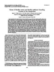
FIG. 1. Genetic organization of the sigB operon and current model of sB regulation. (Top) sigB lies in an eight-gene operon with seven other genes whose products posttranslationally regulate sB activity (4, 5, 9, 13, 20, 34, 37); these genes are termed rsb (for regulator of sigma B). The hatched rectangles denote the sequence similarity shared by the predicted products of rsbS and rsbV, whereas the shaded rectangles signify the resemblance of the predicted products of rsbT and rsbW (see Fig. 3). The sigB operon has two promoters, the sA-like promoter PA and an internal, sB-dependent promoter PB (20, 37). In order to conduct some of the experiments described here, we eliminated the normal autocatalytic induction of sigB expression by using a strain in which PB has been replaced by the inducible Pspac promoter (9, 20). Previous work has shown that transcription from PA into the downstream four genes of the sigB operon is not required for induction of sB activity in response to stress (37). Therefore, the Pspac construction would not be expected to disrupt sB regulation. Furthermore, most of the experiments described here used either point mutations or large, in-frame deletions in the rsb genes that would not be expected to have polar effects on expression of downstream members of the operon. (Bottom) Current model of the sB regulatory network, based on the available genetic and biochemical data. In this scheme, the RsbW anti-s factor is the primary regulator of sB activity, and its negative action is countered by the RsbV anti-anti-s factor. RsbW also has a kinase activity which can modify and inactivate its RsbV antagonist. The biochemical activities of the RsbX negative regulator and the RsbU positive regulator are presently unknown, but genetic analysis has shown that RsbX requires RsbU for function and that RsbU requires RsbV for function (34). This analysis places the RsbX and RsbU regulators upstream from the RsbV-RsbW pair in the signal transduction pathway.
alteration, we replaced primer SS59A-2 with primer SS59D-2 (59-CACTTTCG CGATAAAGTCATCAATCATATCCAC-39, in which the original codon for serine is replaced by an aspartate codon, underlined). For constructing a large, in-frame deletion mutation within rsbS, we used primer S3 (59-TTAGAGCAGGGGCTTGAG-39, complementary to residues 1508 to 1525) and primer S2 (59-CTCAAGCCCCTGCTCTAACGGGATTTTCGGAT GTCT-39, in which the 39 half corresponds to nt 1190 to 1207 and the 59 half is complementary to primer S3) with primers SE1 and SE2. This resulted in a construct in which 100 codons internal to the 121-codon rsbS gene are deleted.
For constructing a large, in-frame deletion mutation within rsbT, we used primer T3 (59-TCCGTAGCGGGGGAAGGA-39, complementary to nt 1907 to 1920) and primer T2 (59-TCCTTCCCCCGCTACGGAGGATTGGTCGTTCATTCG39, in which the 39 half corresponds to nt 1553 to 1570 and the 59 half is complementary to primer T3). This construct had 112 codons internal to the 132 codons of the rsbT gene removed. To make a double deletion of both rsbS and rsbT, we used primer T3 and primer ST2 (59-TCCTTCCCCCGCTACGGACGG GATTTTCGGATGTCT-39, in which the 39 half corresponds to nt 1190 to 1207 and the 59 half is complementary to primer T3) together with primers SE1 and
TABLE 1. B. subtilis strains used in this study Strain
Genotype
Reference or construction
PB2 PB198 PB206 PB212 PB213 PB218 PB244 PB423 PB424 PB454 PB458 PB462 PB469 PB470 PB473 PB478 PB486 PB489
trpC2 amyE::ctc-lacZ trpC2 rsbVD1 amyE::ctc-lacZ trpC2 Pspac (rsbV1 rsbW1 sigB1 rsbX1) amyE::ctc-lacZ trpC2 Pspac (rsbV1 rsbWD1 sigB1 rsbX1) amyE::ctc-lacZ trpC2 rsbX::ermC amyE::ctc-lacZ trpC2 rsbU::ermC amyE::ctc-lacZ trpC2 rsbTD1 amyE::ctc-lacZ trpC2 rsbSD1 amyE::ctc-lacZ trpC2 rsbTD1 Pspac (rsbV1 rsbWD1 sigB1 rsbX1) amyE::ctc-lacZ trpC2 rsbTD1 Pspac (rsbV1 rsbW1 sigB1 rsbX1) amyE::ctc-lacZ trpC2 rsbSD1 rsbTD1 amyE::ctc-lacZ trpC2 rsbSD1 rsbVD1 amyE::ctc-lacZ trpC2 rsbSS59A amyE::ctc-lacZ trpC2 rsbSD1 rsbU::ermC amyE::ctc-lacZ trpC2 rsbSS59D Pspac (rsbV1 rsbW1 sigB1 rsbX1) amyE::ctc-lacZ trpC2 rsbTD1 Pspac (rsbV1 rsbW1 sigB1 rsbX::ermC) amyE::ctc-lacZ trpC2 Pspac (rsbV1 rsbW1 sigB1 rsbX::ermC) amyE::ctc-lacZ trpC2
Wild-type Marburg strain 9 9 9 9 9 37 pCK1 3 PB198 pCK2 3 PB198 pAR7 (9) 3 PB423 pCK1 3 PB212 pCK4 3 PB198 pCK2 3 PB206 pCK5 3 PB198 pCK2 3 PB244 pCK6 3 PB212 pCK1 3 PB489 PB218 3 PB212
3848
KANG ET AL.
SE2. The 59 endpoint of this double deletion was therefore the same as that of the rsbS deletion, and the 39 endpoint was the same as that of the rsbT deletion. Each of these PCR-mutagenized products was then cloned into the pCP115 integration vector (29), resulting in plasmids pCK1 (rsbTD1), pCK2 (rsbSD1), pCK4 (D[rsbS-rsbT]1), pCK5 (rsbSS59A), and pCK6 (rsbSS59D). The relevant coding region of each of these plasmids was sequenced to ensure the presence of the desired alterations and the absence of any additional, PCR-generated mutations. We introduced these deletion and point mutations into the homologous copies of the rsbS and rsbT genes on the B. subtilis chromosome using the two-step allele replacement method of Stahl and Ferrari (31). Following plasmid insertion and excision from the chromosome of wild-type strain PB2, PCR amplification and direct DNA sequencing (Sequenase PCR Sequencing Kit; United States Biochemicals) confirmed the presence of the desired mutation on the chromosome of each recipient strain. To monitor sB activity in these mutant strains, we inserted a sB-dependent ctc-lacZ transcriptional fusion (9) in single copy at the amyE locus of each. Enzyme assays. Strains were grown in buffered Luria broth (LB) medium (7) until the cultures were in the exponential growth phase, at which time they were diluted 1:25 into fresh buffered LB medium. For stress experiments, NaCl was added to one of two parallel cultures in the early exponential phase, to yield a final concentration of 0.3 M. For stationary-phase experiments, samples were collected during both exponential and stationary phases. b-Galactosidase assays were done essentially as described by Miller (25). Cells were washed with Z buffer (25) and permeabilized by using sodium dodecyl sulfate and chloroform. Protein levels were determined with whole-cell samples by using the Bio-Rad Protein Assay reagent (Bio-Rad Laboratories, Richmond, Calif.). Activity was defined as DA420 3 1,000 per minute per milligram of protein.
RESULTS AND DISCUSSION In vivo function and hierarchical relationship of the RsbS and RsbT regulators of sB activity. In order to determine the possible regulatory roles of rsbS and rsbT, we made large inframe deletions of each gene and substituted them for the chromosomal copies by a two-step allele replacement technique (31). To provide an assay for sB activity, each of the mutant strains also carried a single-copy transcriptional fusion between the well-characterized sB-dependent ctc promoter and an E. coli lacZ reporter gene (9, 19, 27). sB is activated both by signals of environmental stress and by signals of energy depletion (2, 4, 6, 7, 9, 35, 36). Because distinct parts of the sB regulatory network are thought to be responsible for the differential transmission of these two classes of stress signals (35), we used addition of salt to exponentially growing cells to represent environmental stress and entry into stationary phase in rich medium to represent energy stress. As shown in Fig. 2, loss of rsbS function caused a dramatic increase in sB activity in exponentially growing cells, and this activity was not further increased by salt addition (Fig. 2A). Notably, the rsbS null mutant also manifested a small-colony phenotype. This small-colony phenotype was apparently due to a deleterious increase in sB activity, because colony size was restored to the wild type in an rsbS-sigB double null mutant (data not shown). On the basis of these results, we conclude that RsbS is formally a negative regulator of sB activity. In contrast, loss of rsbT function caused a complete loss of response to salt addition in exponentially growing cells (Fig. 2B). Therefore, RsbT is formally a positive regulator of sB activity in response to salt stress. Because at least one other positive regulator of sB activity—RsbU—affects response to environmental but not energy stress (35), we also tested the effect of the rsbT mutation on sB activity as cells entered stationary phase. As shown in Fig. 2C, loss of rsbT function had no effect on sB activity under these conditions. We conclude that loss of rsbT function has a dramatic effect on response to environmental stress and no substantial effect on response to energy stress. Because neither the rsbS nor the rsbT null mutation had any impact on expression of a sF-dependent spoIIIG-lacZ fusion (32) or a sH-dependent minCD-lacZ fusion (22), it appears that RsbS and RsbT specifically regulate sB (data not shown).
J. BACTERIOL.
We then determined that the RsbS regulator requires RsbT function in order for RsbS to exert its negative regulatory action on sB. With respect to the level of sB activity observed in exponentially growing cells both before and after salt stress, the phenotype of the rsbS-rsbT double mutant was the same as that of the rsbT single mutant (Fig. 2D). Furthermore, with respect to colony size, the rsbS-rsbT double mutant manifested wild-type colony morphology, the same as that of the rsbT single mutant, and not the small-colony phenotype characteristic of the rsbS null allele (data not shown). Therefore, loss of rsbT function completely overrides loss of rsbS function. The simplest interpretation of these results is summarized in the following genetic formula, in which RsbT acts as a positive regulator of sB activity, either directly or indirectly, and RsbS acts as a negative regulator of sB activity by countering the positive function of RsbT: RsbS O RsbT 3 sB. Notably, this model of RsbS-RsbT action closely resembles the mode of action of the sB regulators RsbV-RsbW (Fig. 1) and of the sF regulators SpoIIAA-SpoIIAB (30), but the RsbS-RsbT pair exerts opposite regulatory effects. That is, whereas RsbT is a positive regulator that activates sB, both RsbW and SpoIIAB are negative regulators that inhibit activity of their cognate s factors (2, 4, 5, 9, 15, 26, 30). Phenotypes associated with alterations within rsbS suggest that modification by phosphorylation is important for RsbS function. Inspection of the predicted amino acid sequences of the RsbV and SpoIIAA anti-anti-s factors reveals substantial sequence similarity between these proteins and RsbS (37) (Fig. 3A). Moreover, inspection of the predicted amino acid sequences of the RsbW and SpoIIAB anti-s factors reveals a resemblance between these proteins and RsbT in residues thought to be important for ATP binding and, therefore, for kinase function (Fig. 3B). In addition to these sequence similarities, the genetic model of RsbS-RsbT action resembles the mode of action of the RsbV-RsbW and the SpoIIAA-SpoIIAB protein pairs, each of which controls the activity of their cognate s factors by direct protein-protein interactions (1, 2, 5, 11, 13, 15). The binding ability of the anti-s factor in each system is substantially controlled by a phosphorylation event (2, 11, 13, 14, 23). In addition to their specific anti-s factor activity, the RsbW and SpoIIAB proteins each possess a kinase activity directed toward their respective antagonist proteins (2, 11, 13, 26). In the phosphorylated state, neither the RsbV nor the SpoIIAA antianti-s protein can bind its cognate RsbW or SpoIIAB anti-s factors, which are therefore free to bind their target s factors and prevent their association with RNA polymerase core enzyme (1, 2, 11, 13, 14, 23). For the SpoIIAA anti-anti-s factor, the sole site of phosphorylation has been biochemically determined to be serine residue 58 (28). Genetic studies have shown that serine 58 is important for SpoIIAA function both in vitro and in vivo (11). Alteration of serine 58 to aspartate, which is thought to mimic the serine in its phosphorylated state, produces a mutant SpoIIAA protein that cannot complex SpoIIAB in vitro and has no anti-anti-s function in vivo. In contrast, alteration of serine 58 to alanine, which is not subject to phosphorylation, produces a mutant SpoIIAA protein that has constitutively active antianti-s function both in vitro and in vivo. These results with the SpoIIAA-SpoIIAB regulatory pair provided the basis of our genetic approach to infer whether modification by phosphorylation was also important for RsbS and RsbT function. Because the residue corresponding to serine 58 of SpoIIAA appears to be conserved in both RsbV and RsbS (Fig. 3A), we hypothesized that phosphorylation of RsbS on serine residue
VOL. 178, 1996
HOMOLOGOUS PAIRS OF REGULATORS CONTROL B. SUBTILIS sB
3849
FIG. 2. Regulatory effects of single and double null mutations in rsbS and rsbT. (A) Effect of an rsbS null mutation on b-galactosidase activity of a sB-dependent ctc-lacZ transcriptional fusion in response to salt stress. Two parallel cultures of each strain tested were grown in buffered LB medium until early exponential phase. At time zero, sufficient NaCl was added to one of the two cultures to yield a final concentration of 0.3 M. F and E, PB198 (wild type) with and without salt addition, respectively; å and Ç, PB424 (rsbSD1) with and without salt addition, respectively. (B) Effect of an rsbT null mutation on b-galactosidase activity in response to salt stress. F and E, PB198 (wild type) with and without salt addition, respectively; ■ and h, PB423 (rsbTD1) with and without salt addition, respectively. (C) Effect of an rsbT null mutation on b-galactosidase activity in response to energy stress. Each strain tested was grown in buffered LB medium and sampled periodically during the exponential and stationary growth phases. E, activity of PB198 (wild type); h, activity of PB423 (rsbTD1); Q, growth of PB198 (wild type). Values on the right axis show Klett units. (D) Effect of an rsbS-rsbT double null mutation on b-galactosidase activity. h, PB423 (rsbTD1) without salt addition; Ç, PB424 (rsbSD1) without salt addition; {, PB462 (rsbSD1 rsbTD1) without salt addition. Activity with salt addition was essentially the same for all three strains (data not shown).
59 was important for its regulatory role. If this were the case, we would predict that alteration of serine 59 of RsbS to either alanine (S59A) or aspartate (S59D) would produce opposite regulatory effects on sB activity in vivo, much like similar alterations of serine residue 58 of SpoIIAA produce in the sF system. As shown in Fig. 4A, the result of the rsbSS59D substitution was to increase sB activity in vivo, paralleling the effect of the rsbS null mutation shown in Fig. 2A. In contrast, as shown in Fig. 4B, the phenotype of the mutant with the rsbSS59A substitution was the opposite of that of the rsbS null mutant (Fig. 2A) and in fact resembled the phenotype of the rsbT null mutant (Fig. 2B), insofar as rsbSS59A had largely lost the ability to respond to salt stress. From these results, we conclude that serine 59 of RsbS is important for its regulatory role in vivo. Because similar substitutions in serine residue 58 of the SpoIIAA anti-anti-s factor also have opposite regulatory ef-
fects in vivo (11), and because serine 58 is the site of phosphorylation of SpoIIAA by the SpoIIAB anti-s factor (28), these data suggest that RsbS activity is also controlled by phosphorylation, presumably due to a kinase activity borne by RsbT. The action of RsbV is also known to be controlled by phosphorylation directed by the kinase activity of RsbW, although the residue at which this modification occurs has not yet been identified (1, 13). In view of the finding that both the SpoIIAA-SpoIIAB pair and the RsbV-RsbW pair function by direct protein-protein interaction, and that these interactions are controlled by the phosphorylation state of SpoIIAA and RsbV, respectively (1, 2, 11, 13, 14, 23), we propose that RsbS and RsbT also function by direct protein-protein interaction that is controlled by the phosphorylation state of RsbS. One anomaly of our results deserves comment. We stated above that a large, in-frame deletion of rsbS causes a smallcolony phenotype, presumably due to the deleterious effects of
3850
KANG ET AL.
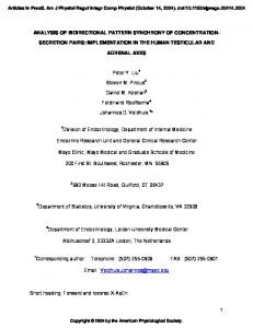
FIG. 3. Sequence similarities among s-factor regulators. (A) Clustal alignment (17) of RsbS with the SpoIIAA and RsbV anti-anti-s factors. Residues identical in all three proteins are indicated by a ? symbol, and conserved substitutions (10) are indicated by a : symbol. The arrow at SpoIIAA serine residue 58 denotes the S58A and S58D substitutions that have opposite regulatory effects on sF activity in vivo (11). The arrow at RsbS serine residue 59 denotes the S59A and S59D substitutions we introduced into the chromosomal coding sequence of rsbS. (B) Alignment of RsbT with the SpoIIAB and RsbW anti-s factors and six bacterial histidine protein kinases. This alignment is adapted from Fig. 3 of Min et al. (26) and revised to include B. subtilis KinB (33) and KinC (21). Although RsbW and SpoIIAB lack the conserved histidine residue and the autophosphorylation mechanism characteristic of histidine protein kinases, they nonetheless possess a kinase activity directed toward their antagonist proteins, RsbV and SpoIIAA, and their predicted sequences closely resemble histidine protein kinases in conserved residues that are thought to be important for ATP binding (1, 2, 11, 26). These conserved residues are indicated in boldface; parentheses enclose the number of omitted residues that lie between the motifs shown.
J. BACTERIOL.
substantially increased sB activity in exponentially growing cells. However, we were easily able to recover this rsbS deletion in the chromosome of a strain which bore an otherwise wildtype sigB operon and was therefore subject to the autocatalytic induction directed by the sB-dependent internal promoter of the sigB operon, PB (20, 37). In contrast, and despite repeated attempts, we were unable to recover the rsbSS59D allele in the chromosome of any strain containing the wild-type PB promoter. Therefore, the experiment shown in Fig. 4A was necessarily conducted with a strain in which PB had been exactly replaced by the inducible Pspac promoter (Fig. 1). This result suggests the possibility that the rsbSS59D allele is more deleterious than the large rsbS deletion. If this were the case, one possible explanation would be that in addition to its regulatory function, RsbS also has a structural role in a complex with other sB regulatory proteins. Genetic relationship of the RsbS and RsbT regulators to other known regulators of sB activity. Previous analysis of the phenotypes of single and double mutants has shown that the known regulators of sB form a hierarchical signal transduction pathway (4, 9, 34), with RsbV and RsbW acting downstream from RsbX and RsbU (Fig. 1). What is the relationship of the newly identified regulators RsbS and RsbT to these previously identified elements? In order to determine the relationship between the RsbSRsbT pair and their presumed RsbV-RsbW homologs, we combined within a single strain two null mutations which produced opposite regulatory phenotypes and analyzed their effects on sB-dependent gene expression. As demonstrated in Fig. 5A, the rsbS-rsbV double null mutant had the same phenotype as an rsbV single null mutant in that no elevated sB activity was seen in exponentially growing cells either before or after salt addition. And with respect to colony size, the rsbSrsbV double null mutant had wild-type colony morphology, the same as that of the rsbV single null mutant, and not the smallcolony phenotype of the rsbS single null mutant (data not shown). Similarly, as demonstrated in Fig. 5B, the rsbT-rsbW
FIG. 4. Serine 59 is important for RsbS function. (A) b-Galactosidase activity of a mutant bearing the rsbSS59D substitution and a sB-dependent ctc-lacZ transcriptional fusion. Because of the lethal nature of the rsbSS59D allele in an otherwise wild-type sigB operon, this experiment was conducted with a strain in which the internal, sB-dependent promoter PB was replaced by the isopropyl-b-D-thiogalactopyranoside (IPTG)-inducible promoter Pspac (Fig. 1). This construction interrupted the autocatalytic induction of sB synthesis normally seen to occur in the sigB operon (9, 20) and led to significantly reduced b-galactosidase accumulation (compare panel A with panel B). Both strains tested were grown in buffered LB medium to early exponential phase, when IPTG was added at time zero. E, PB212 (rsbS1 Pspac [rsbV1 rsbW1 sigB1 rsbX1]); Ç, PB478 (rsbSS59D Pspac [rsbV1 rsbW1 sigB1 rsbX1]). (B) b-Galactosidase activity of a mutant bearing the rsbSS59A substitution. Two parallel cultures of each strain tested were grown in buffered LB medium until early exponential phase. At time zero, sufficient NaCl was added to one of the two cultures to yield a final concentration of 0.3 M. F and E, PB198 (wild type) with and without salt addition, respectively; ■ and h, PB470 (rsbSS59A) with and without salt addition, respectively.
VOL. 178, 1996
HOMOLOGOUS PAIRS OF REGULATORS CONTROL B. SUBTILIS sB
3851
FIG. 5. Phenotypes of rsbS-rsbV and rsbT-rsbW double mutants. (A) Effect of an rsbS-rsbV double null mutation on b-galactosidase activity of a sB-dependent ctc-lacZ transcriptional fusion in response to salt stress. The predicted products of rsbS and rsbV have significant sequence similarity (Fig. 3A), but null mutations within the coding sequence of each have opposite regulatory effects. The three strains tested were grown in buffered LB medium to early exponential phase. At time zero, sufficient NaCl was added to each culture to yield a final concentration of 0.3 M. å, PB424 (rsbSD1); ■, PB206 (rsbVD1); }, PB469 (rsbSD1 rsbVD1). (B) Effect of an rsbT-rsbW double null mutation on b-galactosidase activity of a sB-dependent ctc-lacZ transcriptional fusion. The predicted products of RsbT and RsbW bear sequence resemblance (Fig. 3B), but null mutations within the coding sequence of each have opposite regulatory effects. Because the rsbW null allele is lethal in an otherwise wild-type sigB operon (9), this experiment was conducted with strains in which the internal, sB-dependent promoter PB had been replaced by the isopropyl-b-Dthiogalactopyranoside (IPTG)-inducible promoter Pspac. The three strains tested were grown in buffered LB medium to early exponential phase, when IPTG was added at time zero. h, PB458 (rsbTD1 Pspac [rsbV1 rsbW1 sigB1 rsbX1]); Ç, PB213 (rsbT1 Pspac [rsbV1 rsbWD1 sigB1 rsbX1]); {, PB454 (rsbTD1 Pspac [rsbV1 rsbWD1 sigB1 rsbX1]).
double null mutant had the same phenotype as an rsbW single null mutant, with an exceedingly high level of sB activity. Because loss of rsbV function completely overrides loss of rsbS and because loss of rsbW function completely overrides loss of rsbT, we conclude that, under the conditions tested, the RsbSRsbT proteins require the RsbV-RsbW proteins in order to regulate sB activity. These genetic data are consistent with the model of a linear signal transduction pathway shown in Fig. 6. In order to determine the relationship between the RsbSRsbT pair and the previously identified RsbX and RsbU regulators, we performed further phenotypic analyses of selected single and double mutants. As shown in Fig. 7A, with regard to sB activity, the phenotype of an rsbS-rsbU double null mutant was the same as that of an rsbU single mutant, suggesting that RsbS requires RsbU function in order to influence sB activity. These expression data were supported by the colony morphol-
FIG. 6. Genetic model of relationships among sB regulators. The model shown positions RsbS and RsbT upstream from RsbV and RsbW in a linear signal transduction pathway. RsbT requires RsbW function in order to positively regulate sB activity, either directly or indirectly (indicated by the double arrow). RsbW functions as an anti-s factor which directly binds and inactivates sB, and it is thought to exist as a homodimer (1, 5). Considering only the phenotype of an rsbT-rsbW double null mutant, it remains formally possible that RsbT could form a heterodimer with RsbW and thereby abrogate the anti-s activity of RsbW. This interpretation would suggest a branched signal transduction pathway converging on RsbW. However, considering the strength of the RsbS regulation, and the fact that RsbS absolutely requires both RsbT and RsbV functions in order to negatively regulate sB activity, we favor the linear pathway shown. In addition to its anti-s activity, RsbW is known to bear a kinase activity that modifies and inactivates RsbV (13). On the basis of the amino acid sequence homology noted between RsbS and RsbV and between RsbW and RsbT (Fig. 3) and the opposite regulatory phenotypes of the S59A and S59D substitutions in conserved serine 59 of RsbS (Fig. 4), we argue that RsbT also inactivates RsbS by a similar molecular mechanism.
ogy of the rsbS-rsbU double null mutant, which had the wildtype morphology characteristic of the rsbU null mutant rather than the small-colony morphology of the rsbS null mutant (data not shown). And, as shown in Fig. 7B, with regard to sB activity, the phenotype of an rsbX-rsbT double null mutant was the same as that of an rsbT single mutant, suggesting that RsbX requires RsbT function in order to regulate sB. These expression data were consistent with the colony morphology of the rsbX-rsbT double null mutant, which had the wild-type morphology of the rsbT null mutant rather than the small-colony morphology of an rsbX null mutant (data not shown). A model of sB regulation which summarizes these genetic data is shown in Fig. 8. Signal transduction in the sB regulatory network. Our results show that RsbS and RsbT are members of the signal transduction pathway that activates the general stress transcription factor sB in response to environmental signals. The simplest interpretation of the double-mutant analysis is that the rsbV-rsbW pair acts in the genetic pathway between rsbSrsbT and sigB. This argument positions RsbS and RsbT as dependent upon RsbV and RsbW in a regulatory hierarchy and is the basis of the model shown in Fig. 8. Because the phenotype of an rsbV null mutant completely overrides that of an rsbS null mutant (Fig. 5A) and because the phenotype of an rsbW null mutant completely overrides that of an rsbT null mutant (Fig. 5B), our data are not consistent with models of parallel or converging signal transduction pathways. Additional double-mutant analysis indicates that RsbS depends upon RsbU action in order to exert its regulatory effect, suggesting that RsbU serves as the link between the RsbSRsbT pair and the RsbV-RsbW pair. In this regard, the significant sequence similarity between RsbU and the SpoIIE phosphatase of the sF regulatory pathway leads to the intriguing speculation that RsbU might function as a phosphatase in the sB network (14). From the deduced position of RsbU in the signal transduction pathway, we would further speculate that
3852
KANG ET AL.
J. BACTERIOL.
FIG. 7. Phenotypes of rsbS-rsbU and rsbT-rsbX double mutants. (A) Effect of an rsbS-rsbU double null mutation on b-galactosidase activity of a sB-dependent ctc-lacZ transcriptional fusion in response to salt stress. The three strains tested were grown in buffered LB medium to early exponential phase. At time zero, sufficient NaCl was added to each culture to yield a final concentration of 0.3 M. å, PB424 (rsbSD1); ■, PB244 (rsbU::ermC); }, PB473 (rsbSD1 rsbU::ermC). (B) Effect of an rsbT-rsbX double null mutation on b-galactosidase activity of a sB-dependent ctc-lacZ transcriptional fusion. Because an rsbX null allele produces a slow-growth phenotype in an otherwise wild-type sigB operon (19, 20), this experiment was conducted with strains in which the internal, sB-dependent promoter PB had been replaced by the isopropyl-b-D-thiogalactopyranoside (IPTG)-inducible promoter Pspac. The three strains tested were grown in buffered LB medium to early exponential phase, when IPTG was added at time zero. h, PB458 (rsbTD1 Pspac [rsbV1 rsbW1 sigB1 rsbX1]); Ç, PB489 (rsbT1 Pspac [rsbV1 rsbW1 sigB1 rsbX::ermC]); {, PB486 (rsbTD1 Pspac [rsbV1 rsbW1 sigB1 rsbX::ermC]).
phosphorylated RsbV is the target of the presumed dephosphorylation activity of RsbU and that the signal controlling this dephosphorylation activity is transmitted to RsbU via RsbS and RsbT. The model depicted in Fig. 8 therefore offers an attractive explanation of how the upstream elements of the regulatory network could communicate signals of environmental stress to the principal downstream elements, the RsbV anti-anti-s factor and the RsbW anti-s factor. In addition to the linear model shown in Fig. 8, there are other formal explanations for the observed double-mutant phenotypes. For example, some of the Rsb proteins could exist in a complex whose activity or stability depended on the concentration of at least one critical Rsb protein within the complex; if this protein were absent because of deletion of its
structural gene, the function of the entire complex would be disrupted. We also remain mindful that the functions of additional potential regulators of sB, such as rsbR, remain to be discovered. Nonetheless, our analysis has generated strong inferences regarding the biochemical activities of the known regulators of sB activity, and experiments to test these predictions are in progress. ACKNOWLEDGMENTS We thank Richard Losick and William Haldenwang for stimulating discussions and for sharing unpublished data, Alan Grossman and Mitchell Singer for their constructive comments on the manuscript, Patrick Stragier for providing the spoIIIG-lacZ fusion, and Hsu Ling Tan for her assistance in constructing the rsbS and rsbT mutants.
FIG. 8. Model of sB regulation. The relationship among RsbU, the RsbV anti-anti-s factor, the RsbW anti-s factor, and sB is derived from previous genetic and biochemical analysis (4, 5, 9, 13, 34). The double-mutant analysis we present in Fig. 5 and 7 is consistent with the linear signal transduction pathway shown, wherein RsbX is an activator of RsbS, which in turn inhibits the function of RsbT. We suggest that RsbT activates RsbU, which therefore serves as the link between the upstream and downstream homologous regulators RsbS-RsbT and RsbV-RsbW. Earlier, Voelker et al. (35) discovered that the RsbV-RsbW pair is required for the transmission of both environmental and energy stress signals, whereas RsbU is required only for the transmission of environmental signals. On the basis of the data presented in Fig. 2, we can now extend the RsbU portion of the signal transduction pathway to show that RsbT is also required only for the transmission of environmental signals, as indicated by the open vertical arrows. Because RsbX and RsbS are negative regulators of sB activity, null mutations in their structural genes elicit high-level sB activity even in the absence of stress. Therefore, it is more difficult to test the possible role of RsbX and RsbS in conveying environmental stress signals. Furthermore, our genetic analysis does not allow us to unequivocally determine the order of action of RsbX and RsbS or of RsbT and RsbU. However, the order shown is supported by the fact that the RsbV-RsbW protein pair functions by a direct protein-protein interaction which is controlled by the phosphorylation state of RsbV (1, 5, 13), together with the strong inference presented here that the RsbS-RsbT protein pair functions by a similar molecular mechanism (Fig. 3 and 4). We therefore presume that the RsbX and RsbS regulators will also prove to be involved in the transmission of environmental stress signals to sB.
HOMOLOGOUS PAIRS OF REGULATORS CONTROL B. SUBTILIS sB
VOL. 178, 1996
This research was supported by Public Health Service grant GM42077 from the National Institute of General Medical Sciences. 20. REFERENCES 1. Alper, S., A. Dufour, D. A. Garsin, L. Duncan, and R. Losick. Role of adenosine nucleotides in the regulation of a stress response transcription factor in Bacillus subtilis. J. Mol. Biol., in press. 2. Alper, S., L. Duncan, and R. Losick. 1994. An adenosine nucleotide switch controlling the activity of a cell type-specific transcription factor in B. subtilis. Cell 77:195–205. 3. Antelmann, H., J. Bernhardt, R. Schmid, and M. Hecker. 1995. A gene at 3338 on the Bacillus subtilis chromosome encodes the newly identified sBdependent general stress protein GspA. J. Bacteriol. 177:3540–3545. 4. Benson, A. K., and W. G. Haldenwang. 1992. Characterization of a regulatory network that controls sB expression in Bacillus subtilis. J. Bacteriol. 174:749–757. 5. Benson, A. K., and W. G. Haldenwang. 1993. Bacillus subtilis sB is regulated by a binding protein (RsbW) that blocks its association with core RNA polymerase. Proc. Natl. Acad. Sci. USA 90:2330–2334. 6. Benson, A. K., and W. G. Haldenwang. 1993. The sB dependent promoter of the Bacillus subtilis sigB operon is induced by heat shock. J. Bacteriol. 175:1929–1935. 7. Boylan, S. A., A. R. Redfield, M. S. Brody, and C. W. Price. 1993. Stressinduced activation of the sB transcription factor of Bacillus subtilis. J. Bacteriol. 175:7931–7937. 8. Boylan, S. A., A. R. Redfield, and C. W. Price. 1993. Transcription factor sB of Bacillus subtilis controls a large stationary-phase regulon. J. Bacteriol. 175:3957–3963. 9. Boylan, S. A., A. Rutherford, S. M. Thomas, and C. W. Price. 1992. Activation of Bacillus subtilis transcription factor sB by a regulatory pathway responsive to stationary-phase signals. J. Bacteriol. 174:3695–3706. 10. Dayhoff, M. D., R. M. Schwartz, and B. C. Orcutt. 1978. A model of evolutionary change in proteins, p. 345–352. In M. D. Dayhoff (ed.), Atlas of protein sequence and structure, vol. 5, suppl. 3. National Biomedical Research Foundation, Silver Spring, Md. 11. Diederich, B., J. Wilkinson, T. Magnin, S. M. A. Najafi, J. Errington, and M. Yudkin. 1994. Role of the interactions between SpoIIAA and SpoIIAB in regulating cell-specific transcription factor sF of Bacillus subtilis. Genes Dev. 8:2653–2663. 12. Dubnau, D., and R. Davidoff-Abelson. 1971. Fate of transforming DNA following uptake by competent Bacillus subtilis. J. Mol. Biol. 56:209–221. 13. Dufour, A., and W. G. Haldenwang. 1994. Interactions between a Bacillus subtilis anti-sigma factor (RsbW) and its antagonist (RsbV). J. Bacteriol. 176:1813–1820. 14. Duncan, L., S. Alper, F. Arigoni, R. Losick, and P. Stragier. 1995. Activation of cell-specific transcription by a serine phosphatase at the site of asymmetric division. Science 270:641–644. 15. Duncan, L., and R. Losick. 1993. SpoIIAB is an anti-sigma factor that binds to and inhibits transcription by regulatory protein sF from Bacillus subtilis. Proc. Natl. Acad. Sci. USA 90:2325–2329. 16. Engelmann, S., C. Lindner, and M. Hecker. 1995. Cloning, nucleotide sequence, and regulation of katE encoding a sB-dependent catalase in Bacillus subtilis. J. Bacteriol. 177:5598–5605. 17. Higgins, D. G., and P. M. Sharp. 1988. Clustal: a package for performing multiple sequence alignment on a microcomputer. Gene 73:237–244. 18. Ho, S. N., H. D. Hunt, R. M. Horton, J. K. Pollen, and L. R. Pease. 1989. Site-directed mutagenesis by overlap extension using the polymerase chain reaction. Gene 77:51–59. 19. Igo, M., M. Lampe, C. Ray, W. Schafer, C. P. Moran, and R. Losick. 1987.
21.
22.
23.
24.
25. 26.
27.
28.
29.
30.
31.
32.
33.
34.
35.
36.
37.
3853
Genetic studies of a secondary RNA polymerase sigma factor in Bacillus subtilis. J. Bacteriol. 169:3464–3469. Kalman, S., M. L. Duncan, S. M. Thomas, and C. W. Price. 1990. Similar organization of the sigB and spoIIA operons encoding alternate sigma factors of Bacillus subtilis RNA polymerase. J. Bacteriol. 172:5575–5585. LeDeaux, J. R., and A. D. Grossman. 1995. Isolation and characterization of kinC, a gene that encodes a sensor kinase homologous to the sporulation sensor kinases KinA and KinB of Bacillus subtilis. J. Bacteriol. 177:166– 175. Lee, S., and C. W. Price. 1993. The minCD locus of Bacillus subtilis lacks the minE determinant that provides topological specificity to cell division. Mol. Microbiol. 7:601–610. Magnin, T., M. Lord, J. Errington, and M. D. Yudkin. 1996. Establishing differential gene expression in sporulating Bacillus subtilis: phosphorylation of SpoIIAA (anti-anti-sF) alters its conformation and prevents formation of a SpoIIAA/SpoIIAB/ADP complex. Mol. Microbiol. 19:901–907. Maul, B., U. Vo¨lker, S. Riethdorf, S. Engelmann, and M. Hecker. 1995. sB-dependent regulation of gsiB in response to multiple stimuli in Bacillus subtilis. Mol. Gen. Genet. 248:114–120. Miller, J. H. 1972. Experiments in molecular genetics. Cold Spring Harbor Laboratory, Cold Spring Harbor, N.Y. Min, K.-T., C. M. Hilditch, B. Diederich, J. Errington, and M. D. Yudkin. 1993. sF, the first compartment-specific transcription factor of B. subtilis, is regulated by an anti-s factor that is also a protein kinase. Cell 74:735–742. Moran, C. P., Jr., W. C. Johnson, and R. Losick. 1982. Close contacts between s37-RNA polymerase and a Bacillus subtilis chromosome promoter. J. Mol. Biol. 162:709–713. Najafi, S. M. A., A. C. Willis, and M. Yudkin. 1995. Site of phosphorylation of SpoIIAA, the anti-anti-sigma factor for sporulation-specific sF of Bacillus subtilis. J. Bacteriol. 177:2912–2913. Price, C. W., and R. H. Doi. 1985. Genetic mapping of rpoD implicates the major sigma factor of Bacillus subtilis RNA polymerase in sporulation initiation. Mol. Gen. Genet. 201:88–95. Schmidt, R., P. Margolis, L. Duncan, R. Coppolecchia, C. P. Moran, and R. Losick. 1990. Control of developmental transcription factor sF by sporulation regulatory proteins SpoIIAA and SpoIIAB in Bacillus subtilis. Proc. Natl. Acad. Sci. USA 87:9221–9225. Stahl, M. L., and E. Ferrari. 1984. Replacement of the Bacillus subtilis subtilisin structural gene with an in vitro-derived mutation. J. Bacteriol. 158: 411–418. Sun, D., R. M. Cabrera-Martinez, and P. Setlow. 1991. Control of transcription of the Bacillus subtilis spoIIIG gene, which codes for the foresporespecific transcription factor sG. J. Bacteriol. 173:2977–2984. Trach, K. A., and J. A. Hoch. 1993. Multisensory activation of the phosphorelay initiating sporulation in Bacillus subtilis: identification and sequence of the protein kinase of the alternate pathway. Mol. Microbiol. 8: 69–79. Voelker, U., A. Dufour, and W. G. Haldenwang. 1995. The Bacillus subtilis rsbU gene product is necessary for RsbX-dependent regulation of sB. J. Bacteriol. 177:114–122. Voelker, U., A. Voelker, B. Maul, M. Hecker, A. Dufour, and W. G. Haldenwang. 1995. Separate mechanisms activate sB of Bacillus subtilis in response to environmental and metabolic stresses. J. Bacteriol. 177:3771–3780. Vo ¨lker, U., S. Engelmann, B. Maul, S. Riethdorf, A. Vo¨lker, R. Schmid, H. Mach, and M. Hecker. 1994. Analysis of the induction of general stress proteins of Bacillus subtilis. Microbiology 140:741–752. Wise, A. A., and C. W. Price. 1995. Four additional genes in the sigB operon of Bacillus subtilis that control activity of the general stress factor sB in response to environmental signals. J. Bacteriol. 177:123–133.