Biochemistry. Functional conversion of the homologous proteins a-lactalbumin and lysozyme by exon exchange. (c-type lysozyme/exon/protein engineering).
Proc. Nail. Acad. Sci. USA Vol. 89, pp. 5887-5891, July 1992 Biochemistry
Functional conversion of the homologous proteins a-lactalbumin and lysozyme by exon exchange (c-type lysozyme/exon/protein engineering)
IZUMI KUMAGAI*, SHIGEKI TAKEDA, AND KIN-ICHIRO MIURAt Department of Industrial Chemistry, Faculty of Engineering, The University of Tokyo, Hongo, Tokyo 113, Japan
Communicated by Motoo Kimura, March 20, 1992
Exons of eukaryotic genes that encode proABSTRACT teins frequently appear to encode structural and/or functional protein units [Gilbert, W. (1978) Nature (London) 271, 501; Blake, C. C. F. (1979) Nature (London) 277, 598]. a-Lactalbumin and c-type lysozyme are functionally quite different but structurally highly homologous proteins. Their gene organizations have been shown to be virtually the same and their exon structures are identical. The exon 2 region of hen lysozyme contains most of the amino acid residues that make up its catalytic cleft. In this study, we engineered a hybrid protein in which the exon 2 region of goat a-lactalbumin was replaced with that of hen lysozyme. This conferred catalytic activity on the a-lactalbumin, which is a nonenzymatic protein in its native structural form.
is located on exon 2 of its gene, encoding residues from Trp-28 to Ala-82, which include catalytically active residues and a cluster of residues that bind rings B, C, D, E, and F of the oligosaccharide substrate. The organizations of the rat, guinea-pig, human, and goat a-lactalbumin genes have been demonstrated to be closely related to the organization of hen egg-white lysozyme (13-16). The a-lactalbumin and c-type lysozyme genes contain three introns at exactly the same positions, although the structures of the introns and of the fourth exon differ slightly (Fig. 1). Therefore, it is quite likely that these two proteins may have diverged from a common ancestor by gene duplication during evolution (8, 17, 18) and they may prove useful for studying the evolution of structural and functional relationships of proteins.
The discovery of the exon and intron structures of eukaryotic genes has provided valuable information that has led to further clarification of the evolution of genes. Gilbert (1) hypothesized that exons are functional units of protein molecules and that new functional proteins have evolved from a relatively small number of exon modules by selection of various combinations of the functional units produced by unequal crossovers on introns. To perform a biological function, a protein must possess a stable specific conformation. Blake (2) hypothesized that if exons encode both structural and functional units, then combinations of such exons would be expected to be capable of producing stable functional proteins. Go (3, 4) proposed a "module" structure as a compact protein structural unit, which was identified by plotting the distances between the a-carbons of the residues and corresponded well to the exon region of a gene. Such a close relationship between a gene exon and the module protein structure lends support to Blake's view. a-Lactalbumin is a mammalian milk whey protein and a regulatory subunit of lactose synthase (EC 2.4.1.22), which is produced specifically in the lactating mammary gland. When this protein binds to the enzyme, galactosyltransferase, that catalyzes the transfer of galactose to N-acetyl-D-glucosamine (GlcNAc), lactose is synthesized efficiently from uridine 5'-diphosphogalactose (UDP-galactose) and glucose (5). a-Lactalbumin also binds calcium strongly (6). The primary (7, 8) and three-dimensional (9, 10) structures of a-lactalbumin have been demonstrated to be quite homologous with those of the c-type lysozymes, although these proteins are functionally quite different. Hen egg-white lysozyme (EC 3.2.1.17) catalyzes the hydrolysis of the f-1,4-glycosidic linkage between GlcNAc and N-acetyl-D-muramic acid (MurNAc) in polysaccharides and is the most studied, and consequently best known, of this type of enzyme. Its structural and functional relationships (11) and gene organization (12) have been elucidated. The catalytic site of the lysozyme
MATERIALS AND METHODS Materials. Goat a-lactalbumin was a generous gift from S. Sugai and K. Kuwajima (Hokkaido University, Sapporo, Japan); Micrococcus lysodeikticus cells and bovine galactosyltransferase were purchased from Sigma; UDP-[14C]galactose (1.11 GBq/mmol) was obtained from Amersham; and
p-nitrophenyl penta-N-acetyl-,8-chitopentaoside [(GlcNAc)5pNP)] and hen egg-white lysozyme were purchased from Seikagaku Kogyo (Tokyo). p-Nitrophenyl N-acetyl-P3-Dglucosaminide (GlcNAc-pNP), p-nitrophenyl di-N-acetyl-,8chitobioside [(GlcNAc)2-pNP], p-nitrophenyl tri-N-acetyl-j3chitotrioside [(GlcNAc)3-pNP], and p-nitrophenyl tetra-Nacetyl-f8-chitotetraoside [(GlcNAc)4-pNP] were generous gifts from K. Sakai (Yaizu Suisan Kagaku Industry, Yaizu, Japan). The high-performance liquid chromatography (HPLC) column (YMC-Pac A-014, 6 x 300 mm) used was from Nishio Kogyo (Tokyo), DEAE-Sepharose Fast Flow and Sephacryl S-200 were from Pharmacia, and the DNA sequencing kits were purchased from Toyobo (Osaka). All other chemicals used were of reagent grade appropriate for biochemical use. Plasmid. We used the Escherichia coli expression plasmid pAKTS-1EX for goat a-lactalbumin, in which the mature coding region of goat a-lactalbumin was fused with the NH2-terminal half of porcine adenylate kinase cDNA, and the fused protein was expressed under the control of the tac promoter, as described previously (19). Plasmids were propagated in E. coli JM109 grown in LB medium containing ampicillin (50 ,ug/ml). Site-Directed Mutagenesis and Plasmid Construction. Four plasmids were constructed from pAKTS-1EX and used for Abbreviations: a-LA(X), mutant a-lactalbumin (X indicating the particular mutation); (GlcNAc)5-pNP, p-nitrophenyl penta-N-acetylP-chitopentaoside (tetramer, trimer, dimer, and monomer are abbreviated similarly). *To whom reprint requests should be addressed. tPresent address: Institute for Biomolecular Science, Gakushuin
The publication costs of this article were defrayed in part by page charge payment. This article must therefore be hereby marked "advertisement" in accordance with 18 U.S.C. §1734 solely to indicate this fact.
University, Mejiro, Tokyo 171, Japan.
5887
5888
Biochemistry: Kumagai et al.
Proc. Natl. Acad. Sci. USA 89
(1992)
Goat a-lactalbumin gene Exon I
76
int Exon2 int2 Exon3 int3 Exon4 GAGTT-----TAG3 TGAG----AG3_ TGAG-----CAG
327
159
474
76
2303
58
Hen lysozyme gene Exon1 -3TA
82
intl Exon2 int2 Exon3 int3 Exon4 TAAGT------CAG G TGAG----AG _GTGAG-----CAG o -0 0- -o4 - -4 I 162 1810 1270 64 79 79
FIG. 1. Comparison of organizations of goat a-lactalbumin and hen egg-white lysozyme genes (12, 15). The solid boxes represent the regions encoding exons and the numbers represent the nucleotide base pairs; int, intron. For exons 1 and 4, only the mature protein coding regions are represented in this figure.
the expression of mutant a-lactalbumins. The Tyr-107 residue of a-lactalbumin is considered to block binding of a lysozyme substrate to the a-lactalbumin cleft region (20, 21). Trp-62 may be essential for the activity of hen lysozyme (22-24), but in goat a-lactalbumin the amino acid residue corresponding to the 62nd position of hen lysozyme is Ile. The Tyr-107 and Ile-62 residues of a-lactalbumin were converted to Ala and Trp, respectively, by site-directed mutagenesis with 5'-GAATTAACGCGTGGTTGGC-3' (from Tyr-107 to Ala and for creation of the Mlu I restriction site) and 5'-ATAATAAATGGTGGTGCAA-3' (from Ile-62 to Trp) as a mutation primer. The double mutant protein thus obtained was designated a-LA(2). The restriction sites of two Rsa I sites in goat a-lactalbumin cDNA were used to exchange the cDNA sequence of goat a-lactalbumin, which corresponds to the region from Thr-31 to Ile-43, for two 41-mer synthetic DNAs (5'-GCCGCAAAATTCGAGAGTAACTTCAACACCCAGGCTACAGT-3' as a coding strand and 5'-ACTGTAGCCTGGGTGTTGAAGTTACTCTCGAATTTTGCGGC-3' as a complementary strand). In addition, Glu-52 was converted to Asp, which is one of the catalytic residues in hen egg-white lysozyme, by site-directed mutagenesis with 5'-AGCACAGATTATGGACT-3'. This mutant protein, which contained most of the substrate binding site residues and both catalytic residues of the hen lysozyme (Glu-35 and Asp-52), was designated a-LA(10). The restriction sites of Taq I and Sac I or Mlu I of hen lysozyme cDNA were used to exchange the cDNA sequences from Phe-34 to Asp-86 or from Phe-34 to Trp-108 of a-LA(10) for those of the hen lysozyme. The corresponding Sac I site on the a-LA(10) DNA sequence was created by site-directed mutagenesis with 5'-TCAGTAAGATCTGAGCTCAGGAACTTGTC-3' as a mutation primer. The amino acid sequences from Trp-28 to Lys-33 are the same in both a-LA(10) and hen lysozyme and, therefore, the former hybrid protein contains an amino acid sequence of hen lysozyme from Trp-28 to Ser-86. This exchanged region corresponded almost exactly to the exon 2 coding region (from Trp-28 to Ala-82) of hen lysozyme except for four residues (from Leu-83 to Ser-86), and it was designated a-LA(EX2). The a-LA(EX2,3) hybrid protein had the same amino acid sequence as that from Trp-28 to Trp-108 of hen lysozyme, which corresponds to the exon 2 and exon 3 coding regions. Therefore, to isolate the a-lactalbumin portion from the expressed fusion protein in E. coli by CNBr degradation, as a purification step, Met-105 of the mutant a-lactalbumin was exchanged for Ile by site-directed mutagenesis with 5'GAAACGGCATCAACGCGT-3'. The resultant protein was designated a-LA(EX2,3). All the site-directed mutagenesis processes were conducted using a double-strand plasmid (25), and all the oligodeoxyribonucleotides used as mutation primers were synthesized on a DNA synthesizer (Applied
Biosystems model 381A) by cyanoethyl phosphoramidite chemistry. Protein Expression and Purification. The mutant proteins were expressed in E. coli JM109 as proteins fused with the N-terminal half of porcine adenylate kinase and they were extracted from an insoluble fraction of the E. coli transformant cells as described by Kumagai et al. (19). The resultant insoluble proteins were subjected to DEAE-Sepharose FF column chromatography in the presence of 8 M urea in 50 mM Tris'HCI, pH 7.5, and eluted with a solution consisting of 8 M urea in 50 mM Tris HC1, pH 7.5/0.5 M NaCI. The mutant a-lactalbumins were isolated from the purified fusion proteins by treatment with CNBr in 70% (vol/vol) formic acid and then purified by DEAE-Sepharose column chromatography using a NaCI linear gradient (0-0.5 M) in the presence of 8 M urea/50 mM Tris HCI, pH 7.5, and by subsequent gel filtration on Sephacryl S-200 in 1 M urea/50 mM Tris HCI, pH 7.5. The homogeneity of the purified proteins was confirmed by sodium dodecyl sulfate/polyacrylamide gel electrophoresis (SDS/PAGE) (26) and amino acid sequence analysis of the NH2-terminal regions by a gas-phase protein sequencer (Applied Biosystems model 473A). The purified mutant proteins were redissolved in 10 mM Tris HCI, pH 7.5, which contained 50 mM dithiothreitol and 8 M urea and the solutions were dialyzed against 10 mM Tris HC1, pH 7.5, which contained 4 mM cysteine, 0.4 mM cystine, and 1 M urea, and then against 50 mM sodium phosphate buffer, pH 6.2, at room temperature. Enzymatic Assays. The lytic activity of each protein was determined by measuring the decrease in the turbidity of a Micrococcus lysodeikticus cell suspension. The rate of decrease in turbidity of cells in 1 ml of 50 mM sodium phosphate buffer, pH 6.2, with three different protein concentrations was monitored at 540 nm and 25°C (24). Hydrolysis of the glycosidic bond by the a-lactalbumin mutant containing the exon 2 of hen lysozyme was monitored by measuring the hydrolysis of (GlcNAc)5-pNP at 300 nm after HPLC on a YMC-Pac A-014 column (6 x 300 mm), which was developed with 80% (vol/vol) CH3CN at a flow rate of 1 ml/min. The reaction mixture contained 20 mM CH3COONa at pH 5.0,0.26 mM (GlcNAc)5-pNP, and 2,ug of protein in 100 ,ul and was incubated 37°C, after which 3-,l aliquots were subjected to HPLC analysis after the appropriate incubation times (27). The activity of a-lactalbumin was determined by measuring its lactose synthetic activity with 0.004 unit of galactosyltransferase and 270 Bq of UDP-[14C]galactose, as described by Brew et al. (28).
RESULTS AND DISCUSSION Construction and Preparation of Mutant a-Lactalbumins. The presence of a Tyr residue at position 107 of a-lactalbumin [in this paper, the residue numbering is based on that of hen
Proc. Natl. Acad. Sci. USA 89 (1992)
Biochemistry: Kumagai et al. egg-white lysozyme (20)] is considered to block access of the substrate to the region corresponding to subsite B of the active site cleft of the hen lysozyme (20, 21), and Trp-62 may be essential for substrate binding to the active site of hen lysozyme (22-24), whereas in goat a-lactalbumin the residue at the 62nd position is Ile. The a-LA(2) protein we produced is a double mutant of goat a-lactalbumin, in which Ile-62 and Tyr-107 were replaced with Trp and Ala, respectively. The a-LA(10) mutant protein possesses 10 mutations (on Thr-31 to Ala, Phe-33 to Lys, His-34 to Phe, Thr-35 to Glu, Gly-37 to Asn, Tyr-38 to Phe, Ile43 to Thr, Glu-52 to Asp, Ile-62 to Trp, and Tyr-107 to Ala) and contains most of the substrate binding site residues and both catalytic residues of hen lysozyme (Glu-35 and Asp-52). The amino acid sequence of a-LA(EX2) is the same as that from Trp-28 to Ser-86 of hen lysozyme, which almost corresponds to the hen lysozyme exon 2 coding region (from Trp-28 to Ala-82) and the replacement of Tyr-107 with Ala. The amino acid sequence of a-LA(EX2,3) is identical to that from Trp-28 to Trp-108 of hen lysozyme and corresponds to the exon 2 and 3 coding regions of hen lysozyme with the exception of the amino acid residue at position 105, which was converted from Met to Ile for CNBr degradation of the fusion protein. As the residue at this position in human lysozyme also is Ile, this mutation would not be expected to hinder lysozymal activity. For the renaturation process, the mutant proteins were dissolved in 10 mM Tris-HCI, pH 7.5/50 mM dithiothreitol/8 M urea and dialyzed first against 10 mM Tris-HCI, pH 7.5/4 mM cysteine/0.4 mM cystine/1 M urea and then against 50 mM sodium phosphate buffer, pH 6.2, at room temperature. After the second dialysis, which took over 30 hr, bacteriolytic activity was observed with the a-LA(EX2) and a-LA(EX2,3) but not the a-LA(2) or a-LA(10) proteins. Enzymatic Characterizations of Mutant a-Lactalbumins. Although a-LA(10) contains most of the amino acid residues that make up the catalytic cleft of hen lysozyme, it demonstrated no activity after renaturation under the conditions described above. However, the reduced and denatured hen lysozyme did not recover its full activity under these conditions (see Fig. 4). Therefore, better renaturation conditions may lead to greater expression of the catalytic activity of refolded proteins. The two a-lactalbumin mutant proteins whose exons had been replaced with the corresponding hen lysozyme exons demonstrated slight but significant bacteri-
5889
olytic activity (2% of that of the wild-type hen lysozyme), shown in Fig. 2, but no a-lactalbumin activity. The bacteriolytic activity assay may not reflect the catalytic processes of these proteins directly. To establish whether these a-lactalbumin mutants possessed hydrolytic activity at the glycosidic bond, a lysozyme substrate, (GlcNAc)5-pNP, was used for the enzymatic assay. Native hen lysozyme hydrolyzed (GlcNAc)5-pNP to predominantly chitotetraose [(GlcNAc)4] and GlcNAc-pNP, which were detected and separated by HPLC (27), whereas no hydrolysis was observed when the substrate was incubated with native goat a-lactalbumin. However, GlcNAc-pNP was detected when a-LA(EX2) was incubated with the substrate (Fig. 3), which indicates not only that a-LA(EX2) possessed hydrolytic activity at the glycosidic bond but also that the initial hydrolytic pattern [the major digestion products were (GlcNAc)4 and GlcNAc-pNP] appeared to be identical to that of hen lysozyme. The hydrolytic activity of a-LA(EX2)
goat a-lactalbumin
I-
(300min)
E
CD r_ 0
a-LA(10) (300min)
CD 0 a) CI
Cn
D0
a-LA(EX2) (300mi)
hen lysozyme (30min)
6-
x
(1)
(3)
I c
0
.
.5
I
5 10 Retention Time, min
FIG. 3. Hydrolysis of the glycosidic bond by the a-lactalbumin
0
m) 20
Protein, ,ug FIG. 2. Bacteriolytic activity assay of engineered mutant a-lactalbumin proteins. The lytic activity of each protein was measured according to the decrease in turbidity of a Micrococcus lysodeikticus cell suspension. The rate of decrease in turbidity of cells in 1 ml of 50 mM sodium phosphate buffer, pH 6.2, was monitored at 540 nm and 25°C at three different protein concentrations.
mutant a-LA(EX2), which contains the hen lysozyme exon 2. Hydrolysis of (GlcNAc)s-pNP with a-LA(EX2) was detected by HPLC on a YMC-Pac A-014 column (6 x 300 mm) that was developed with 80% CH3CN at a flow rate of 1 ml/min; the absorbance unit on the full scale was 0.02 at 300 nm. The reaction conditions were 20 mM CH3COONa at pH 5.0, 0.26 mM (GIcNAc)spNP, and 2 ,ug of protein in 100 Al of reaction mixture. At the appropriate incubation times (indicated in parentheses), 3-,ul aliquots of the reaction mixture were subjected to HPLC analysis. All peaks noted during incubation with goat a-lactalbumin could also be
detected when only (GIcNAc)5-pNP was analyzed. Peak 1, GlcNAcpNP; peak 2, (GlcNAc)-pNP; and peak 3, (GlcNAc)s-pNP.
5890
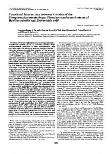
Biochemistry: Kumagai et al. K1
Native hen lysozyme
exonl
W28 I
@
Proc. Natl. Acad. Sci. USA 89 (1992)
exon2
A82
e)
I
W62
exon3
W108 exon4 L129
A107
a-Lactalbumin Bacteriolytic Hydrolysis Substrate interaction residues* activity (%) activity (/) activity (%) 0
100
100
13/13
Refolded hen lysozyme t
not done
i~I i
36
notdone
0
2
4
12/13
0 I~~~~~~~~
2
4
12/13
a-LA(10)
0
0
0
12/13
a-LA(2)
5
0
0
7/13
12
0
a-LA(EX2,3)
El
a-LA(EX2)
Recombinant goat a-lactalbumnin t i
!i
5/13
Native goat
a-lactalbunin
noat done}
I-
I .--
100
0
0
FIG. 4. Summary of the relative activities of engineered mutant a-lactalbumin proteins. In the maps on the left, the numbers represent the amino acid residues and those enclosed in circles (35 and 52) denote the catalytic residues of hen lysozyme. The solid boxes show the regions of the hen lysozyme sequence and the unbroken lines show those of goat a-lactalbumin. a-Lactalbumin activity was measured according to its lactose synthetic activity with 0.004 unit of galactosyltransferase and 270 Bq of UDP-[14C]galactose, as described by Brew et al. (28). Bacteriolytic activity was measured by using Micrococcus Iysodeikticus cells, as described in the legend to Fig. 2, and hydrolysis ofthe glycosidic bonds was measured with (GlcNAc)5-pNP as described in the legend to Fig. 3. The initial hydrolysis rate of (GIcNAc)5-pNP with a-LA(EX2,3) was about 4% of that of hen lysozyme, but no reaction was observed after 90 min, possibly due to protein denaturation. For exons 1 and 4, only the mature protein regions are represented in this figure. *Number of amino acid residues in the proteins among the 13 residues involved in substrate interaction of hen lysozyme [Phe-34, Glu-35, Asn-37, Asn-44, Asp-52, Gln-57, Asn-59, Trp-62, Trp-63, Asp-101, Ala-107, Trp-108, and Arg-114 (22)]. tNative hen lysozyme that had been reduced and denatured was refolded as described in the text. tGoat a-lactalbumin produced in the E. coli expression system and refolded as described in the text.
towards (GlcNAc)5-pNP was estimated to be about 4% that of hen lysozyme, whereas the bacteriolytic activity of a-LA(EX2) was about 2% that of hen lysozyme. Although hen lysozyme and a-lactalbumin are structurally quite homologous, the lysozyme is a very basic protein, whereas a-lactalbumin is acidic. The mutant proteins a-LA(EX2) and a-LA(EX2,3) constructed in this study appeared to be almost neutral. Although the lytic activity of a-LA(EX2,3) was almost identical to that of a-LA(EX2), a-LA(EX2,3) was unstable and precipitated readily at neutral pH, possibly due to its ionic nature. The cell surface of Micrococcus lysodeikticus, a suspension of which was used as the substrate in the bacteriolytic assay, appears to be highly negatively charged. Therefore, when the hen lysozyme binds to the substrate cells, there may be a marked electrostatic interaction between the positively charged lysozyme and the negatively charged cell surface, and thus efficient hydrolysis of the -GlcNAcl1-4MurNAc- linkage in the peptidoglycan may be facilitated and may reflect the lytic process directly. The neutral protein, a-LA(EX2) may, therefore, demonstrate less bacterial cell lytic activity than hydrolytic activity against chitopentaose because it possesses no electric charge. These results are summarized in Fig. 4. Recently, we reported the expression of hen lysozyme in Saccharomyces cerevisiae and the results of site-directed mutagenesis studies with this enzyme (24, 29). A mutant lysozyme in which Glu-35 was replaced with Asp had only slight hydrolytic activity towards (GlcNAc)5-pNP, even though the structural difference between the wild-type and mutant enzyme was only one methylene moiety (I.K., F.S., and K.M., unpublished data). A fusion protein in which exon 2 of hen lysozyme was linked with an unrelated protein, ,B-galactosidase, exhibited very slight lysozyme activity (1/
40,000 that of the native enzyme) when assayed with a synthetic substrate (30). Therefore, the significant hydrolytic activity towards (GlcNAc)5-pNP observed with a-LA(EX2) indicates that the framework constructed by exons 1, 3, and 4 of a-lactalbumin may incorporate exon 2 of hen lysozyme as a functionally active structure. The observation that a-LA(10) exhibited no lysozyme activity, although it contained the same set of amino acid residues involved with substrate binding and catalytic processes of hen lysozyme as did a-LA(EX2) (Fig. 4), indicates that additional replacements introduced into a-LA(EX2) may be important for producing the catalytic cleft structure. A structural unit corresponding to the exon 2 of hen lysozyme would support a unique tertiary structure required for biological function, so that introduction of the entire exon 2 of this lysozyme into a-lactalbumin successfully conferred some lysozyme function upon it. Molecular Evolution and Protein Function. a-Lactalbumin and c-type lysozyme may have evolved from the same primordial gene by gene duplication, with each corresponding exon region having the same size. The identities of the amino acid sequences between the coding regions of exons 1, 2, 3, and 4 of goat a-lactalbumin and hen lysozyme are 29.6%, 50.9%, 46.2%, and 28.6%, respectively, in the mature protein coding region. In the light of our results, it is evident also that even after evolution of the a-lactalbumin molecule to give rise to new functions, exons 1, 3, and 4 of the protein still retain the structural features of the primordial protein. It would be difficult to conduct protein functional conversion by introducing only active site and ligand binding site residues. If acceptable framework structures for the exons encoding functional modules can be identified either from an
Biochemistry: Kumagai et al. evolutionary standpoint or by structural analysis, conversion of protein function by exon exchange should be possible. The authors thank Prof. S. Sugai and Dr. K. Kuwajima for kindly providing the authentic goat a-lactalbumin and Dr. K. Sakai for providing the synthetic lysozyme substrate (GlcNAc)n-pNP (n = 1-5). This work was supported, in part, by Grants-in-Aid for Scientific Research from the Ministry of Education, Science and Culture of Japan. 1. 2. 3. 4. 5. 6. 7.
8. 9. 10.
11. 12. 13. 14.
Gilbert, W. (1978) Nature (London) 271, 501. Blake, C. C. F. (1979) Nature (London) 277, 598. G6, M. (1981) Nature (London) 291, 90-92. G6, M. (1983) Proc. Natl. Acad. Sci. USA 80, 1964-1968. Brew, K. & Hill, R. L. (1975) Rev. Physiol. Biochem. Pharmacol. 72, 105-157. Hiraoka, Y., Segawa, T., Kuwajima, K., Sugai, S. & Murai, N. (1980) Biochem. Biophys. Res. Commun. 95, 1098-1104. Brew, K., Vanaman, T. C. & Hill, R. L. (1967) J. Biol. Chem. 242, 3747-3749. Nitta, K. & Sugai, S. (1989) Eur. J. Biochem. 182, 111-118. Acharya, K. R., Stuart, D. I., Walker, N. P. C., Lewis, M. & Phillips, D. C. (1989) J. Mol. Biol. 208, 99-127. Acharya, K. R., Ren, J., Stuart, D. I., Phillips, D. C.& Fenna, R. E. (1991) J. Mol. Biol. 221, 571-581. Jolles, P. & Jolles, J. (1984) Mol. Cell. Biochem. 63, 165-189. Jung, A., Sippel, A. E., Grez, M. & Schutz, G. (1980) Proc. Natl. Acad. Sci. USA 77, 5759-5763. Qasba, P. K. & Safaya, S. K. (1984) Nature (London) 308, 377-380. Hall, L., Emery, D. C., Davies, M. S., Parker, D. & Craig, R. K. (1987) Biochem. J. 242, 735-742.
Proc. Natl. Acad. Sci. USA 89 (1992)
5891
15. Vilotte, J. L., Soulier, S., Printz, C. & Mercier, J. C. (1991) Gene 98, 271-276. 16. Laird, J. E., Jack, L., Hall, L., Boulton, A. P., Parker, D. & Craig, R. K. (1988) Biochem. J. 254, 85-94. 17. Shewale, J. G., Sinha, S. K. & Brew, K. (1984) J. Biol. Chem. 259, 4947-4956. 18. Prager, E. M. & Wilson, A. C. (1988) J. Mol. Evol. 27, 326-335. 19. Kumagai, I., Takeda, S., Hibino, T. & Miura, K. (1990) Protein Eng. 3, 449-452. 20. Brown, W. J., North, A. C. T., Phillips, D. C., Brew, K., Vanaman, T. C. & Hill, R. L. (1969) J. Mol. Biol. 42, 65-86. 21. Warme, P. K., Momany, F. A., Rumball, S. V., Tuttle, R. W. & Scheraga, H. A. (1974) Biochemistry 13, 768-782. 22. Imoto, T., Johnson, L. N., North, A. C. T., Phillips, D. C. & Rupley, J. A. (1972) in The Enzymes, ed. Boyer, P. D. (Academic, New York), Vol. 7, pp. 665-868. 23. Blake, C. C. F., Johnson, L. N., Mair, G. A., North, A. C. T., Phillips, D. C. & Sarma, V. R. (1967) Proc. R. Soc. London Ser. B 167, 378-388. 24. Kumagai, I., Kojima, S., Tamaki, E. & Miura, K. (1987) J. Biochem. 102, 733-740. 25. Morinaga, Y., Franceschini, T., Inouye, S. & Inouye, M. (1984) Biotechnology 2, 636-639. 26. Laemmli, U. K. (1970) Nature (London) 227, 680-685. 27. Nanjo, F., Sakai, K. & Usui, T. (1988) J. Biochem. 104, 255-258. 28. Brew, K., Vanaman, T. C. & Hill, R. L. (1968) Proc. NatI. Acad. Sci. USA 59, 491-497. 29. Kumagai, I. & Miura, K. (1989) J. Biochem. 105, 946-948. 30. Kuchinke, W. (1989) Biochem. Biophys. Res. Commun. 159, 927-932.