the brachial plexus. •Be able to list the spinal segments that course through each
brachial plexus branch. ... Please Read: Interactive PowerPoint functions like a
web-page. Click on the underlined .... Brachial Plexus Block. •The injection of an
...
Maximizing the Flexibility of Powerpoint
Dr. Jamie Wikenheiser Assistant Professor Director of Medical Gross Anatomy Director of Surgical Anatomy University of California, Irvine School of Medicine
UCI Medical Gross Anatomy • Most lectures are followed up by a lab • 36 total dissection labs • Online dissector with videos loaded onto the iPads • 5 color groups rotate (each student however dissects 8 times) • 2 people per donor • The group that dissects presents their dissection with an emphasis on the clinical correlates • Lectures use interactive powerpoints • A main lecture with built in review/practice • Hyperlink slide bank
Interactive Powerpoint Format • • • •
Title Learning Objectives Main Menu Chapter Content • Slides with internal hyperlinks • Thank You and Contact Slide • References • Hyperlink Slide Bank • All slides from internal hyperlinks
Learning Objectives •Be able to describe the 3 parts of the axillary artery and the branches that come off each part. •Be able to describe the roots, trunks, divisions, cords and terminal branches of the brachial plexus. •Be able to list the spinal segments that course through each brachial plexus branch. •Be able to describe the clinical correlates associated with injuries to the brachial plexus.
Main Menu
Main Menu I. Vasculature of the Axilla II. Brachial Plexus III. Arm Muscles & Neurovasculature
Please Read: Interactive PowerPoint functions like a web-page. Click on the underlined text to go directly to that topic. You must be in Slide Show mode for the hyperlinks and highlights to work. All images are copyrighted by the authors listed on the “References” slide. DO NOT DISTRIBUTE! For educational purposes only. Nongraphical content & organization Copyright 2013 Dr. Jamie Wikenheiser.
Main Menu
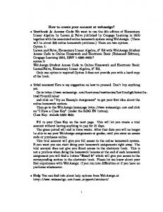
I. Vasculature of the Axilla •The axillary artery extends from the lateral edge of the 1st rib to the inferior border of the teres major muscle. The axillary artery begins at the termination of the subclavian artery and ends as the brachial artery. •The axillary artery has 3 parts. •The axillary vein is formed by the union of the brachial veins & basilic vein and terminates as the subclavian vein.
Axillary Vasculature
Main Menu
•The axillary artery extends from the lateral edge of the 1st rib to the inferior border of the teres major muscle. The axillary artery begins at the termination of the subclavian artery and ends as the brachial artery. •It has 3 parts: •1st part: located between the lateral 1st rib & medial border of pectoralis minor. It is enclosed by in the axillary sheath and has 1 branch.
1 2 3
•2nd part: located posterior to the pectoralis minor has 2 branches. •3rd part: located between the lateral border of pectoralis minor & inferior border of teres major muscle. It has 3 branches.
Pectoralis minor
Anterior View
Axillary Artery Branches
Main Menu
•The axillary artery has 3 parts: •1st part has 1 branch: •Superior thoracic artery. •2nd part has 2 branches: •Thoracoacromial artery/trunk •Lateral thoracic artery •3rd part has 3 branches: •Subscapular •Anterior circumflex humeral •Posterior circumflex humeral
1st part of axillary 2nd part of axillary 3rd part of axillary Supreme thoracic Thoracoacromial (has 4 branches) Lateral thoracic Subscapular Anterior humeral circumflex Posterior humeral circumflex Anterior View
Shoulder: Axillary Arteriogram
Main Menu
1st part of axillary 2nd part of axillary 3rd part of axillary Supreme thoracic
Thoracoacromial Lateral thoracic Subscapular Anterior humeral circumflex Posterior humeral circumflex Brachial
Left Shoulder/Anterior View
Transverse Section (T2 vertebral level)
Main Menu
CT
Trachea Brachiocephalic trunk (artery)
MRI
L common carotid artery L subclavian artery L brachiocephalic vein R brachiocephalic vein
Main Menu
II. Brachial Plexus
Brachial Plexus
Main Menu
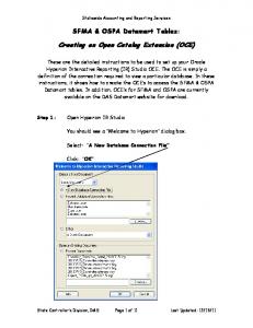
•The brachial plexus is a major network of somatic nerves supplying the upper limb.
3 Trunks (superior, middle & inferior)
•Formed by the union of the ventral rami of C5-T1 nerves and constitute the roots of the brachial plexus. •The brachial plexus is divided into: •5 roots •3 trunks •6 divisions (3 anterior & posterior divisions each) •3 cords •5 terminal branches (musculocutaneous, axillary, median, ulnar & radial)
Clinical Correlate 1
Clinical Correlate 2
Ventral Roots (C5-T1)
3 anterior divisions
Musculocutaneous
Axillary
Radial
3 posterior divisions 3 Cords (lateral, posterior & medial)
Median Ulnar
Posterior Cord Branches
Main Menu
•All branches associated with the posterior cord of the brachial plexus are listed with the roots/spinal segments that contribute to that particular nerve branch.
C5
•Note: the specific root/spinal segment contributions of these nerves will vary slightly from various sources.
C6 C7
C8
AD
Thoracodorsal (C6-C8) Lower subscapular (C5, C6)
T1 PD
Ansa pectoralis Axillary (C5, C6)
Radial (C5-C8, T1) Upper subscapular (C5, C6)
Test Yourself
Main Menu
C5
•Identify the nerve and spinal segments that course through it. C6 C7
C8
AD
Identify Identify
T1 PD
Ansa pectoralis Identify
Identify Identify
Main Menu
III. Arm Muscles & Neurovasculature
Muscles of the Anterior Arm
Main Menu
•The biceps brachii functions in flexion & supination of the forearm and flexes the arm. It has a long & short muscle belly. •The brachialis functions in flexion of the arm. •The coracobrachialis functions in flexion & adduction of the arm. •All three muscles are innervated by the musculocutaneous nerve. At the elbow it continues as the lateral cutaneous nerve of the forearm.
Coracobrachialis
Musculocutaneous nerve
Biceps brachii Brachialis
Bicipital Myotatic Reflex Lateral cutaneous nerve of forearm
Main Menu
Flashcard vs. Highlight with a Question
Biceps brachii
Biceps brachii (w/bicipital aponeurosis)
What is the innervation of the bicep brachii muscle?
Answer: Musculocutaenous nerve
Orbital Muscles: Muscle Function Test
Main Menu
Elevators
Medial Rotation
Lateral Rotation
Adductors
Abductors
Lateral
Medial
Depressors Left Eye
In-Lecture Question
Main Menu
A radiograph of a 45-year-old woman reveals a perforation in the posterior wall of the stomach in which the gastric contents have spilled into the lesser sac. The surgeon notices the gastric juices have eroded parts of the gastrosplenic ligament. Which arteries are at risk of being damaged?
Answer
Left gastro-omental (or epiploic) and Short gastric
In-Lecture Question
Main Menu
A radiograph of a 45-year-old woman reveals a perforation in the posterior wall of the stomach in which the gastric contents have spilled into the lesser sac. The surgeon notices the gastric juices have eroded parts of the gastrosplenic ligament. Which arteries are at risk of being damaged?
Answer
Templates Structure 1
Structure 2
Question: ???
Answer: ???
CT Structure 3 (CT vs MRI)
MRI
Main Menu
THE END! If you have any questions after using this Interactive PowerPoint please let me know! Dr. Jamie Wikenheiser email:
[email protected]
Main Menu
References The images were adapted and modified from the following sources:
Moore, KL, Agur, AMR and Dalley, AF. (2011). Essential Clinical Anatomy. 4th Edition, Baltimore, MD: Lippincott Williams & Wilkins. Moore, KL and Dalley, AF. (1999). Clinically Orientated Anatomy. Baltimore, MD: Lippincott Williams & Wilkins. Schuenke, M, Schulte, E and Schumacher, U. (2007). Thieme Atlas of Anatomy: General Anatomy and Musculoskeletal System. New York, NY: Thieme. Netter, F. (2006). Atlas of Human Anatomy. Philadelphia, PA. Saunders/Elsevier.
Main Menu
Hyperlink Slide Bank
Practice: Bicep Brachii •Innervation: musculocutaneous nerve •Function: supinates forearm; flexes forearm when it is supinated; flexes arm; short head resists dislocation of shoulder
•Origin: long head: supraglenoid tubercle of scapula; short head: coracoid process of scapula •Insertion: radial tuberosity and fascia of forearm via bicipital aponeurosis
Biceps brachii
Main Menu
BACK
Prefixed versus Postfixed Brachial Plexuses
Main Menu
BACK
•A normal brachial plexus begins at cervical root C5 and ends at thoracic root T1. •The prefixed brachial plexus begins cervical root C4 and ends at cervical root C8.
Postfixed (C6-T2)
Normal (C5-T1)
•The postfixed brachial plexus begins at cervical root C6 and ends at thoracic root T2.
C5
C6
C6
C7
C7
C8
C8
T1 T2
T1
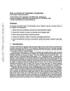
Brachial Plexus Block •The injection of an anesthetic solution into or immediately surrounding the axillary sheath interrupts nerve impulses of the structures supplied by the branches of the cords of the brachial plexus. •This procedure allows surgeons to operate on the upper limb without using a general anesthetic. •Needle approaches include an interscalene, supraclavicular, infraclavicular & axillary approach.
www.frca.co.uk
Main Menu
BACK
Test Yourself
Main Menu
BACK C5
C6 C7
C8
AD
Thoracodorsal (C6-C8)
T1 PD
Ansa pectoralis
Test Yourself
Main Menu
BACK C5
C6 C7
C8
AD
T1
Lower subscapular (C5, C6)
PD
Ansa pectoralis
Test Yourself
Main Menu
BACK C5
C6 C7
C8
AD
T1 PD
Ansa pectoralis Axillary (C5, C6)
Test Yourself
Main Menu
BACK C5
C6 C7
C8
AD
T1 PD
Ansa pectoralis
Radial (C5-C8, T1)
Test Yourself
Main Menu
BACK C5
C6 C7
C8
AD
T1 PD
Ansa pectoralis
Upper subscapular (C5, C6)
Bicipital Myotatic Reflex •The bicipital myotatic reflex or biceps brachii reflex is one of several deeptendon reflexes that are routinely tested during a physical examination. •A normal (positive) response is an involuntary contraction of the biceps, felt as a momentarily tensed tendon with usually a brief jerk-like flexion of the elbow. •A positive response confirms the integrity of the musculocutaneous nerve and the C5 & C6 spinal cord segments.
Main Menu
BACK
Biceps Brachii
Main Menu
•Innervation: musculocutaneous nerve •Function: supinates forearm; flexes forearm when it is supinated; flexes arm; short head resists dislocation of shoulder
•Origin: long head: supraglenoid tubercle of scapula; short head: coracoid process of scapula •Insertion: radial tuberosity and fascia of forearm via bicipital aponeurosis
Biceps brachii
BACK
Brachialis
Main Menu
BACK
•Innervation: musculocutaneous nerve •Function: flexes forearm •Origin: distal 1/2 of anterior surface of humerus
•Insertion: coronoid process and tuberosity of ulna
Brachialis
Coracobrachialis
Main Menu
BACK
•Innervation: musculocutaneous nerve •Function: flex and adduct the arm; resists dislocation of the shoulder joint •Origin: coracoid process of scapula
•Insertion: middle 1/3 of medial surface of humerus
Coracobrachialis
Orbital Muscles: Function (Elevation)
Main Menu
BACK Inferior oblique
Horizontal axis
Medial
Lateral
Superior rectus
Orbital Muscles: Function (Depression)
Main Menu
BACK Superior oblique
Horizontal axis
Medial
Lateral
Inferior rectus
Orbital Muscles: Function (Abduction)
Main Menu
BACK Inferior oblique
Superior oblique
Vertical axis
Lateral rectus
Medial
Lateral
Orbital Muscles: Function (Adduction)
Main Menu
BACK Vertical axis
Medial rectus
Superior rectus
Inferior rectus Medial
Lateral
Orbital Muscles: Function (Lateral Rotation)
Main Menu
BACK Anteroposterior axis
Lateral rotation
Inferior rectus
Inferior oblique Lateral
Medial
Orbital Muscles: Function (Medial Rotation)
Main Menu
BACK Superior rectus
Superior oblique
Medial rotation
Anteroposterior axis
Lateral
Medial