is followed by two epidermal growth factor-like domains, an activation peptide, and ... Therefore the triple helix is not inperfect register, a condition which appears.
Proc. Natl. Acad. Sci. USA Vol. 93, pp. 11068-11073, October 1996 Medical Sciences
Identification of the endothelial cell binding site for factor IX (collagen/coagulation)
WING-FAI CHEUNG*, JACOB VAN DEN BORNt, KLAUS KtJHNt, LENA KJELLEN§, BILLY G. HUDSONT, DARREL W. STAFFORDII
AND
*Department of Medical and Physiological Chemistry, University of Uppsala, the Biomedical Center, Box 575, S-751 23 Uppsala, Sweden; tDivision of Nephrology, University Hospital St. Radboud, 6500 HB Nijmegen, The Netherlands; *Max Planck Institute for Biochemistry, Martinsried 82152, Germany; §Department of Veterinary Medical Chemistry, Swedish University of Agricultural Sciences, the Biomedical Center, Box 575, S-751 23 Uppsala, Sweden; tDepartment of Biochemistry and Molecular Biology, University of Kansas Medical Center, 3901 Rainbow Boulevard, Kansas City, KS 66160-7421; and IDepartment of Biology, University of North Carolina at Chapel Hill, Chapel Hill, NC 27599-3280
Communicated by Kenneth M. Brinkhous, University of North Carolina, Chapel Hill, NC, July 26, 1996 (received for review April 14, 1996)
ABSTRACT We previously demonstrated that the primary region of factor IX and IXa responsible for saturable specific binding to bovine aortic endothelial cells resides in residues 3-11 at the amino terminus of factor IX. We also demonstrated that mutations of lysine to alanine at residue 5, factor IX K5A, or valine to lysine at residue 10, factor IX V10K, resulted in a molecule unable to bind to endothelial cells. Moreover, a mutation with lysine to arginine at residue 5, factor IX K5R, resulted in a factor IX molecule with increased affinity for the endothelial cell binding site. In this paper we report that collagen IV is a strong candidate for the factor IX binding site on endothelial cells. Factor IX and factor IX K5R compete with '251-labeled factor IX for binding to tetrameric collagen IV immobilized on microtiter plates, while factor X, factor VII, and factor IX K5A or V1OK fail to compete. The Kd for wild-type factor IX binding to collagen IV in the presence of heparin was 6.8 ± 2 nM, and the Kd for factor IX K5R was 1.1 + 0.2 nM, which agrees well with our previously published Kd values of 7.4 and 2.4 nM for binding of the same proteins to endothelial cells. Our working assumption is that we have identified the endothelial cell binding site and that it is collagen IV. Its physiological relevance remains to be determined.
complex assembled upon phospholipids. Finally, Brinkman et al. (5) showed that the endothelium may serve as an effective surface for formation of a tenase complex. We (6) determined that the primary region of factor IX and IXa responsible for binding to bovine aortic endothelial cells resides in residues 3-11 at the amino terminus of factor IX. We also demonstrated that mutations of lysine to alanine at residue 5, factor IX K5A, or valine to lysine at residue 10, factor IX V10K, result in a molecule unable to bind to endothelial cells. Moreover, a mutation of lysine to arginine at residue 5, factor IX K5R, results in a factor IX molecule that has a 3-fold increased affinity for the endothelial cell binding site. Our observation that factor IX residues 3-11 are critical for the factor IX endothelial cell interaction have recently been confirmed by Castellino's laboratory (7). They demonstrated that converting the first 11 amino acids of protein C to those of factor IX resulted in a protein C chimera that bound to endothelial cells. In an attempt to identify the molecule on bovine aortic endothelial cells to which factor IX was binding, Rimon et al. (8) cross-linked factor IX to a 140-kDa protein on the surface of endothelial cells. In spite of this initial success, a number of laboratories have failed to identify the molecule to which factor IX binds, probably because the cross-linked factor IX was bound to a proteolytic fragment of collagen IV. In this paper we report that collagen IV is a strong candidate for the factor IX binding site on endothelial cells. Collagen IV is an extracellular matrix (ECM) protein which is one of the major components of the basement membrane of endothelial cells (9, 10). It is found arranged in a two-dimensional array in the lamina densa of basement membranes and is usually isolated from placenta (11) or the mouse tumor cell line EHS (12). The isolation includes a limited pepsin digestion which leads to a significant fraction of tetrameric collagen. Tetrameric collagen IV is composed of four triple-helix collagen molecules joined by their amino-terminal 7S globular domains (13). The triple-helical molecule, which consists of two al chains and one a2 chain, constitutes the collagen-like domain of collagen IV. The collagenous domain of collagen IV differs from fibrillar collagens in that its Gly-Xaa-Yaa sequence is interrupted by noncollagen-type sequences. Therefore the triple helix is not in perfect register, a condition which appears to provide flexibility for supercoiling of triple helices. Moreover, collagen IV has noncollagenous domains at both the amino and carboxyl termini of its constituent polypeptide chains. Collagen IV may also be extracted by limited cleavage with collagenase to yield dimeric collagen. Dimeric collagen is composed of two triple-helical collagen molecules which are attached at their carboxyl termini by their NC1 domains and
Factor IX is the zymogen of a serine protease which is active in normal hemostasis. Mutations in factor IX result in the bleeding disease hemophilia B. Factor IX is activated by factor XIa or factor VIla-tissue factor to yield factor IXa; this reaction results from cleavage between residues 145/146 and 180/181 of the factor IX. Activated factor IX, together with its nonenzymatic cofactor factor VIlla, cleaves factor X to factor Xa. Factor IX, like factors X and VII, is a vitamin K-dependent clotting factor which contains multiple 4-carboxyglutamic acid (Gla) residues at its amino terminus. The factor IX Gla domain is followed by two epidermal growth factor-like domains, an activation peptide, and a serine protease domain. For a recent review see Roberts (1). The binding of factor IX to a specific saturable site on endothelial cells was first described by Heimark et al. (2) and Stem et al. (3). Heimark et al. found that bovine endothelial cells had approximately 280,000 sites per cell with a Kd for factor IX of about 5 nM. The factor IX binding sites on the endothelial cell were subsequently characterized in great detail by Stern et al. (4). They reported about 40,000 sites per cell and a Kd for factor IX or IXa of about 2 nM. Moreover, it was found that factor IXa in the presence of factor VIII and factor X binds with a 10-fold increased affinity. The factor X-cleaving ("tenase") complex on endothelial cells could catalyze the formation of factor Xa with a greater Vmax than a tenase The publication costs of this article were defrayed in part by page charge payment. This article must therefore be hereby marked "advertisement" in accordance with 18 U.S.C. §1734 solely to indicate this fact.
Abbreviations: ECM, extracellular matrix; EHS, Engelbreth-HolmSwarm. 11068
Medical Sciences:
Cheung et al.
which lack the 7S domain (14). A cyanogen bromide fragment from the triple-helical region of collagen IV (15, 16) possesses high-affinity binding sites for a1l31 and a2031 integrins. The major collagen-binding integrin of platelets (17) is gp Ia/IIa, which is a synonym for a2f1. We report here that the triple-helical region of collagen IV possesses all of the attributes previously described for the factor IX binding site on endothelial cells.
MATERIALS AND METHODS Materials. Dulbecco's modified Eagle's medium (DMEM), DMEM/F12, fetal bovine serum, and porcine intestinal mucosa heparin were from GIBCO/BRL. Removable microtiter wells were from Corning. lodobeads were from Pierce. Sodium deoxycholate was from Merck. Bovine aortic endothelial cells were gifts from Charles Esmon and Naomi Esmon of the Oklahoma Medical Research Foundation (Oklahoma City, OK). Human plasma factor IX, factor IXa, factor X, factor VII, and thrombin were from Enzyme Research Laboratories (South Bend, IN). Factor VIII (Recombinate) was from Baxter Laboratories (Glendale, CA). Spectrozyme Xa was from American Diagnostica (Greenwich, CT). Recombinant wild-type factor IX and mutated factor IX were expressed and purified as previously described (6). Human fibronectin and bovine collagen I (from Collagen Corp., Palo Alto, CA) were gifts from Staffan Johansson of the Department of Medical and Physiological Chemistry, Uppsala University, Sweden. Mouse laminin was from Rupert Timpl of the Max-Planck-Institut fur Biochemie, Germany. Recombinant mouse entactin was from Upstate Biotechnology (Lake Placid, NY). Heparan sulfate proteoglycan (HSPG) and lathyritic collagen IV isolated from Engelbreth-Holm-Swarm (EHS) mouse sarcoma and human placenta collagen type V were from Becton Dickinson (Bedford, MA). Tetrameric (18), or dimeric (14) collagen, or the 7S domain (19) and cyanogen bromide fragment (CB3) of human placenta collagen IV (16) were prepared as described in the referenced papers. A different form of dimeric collagen IV, composed of truncated ca1 and a2 chains from bovine testes basement membrane, was prepared as described (20) for lens capsule basement membrane and was characterized by electron microscopy, SDS/ PAGE, and Western blotting (T. Kahsai and B.G.H., unpublished results). Recombinant collagenase, and human collagens types I, III, IV, and V were purchased from Sigma. Rabbit anti-human collagen IV polyclonal antibody was from Chemicon. Binding of Factor IX to ECM. Bovine endothelial cells were grown in 48-well plates in DMEM containing 10% fetal bovine serum. Confluent cell monolayers were solubilized by treating three times for 10 min each with 0.5% sodium deoxycholate/10 mM Tris HCl/1 mM phenylmethylsulfonyl fluoride, pH 8. This was followed by washing the remaining ECM three times with 2 mM Tris.HCl/1 mM phenylmethylsulfonyl fluoride, pH 8 (21). Binding experiments were done by using 0.5 nM 125I_ labeled factor IX (1251-factor X) and 200-fold excess (100 nM) unlabeled proteins. Competition binding of factor IX with other ligands to intact bovine endothelial cells was as described
(6). Binding of Factor IX to Collagen Type IV. Lyophilized collagen IV was dissolved to 3 mg/ml in TBS (10 mM Tris HCl/150 mM NaCl/2 mM CaCl2/1 mM MgCl2, pH 8) and then diluted to 50 ,ug/ml. Removable wells were coated with 0.1 ml of this solution at 4°C overnight. The wells were washed three times with TBS and blocked with 3% ovalbumin for 3 hr at room temperature. Ninety microliters of various concen-
trations of unlabeled factor IX (or other competitors) in the presence (1 ,ug/ml) or absence of heparin was then added to each well. Ten microliters of 125I-factor IX (final 0.5 nM) was added to the wells and incubated at 4°C for 2.5 hr; the wells
Proc. Natl. Acad. Sci. USA 93 (1996)
11069
were then washed six times and bound radioactive material was measured in a y counter (Packard). Other ECM proteins were used to coat the wells as described above and tested for binding with factor IX. Heat Denaturation and Sensitivity to Dithiothreitol. To test for the dependency of factor IX binding to the triple-helical conformation of collagen IV,tetrameric type IV collagen was denatured by heating at 60°C for 30 min and/or treatment with 10 mM dithiothreitol before coating. The binding study was conducted as described above by incubating 0.5 nM 1251-factor IX with a 200-fold excess of unlabeled factor IX. Elimination of Factor IX Binding to Collagen IV by Collagenase Treatment. Five micrograms of human placental collagen IV in 100 ,u was added to each well of a 96-well microtiter plate and incubated at 4°C overnight. After three washes with TBS, the coatings in some wells were digested with 2.5 ,ug of recombinant collagenase-I in 100 ,ul of TBS containing 2 mM CaCl2, .1 mM MgCl2, and 0.2% BSA at 37°C for 2 hr. After three additional washes with TBS, binding experiments were performed at 4°C for 2.5 hr as described above. Factor X Activation on Endothelial Cells and ECMs. Confluent bovine endothelial cells were seeded at 1.5 x 10⁵ cells per well and grown in 24-well plates for 2 days. The ECM of endothelial cells was prepared as described above by removing the cells with 0.5% deoxycholate. Factor IXa (0.5 nM) was incubated with intact endothelial cells and ECM as in a binding experiment, and the tenase activity was measured on both the matrix and washed cells. The reaction was started by the addition of 400 ,lI of a solution containing 5 units/ml factor VIIIa and 0.2 ,uM factor X in 5 mM Tris.HCl/140 mM NaCl/2 mM CaCl2/0.2% BSA, pH 7.5. Factor VIII was preactivated with thrombin at 37°C for 5 min and stored on ice until use. Aliquots (40 ,ul) were collected at timed intervals over 25 min. Each aliquot was added to a well of a 96-well microtiter plate containing 60 gl of 50 mM Tris.HCl/175 mM NaCl/6 mM EDTA/0.05% ovalbumin, pH 7.5. Factor Xa activity was assayed by adding 100 ,ul of 0.2 mM chromogenic substrate, Spectrozyme Xa, and the absorbance at 405 nm was monitored with a plate reader (Labsystems, Chicago) for 3 min at room temperature.
RESULTS AND DISCUSSION Our initial stimulus to examine the role of the ECM in factor IX binding arose from the observation that heparan sulfate and heparin affected the binding of factor IX to endothelial cells. To examine the possible role of the ECM in factor IX binding, endothelial cells were removed by exposure to 0.5% sodium deoxycholate as described in Materials and Methods. Fig. 1 demonstrates that, at the time of measurement, 2 days, more factor IX binding sites are on the matrix than on intact endothelial cells. When the cells reach confluence, however, the situation is reversed. Therefore, although this experiment clearly demonstrates that the ECM binds factor IX, it is likely that collagen IV or an epitope of collagen IV is also present on the surface of endothelial cells. To examine if the ECM maintained the same specificity as that observed previously for endothelial cells we demonstrated that plasma factor IX or factor IX K5R competed for binding, while factor IX with the point mutations previously shown to lack the ability to bind endothelial cells (6) failed to compete (Fig. 2). This observation suggests that the binding site that we have previously characterized on endothelial cells is actually found in the ECM secreted by endothelial cells. We therefore tested a number of known ECM proteins for their ability to bind factor IX. Human fibronectin, mouse laminin, mouse entactin (nidogen), mouse heparan sulfate proteoglycan, and both human and bovine collagens I, III, and V were tested; all failed to bind factor IX (Table 1).
Proc. Natl. Acad. Sci. USA 93
Medical Sciences: Cheung et al.
11070
Binding of Factor DX to Extracellular Matrix
a:
0-03
y
7000
8000 6000
0.02 5p 4000
3000 '4I
0.015
v
0.01
A
2000 0.005k
1000
0
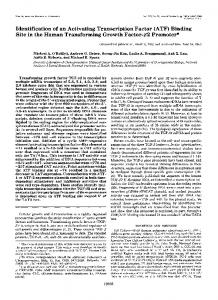
FIG. 1. Binding of 1251-factor IX to intact endothelial cell monolayers or to ECM at 24 hr after seeding. Intact monolayers were used directly or the cells were removed by treating three times, for 10 min each, with 0.5% deoxycholate/10 mM Tris HCl/1 mM phenylmethylsulfonyl fluoride, pH 8, to remove cells. The remaining ECM was washed three times with 2 mM Tris.HCl/1 mM phenylmethylsulfonyl fluoride, pH 8. Binding experiments were done on both the intact cells and the remaining ECM.
In the absence of heparin, factor IX bound to collagen IV, although with lower affinity (Kd = 43 nM) than to the ECM; in the presence of heparin at 1 ,ug/ml, however, collagen IV bound with an affinity similar to that previously determined for endothelial cells (4, 6). Therefore, heparin was used in all subsequent experiments. Fig. 3 shows that the Kd for wild-type factor IX binding to collagen IV in the presence of heparin was 6.8 ± 2 nM and the Kd for factor IX K5R was 1.1 ± 0.2 nM. These values agree well with our previously published Kd values of 7.4 and 2.4 nM for binding of the same factor IX variants to endothelial cells (6). The Kd values were obtained by analyzing three independent experiments simultaneously by the program MK model (Biosoft, Ferguson, MO). There are uncertainties in the determination of the Kd values because the concentration of competitor was frequently insufficient to allow the program to predict the nonspecific background. Therefore, the nonspecific value calculated for the factor IX K5R mutant, which did reach a plateau, was used as a constant value for those experiments. To determine if the specificity of binding observed with the ECM was retained with the purified collagen IV, competition experiments with wild-type and variant factor IXs were done. Fig.4 shows that factor IX and factor IX K5R compete with
(1996)
Table 1. Binding of ECM proteins to factor IX Binding ECM protein No Human fibronectin No Mouse laminin No Mouse nidogen No EHS cell heparan sulfate proteoglycan No Bovine collagen I No Human collagen V No Lathyritic mouse EHS collagen IV Yes Human placental collagen IV tetramer (Kuhn) Yes Human placental collagen IV dimer (Kuhn) No Human placental collagen IV 7S domain (Kuhn) Human placental collagen IV CNBr fragment No (binds to a1l31 and a2p11 integrins) (Kuhn) Yes Human placental collagen IV (Sigma) The major known constituents of the ECM were coated on microtiter plates and tested to determine which was responsible for factor IX binding.
1251-factor IX for binding to tetrameric collagen IV immobilized on microtiter plates, whereas factor X, factor VII, and factor IX K5A or V1OK fail to compete. The relative specificity is exactly that observed for endothelial cells (6). To further examine the interaction between collagen IV and factor IX, monolayers of endothelial cells were incubated with 0.5 nM 1251-factor IX and the competition was done with dimeric or tetrameric collagen IV instead of nonradioactive factor IX. The results for dimeric and tetrameric collagen were similar. Therefore, all four sets of data were analyzed simultaneously by the MK model, and a Kd value of 36 nM was obtained (see Fig. 5). This Kd value approximates the Kd determined for factor IX's binding to collagen IV in the absence of heparin. Therefore, these results appear to confirm the role of collagen IV in binding to endothelial cells. Collagen IV can be dissected into several fragments. Neither the amino-terminal 7S globular domain nor the carboxylterminal noncollagenous domain one (NC1) competed with 0.035
-
0.030
-
0.025 -
*
MU
podpon by nM
Wr FIX, K1
* X K.1
0.020-
Specficity of FIX Binding to Extracellular Matrix amc
t
ir
j
0D3
7000
oxes
i
0.015
-
IL-
i 0.010 -
om2
-m
0.006
-
00.
10
100
0
0015 100
ItSy %0_
_J
PA
300 0
0 Nonne
M
wIFIX
X
X5SR FDX K5A
FIX
V1OK FPt9411
0.1
I
Log FIX Co
ir (nM)
Competitor (200-fold excess) FIG. 3. Competition of factor IX or factor IX K5R for binding of
FIG. 2. Specificity of factor IX binding to the ECM. Confluent bovine endothelial cells were treated as described in the legend to Fig. 1 to remove cells. The remaining ECM was washed three times with 2 mM Tris.HCl/1 mM phenylmethylsulfonyl fluoride, pH 8, and the specificity of the binding was determined. FIX, factor IX; wt, wildtype; FIX 9-11, residues 9-11, Phe-Val-Gln, changed to those of factor X, Met-Lys-Lys.
1251-factor IX to collagen IV. Binding was done by incubating 0.5 nM 1251-factor IX and various concentrations of plasma factor IX as competitor for 2.5 hr at 4°C. Heparin was included in the binding buffer at 1 ,tg/ml. After six washes with cold incubation buffer, the individual wells were counted. In this case the values obtained for binding to tetrameric collagen IV are almost identical to those obtained previously with intact endothelial cells.
Medical Sciences:
Cheung et al.
Proc. Natl. Acad. Sci. USA 93 (1996)
Specficityof FIX Binding to Tetrameri Hunan Placental Coiagen TV
11071
Binding of '15I Factor DX to Collagen IV s500
I
0Q6 i
4000
if
3
I
[
QOm
M
'maS
0QOI
j
GMfl
1000
1000 *l*l
0
Competitor
0 0
t4
FIG. 4. Specificity of factor IX binding to tetrameric collagen IV. Two independent experiments are separated by a space. Lyophilized tetrameric collagen IV was dissolved to 3 mg/ml in 10 mM Tris.HCl/ 150 mM NaCl/2 mM CaCl2/1 mM MgCl2, pH 8, then diluted to 50 ,ug/ml. One hundred microliters of this solution was incubated at 4°C overnight in 96-well microtiter plates, blocked with ovalbumin, and used for binding studies.
endothelial cells for binding to factor IX (Table 1); moreover, both dimeric and tetrameric collagen bind 1251-factor IX. Because dimeric collagen IV lacks the 7S domain while tetrameric collagen IV lacks the NC1 domain, this provides additional positive data that the binding site resides in the collagenous domain of collagen IV. The collagenous domain of collagen IV can be cleaved with cyanogen bromide to give several fragments. One of these, designated CB3, has been shown to bind the integrins alf3l and a2f31 (16). The integrin a2f31 is also known as glycoprotein Ia/Ila, which is a major platelet integrin. Patients with a defect in a2131 are impaired in collagen-stimulated platelet aggregation (22). Furthermore, monoclonal antibodies against a2f31 inhibit specifically the collagen-induced aggregation of platelets (23, 24). Although 0.012
-
0.010
-
cc 0.008
-
* *\
Kd-3 ON
-
Cs
0-006_-
o0
*-
c-I
0xFIX
Treutad
FIG. 6. Treatment of collagen IV with recombinant collagenase eliminates binding of 1251-factor IX. Collagen IV was applied to microtiter plates and a portion of the wells were treated with recombinant collagenase from Sigma. Treatment was with 2.5 p,g of collagenase at 37°C for 2 hr.
the binding site for factor IX does not appear to reside in fragment CB3 of collagen IV (Table 1), it is interesting that factor IX and platelets both have binding sites on this prevalent ECM protein. It is always difficult to prove that the binding one observes is not the result of some minor contaminating protein. However, several lines of evidence point toward collagen IV as the molecule to which factor IX binds. First, a polyclonal antibody to collagen IV inhibited the binding of 1251-factor IX (data not shown). Second, recombinant collagenase treatment of the purified collagen IV totally eliminated binding (Fig. 6). We also treated endothelial cells directly with collagenase and found that factor IX binding was reduced to nearly background levels. More severe treatment of cells with collagenase would result in their release from the plates. Third, collagen IV from several different sources-for example, dimeric or tetrameric collagen IV prepared from human placenta or bovine testesbound factor IX specifically. Finally, none of the other major constituents of the ECM bind factor IX (Table 1). This suggests that it is not an expected contaminant to which factor IX is binding. The evidence that factor IX is not binding to a phospholipid membrane contaminant is shown in Fig. 7. Either heat denaturation or treatment with 10 mM dithiothreitol results in loss of binding of factor IX to collagen IV; this result is consistent with our failure to obtain positive results with ligand blotting experiments. Furthermore, we have found that mutant factor IX K5A or V1OK binds phospholipid membranes normally (Kd = 1.5 and 0.5 ,uM, respectively) while failing to compete with The Effect of Heat Denaturation or DTT Treatment on Collagen IV's FiX Binding
x
0.05
r
0.00411
|
1
0.04
o.oz
0.002
-
0.000
-
't
0.03 0.02
0.1
L
I
10 100 Concentrton of Cologen IV (nM)
1000
IV
0. 0.01
0
None
FIG. 5. Competition by collagen IV for factor IX binding to endothelial cells. In this experiment collagen IV was used as a competitor for the binding of 1251-factor IX to intact endothelial cell monolayers. Heparin was not included and the methods are as described in the legend for Fig. 4.
Heat 60 C
10 mM DrT
10 mM 1 60°C
FIG. 7. Heat denaturation or dithiothreitol (DTT) treatment of tetrameric collagen IV eliminates the binding of 125I-factor IX. The experiments were accomplished as described in the legend to Fig. 4.
Medical Sciences: Cheung et al.
11072
Proc. Natl. Acad. Sci. USA 93
1251-factor IX for binding to endothelial cells (unpublished results). When the ability of the ECM of bovine endothelial cells to function as a surface for tenase activity is compared with that of endothelial cells per se it is found that there is essentially no tenase activity on the ECM (Fig. 8), while the level of tenase activity on intact endothelial cells is similar to that observed by others (4, 5) (about 8 nmol of factor Xa generated per min). While these experiments are qualitative, it is clear that the activity of the matrix is much diminished compared with intact endothelial cells; this may simply reflect the need of a cooperative effect for phospholipid. Preliminary experiments from several industrial laboratories suggest that, while phospholipids do stimulate activity in the presence of collagen IV, the stimulation does not exceed that of phospholipid alone. At present it is difficult to assess the functional significance of these observations. Briet (25) reported that more infused factor IX could be recovered from CRM+ than from CRMpatients (CRM, cross-reacting material). He did mixing experiments to demonstrate that the cellular components did not bind the infused factor IX and postulated that the endothelial cell lining of the blood vessel was the surface to which the infused factor IX was adsorbed. Therefore it is possible that collagen IV functions merely as a storage site and is the "extravascular space" where infused factor IX disappears. This scenario would, at a minimum, put the factor IX where it is needed upon rupture of a vessel. In retrospect it is perhaps not surprising that collagen IV appears to be the factor IX binding site. Collagen IV is the major ECM protein, and one would expect coagulation to proceed after endothelial cell disruption rather than on the endothelial cell surface. Several collagens have been shown to activate platelets (26, 27), and there are a number of experiments implicating the ECM in coagulation (26). Moreover, leech and tick anticoagulants that act by inhibiting the interaction between collagen and platelets have been identified (28, 29). The interactions are complex and are not restricted to collagen IIV; however, the leech anticoagulant protein does inhibit th e binding of platelets to collagen IV (30). Collagen type IV h as also been shown to bind fibrin and cause extensive 1-.
T
,
1_
.1-
1-
I
1-
1_ .
046
0.,
/ /
045t
0
6. Cheung, W. F., Hamaguchi, N., Smith, K. J. & Stafford, D. W.
0
0.1E
0.1
-
./
0
5
10
1S
20
25
30
Time (mAn) Tenase activity of factor IX bound to collagen IV of ECM intact e ndothelial cells. Factor IXa (final 0.5 nM) was bound to endothelialI cells or matrix for 3 hr and washed six times as described in the text . The reaction was started by the addition of 400 ,ul of a solution cc ntaining S units/ml factor Vllla and 0.2 tLM factor X in 5 mM TrisHICl/140 mM NaCl/2 mM CaCl2/0.2% BSA, pH 7.5. Aliquots (40 ,ul) were collected at timed intervals over 25 min. Each aliquot wass added to a well of a 96-well microtiter plate containing 60 jilof50 m A Tris-HCl/175 mM NaCl/6 mM EDTA/0.05% ovalbumin, pH 7.5. FaLctor Xa activity was assayed by adding 100 ,ul of 0.2 mM chromogernic substrate, Spectrozyme Xa, and the absorbance at 405 nm was mc nitored with a plate reader (Labsystems) for 3 min at room temperatuire. A, Tenase activity on endothelial cells; *, tenase activity on the EC],M. FIG. 8.
or
4119-4123.
4. Stern, D. M., Nawroth, P. P., Kisiel, W., Vehar, G. & Esmon, C. T. (1985) J. Biol. Chem. 260, 6717-6722. 5. Brinkman, H. J. M., Mertens, K., Holthuis, J., Zwart-Huinink, L. A., Grijm, K. & van Mourik, J. A. (1994) Br. J. Haematol. 87, 332-342.
/
I
1. Roberts, H. R. (1993) Thromb. Haemostasis 70, 1-9. 2. Heimark, R. L. & Schwartz, S. M. (1983) Biochem. Biophys. Res. Commun. 111, 723-731. 3. Stern, D. M., Drillings, M., Nossel, H. L., Hurlet, Jensen, A., 80,
/
5
structural alterations (31). Another coagulation-related ligand which binds with high affinity to collagen IV is protease nexin-1, which binds specifically to collagen IV with a Kd of approximately 15 nM (32). This interaction decreases protease nexin-l's rate constant for the inhibition of urokinase or plasmin. Because collagen IV appears to have so many properties identical with those of the previously characterized factor IX binding site-that is, the Kd is nearly identical, the specificity of mutant factor IX is identical, and factor IX and IXa bind indistiguishably-it seems highly probable that collagen IV is the factor IX endothelial cell binding site that we have previously characterized. However, we cannot eliminate the possibility that there is yet another binding site for factor IX that resides upon the endothelial cell surface, that has different binding specificity, and that is responsible for the observed tenase activity of endothelial cells. One aspect of our work is in contradiction to the results of Rimon et al. (8), who reported that there was no binding to the plates after cells had been removed by the detergents CHAPS or Triton X-100. In our hands removing cells with either detergent results in a surface with specific factor IX binding activity. In addition, they reported that the binding specificity was maintained by proteins in the cellular extracts. We also observed that partially purified endothelial cell membranes adsorbed to microtiter plates resulted in a factor IX binding surface. We made large-scale membrane preparations from a bovine placenta but found that, while there was binding activity, there was no discrimination between factors IX, X, and VII. These differences remain unexplained. The observation that collagen IV has a specific binding site for factor IX demands a host of additional physiological and biochemical experiments on the interaction between factor IX and the ECM as well as factor IX and collagen IV. Furthermore, it may be profitable to look at the effect of collagen IV on other blood coagulation factors.
LaGamma, K. S. & Owen, J. (1983) Proc. Natl. Acad. Sci. USA
,,
OAI
(1996)
(1992) J. Biol. Chem. 267, 20529-20531. 7. Geng, J.-P., Christiansen, W. T., Plow, E. F. & Castellino, F. J. (1995) Biochemistry 34, 8449-8457. 8. Rimon, S., Melamed, R., Savion, N., Scott, T., Nawroth, P. P. & Stern, D. M. (1987) J. Biol. Chem. 262, 6023-6031. 9. Kuhn, K. (1994) Matrix Biol. 14, 439-445. 10. Hudson, B. G., Reeders, S. T. & Tryggvason, K. (1993) J. Biol. Chem. 268, 26033-26036.
11. Glanville, R. W. & Kuhn, K. (1979) Front. Matrix Biol. 7, 19-26.
12. Timpl, R., Martin, G. R., Bruckner, P., Wick, G. & Wiedemann, H. (1978) Fur. J. Biochem. 84, 43-52. 13. Timpl, R, Wiedemann, H., van Delden, V., Furthmayr, H. & Kuhn, K. (1981) Eur. J. Biochem. 120, 203-211. 14. Dolz, R., Engel, J. & Kuhn, K. (1988) Eur. J. Biochem. 178, 357-366. 15. Eble, J. A., Golbik, R., Mann, K. & Kuhn, K. (1993) EMBO J. 12, 4795-4802. 16. Vandenberg, P., Kern, A., Ries, A., Luckenbill-Edds, L., Mann, K. & Kuhn, K. (1991) J. Cell Biol. 113, 1475-1483. 17. Sixma, J. J. & de Groot, P. G. (1994) Ann. N.Y Acad. Sci. 714, 190-199. 18. Glanville, R. W., Rauter, A. & Fietzek, P. P. (1979) Eur. J. Biochem. 95, 383-389.
Medical Sciences: Cheung et al. 19. Risteli, J., Bachinger, H. P., Engel, J., Furthmayr, H. & Timpl, R. (1980) Eur. J. Biochem. 108, 239-250. 20. Gunwar, S., Noelken, M. E. & Hudson, B. G. (1991) J. Biol. Chem. 266, 14088-14094. 21. Hedman, K., Kurkinen, M., Alitalo, K., Vaheri, A., Johansson, S. & Hook, M. (1979) J. Cell Biol. 81, 83-91. 22. Nieuwenhuis, H. K., Sakariassen, K. S., Houdijk, W. P., Nievelstein, P. F. & Sixma, J. J. (1986) Blood 68, 692-695. 23. Santoro, S. A. (1986) Cell 46, 913-920. 24. Coller, B. S., Beer, J. H., Scudder, L. E. & Steinberg, M. H. (1989) Blood 74, 182-192. 25. Briet, E. (1977) Ph.D. thesis (Univ. of Leiden, Leiden, The
Netherlands).
Proc. Natl. Acad. Sci. USA 93 (1996)
11073
26. Sixma, J. J., van Zanten, G. H., Banga, J. D., Nieuwenhuis, H. K. & de Groot, P. G. (1995) Semin. Hematol. 32, 89-98. 27. Kehrel, B. (1995) Semin. Thromb. Hemostasis 21, 123-129. 28. Connolly, T. M., Jacobs, J. W. & Condra, C. (1992) J. Biol. Chem. 267, 6893-6898. 29. Karczewski, J., Waxman, L., Endris, R. G. & Connolly, T. M. (1995) Biochem. Biophys. Res. Commun. 208, 532-541. 30. van Zanten, G. H., Connolly; T. M., Schiphorst, M. E., de Graaf, S., Slootweg, P. J. & Sixma, J. J. (1995) Arterioscler. Thromb. Vasc. Biol. 15, 1424-1431. 31. Jones, M. & Gabriel, D. A. (1988) J. Biol. Chem. 263, 7043- 7048. 32. Donovan, F. M., Vaughan, P. J. & Cunningham, D. D. (1994) J. Biol. Chem. 269, 17199-17205.