A, Drawing shows anatomy pertinent to nodal classification. ..... manubrium in the superior mediastinum, be- ... rior mediastinal nodes extend caudally to the.
Downloaded from www.ajronline.org by 54.210.20.124 on 11/02/15 from IP address 54.210.20.124. Copyright ARRS. For personal use only; all rights reserved
Imaging-Based Nodal Classification for Evaluation of Neck Metastatic Adenopathy Peter M. Som 1 Hugh D. Curtin 2 Anthony A. Mancuso 3
OBJECTIVE. This study was undertaken to create an imaging-based classification for the lymph nodes of the neck that will be readily accepted by clinicians, result in consistent nodal classification, and be easily used by radiologists. SUBJECTS AND METHODS. Over an 18-month period, the necks of 50 patients with cervical lymphadenopathy were scanned with CT, MR imaging, or both. Imaging anatomic landmarks were sought that would create a nodal classification of these necks similar to the clinically based nodal classifications of the American Joint Committee on Cancer and the American Academy of Otolaryngology–Head and Neck Surgery. Each nodal level was defined to ensure consistent nodal classification and eliminate areas of confusion existing in the clinically based classifications. RESULTS. Necks were classified using the imaging-based classification and then compared with the classification of the same necks using the most common clinically based classifications. The imaging-based nodal classifications of the superficial nodes were the same as the clinically based classifications; however, the deep nodes of eight patients were found only by imaging. The anatomic precision and the level definition afforded by sectional imaging allowed the radiologists to use the imaging-based classification in a consistent manner. CONCLUSION. This imaging-based classification has been endorsed by clinicians who specialize in head and neck cancer. The boundaries of the nodal levels were easily discerned by radiologists and yielded consistent nodal classifications. The reproducibility of this classification will allow it to become an essential component of future classifications of metastatic neck disease.
F
Received June 15, 1999; accepted after revision August 19, 1999. 1 Department of Radiology, Mount Sinai School of Medicine, City University of New York, One Gustave Levy Pl., New York, NY 10029. Address correspondence to P. M. Som. 2
Department of Radiology, Massachusetts Eye and Ear Infirmary, 243 Charles St., Boston, MA 02114.
3 Department of Radiology, Shands Hospital, University of Florida College of Medicine, 1600 Southwest Archer Rd., Gainesville, FL 32610.
AJR 2000;174:837–844 0361–803X/00/1743–837 © American Roentgen Ray Society
AJR:174, March 2000
or nearly four decades, the most commonly used classification for the cervical lymph nodes was that developed by Rouvière [1] in 1938. His work, and earlier works, precisely defined the anatomic location of the lymph nodes and mapped their drainage areas [2, 3]. The landmarks used in those early classifications were those identified by palpation and those noted only at surgery or dissection. In 1981, Shah et al. [4] suggested that the anatomically based terminology be replaced with a simpler classification based on levels. Since then, a number of classifications have been proposed that use such level, region, or zone terminology [5–13]. The purpose of these newer classifications was not to change terminology, but to group the cervical nodes on the basis of the clinical and pathophysiologic information gleaned in the intervening 60 years [8, 14, 15]. The direction of nodal
classification changed from that of a pure anatomic study to a nodal mapping guide for selecting the most appropriate surgical procedure among the various types of neck dissections [15]. The latest and most used of these classifications are the ones of the American Joint Committee on Cancer and the American Academy of Otolaryngology–Head and Neck Surgery [10, 11, 15]. The imagingbased classification created in our study was designed to be compatible with these widely accepted clinically based classifications [16]. Imaging was chosen as the basis of this new classification for several reasons: at least 80% of patients with head and neck cancer undergo CT or MR imaging before treatment and, in general, only those patients with small superficial tumors do not receive such pretreatment imaging; imaging can reveal clinically silent nodes [5, 17–24]; imaging, if properly performed, has the potential to best
837
Downloaded from www.ajronline.org by 54.210.20.124 on 11/02/15 from IP address 54.210.20.124. Copyright ARRS. For personal use only; all rights reserved
Som et al. show precise anatomic landmarks that make the nodal levels reproducible; and nodal information to classify nodes in the neck is no longer obtainable only at surgery. Although both the imaging-based and clinically based classifications are designed as independent classifications, the best possible classification of cervical nodal disease may be accomplished by using both clinical palpation and information provided by imaging. For example, because of the slope of the shoulders, the supraclavicular fossa is not as well defined on axial CT and MR imaging as it is on palpation, especially when Ho’s triangle is used as the defining anatomic plane [11]. Classifying some lymph nodes located at the junction between levels may be diffi-
cult with imaging; however, when combined with clinical assessment, such classification problems are easily resolved. This article presents the imaging-based nodal classification and explains how to use it. Subjects and Methods Prospectively, over an 18-month period, the necks of 50 patients with cervical lymphadenopathy were scanned with CT, MR imaging, or both. The anatomic landmarks of the two most common clinically based classifications were used as initial guidelines for assessing the boundaries of nodal levels. This was done to create imaging definitions that would be consistent with those of the clinically based classifications. Areas of difficulty in both the clinically based and imaging-based classifications were then prospectively evaluated
A
and new imaging anatomic landmarks were sought to resolve any confusion among sites. Radiologists independently classified the cervical lymphadenopathy on the CT and MR imaging studies using the imaging-based classification. The same patients were independently classified by otolaryngologists using the clinically based classifications. Comparison between these neck assessments was then made. The aim of the study was to have each neck similarly classified using the clinically and imaging-based classifications so that clinicians would accept the imaging-based classification. When using the imaging-based classification, a consistent scanning technique must be used to provide reproducible nodal levels. For CT, such consistency includes patient positioning and gantry angulation. Although there is no single method of performing CT of the neck, the following technique is used by many head and neck radiologists, and
B
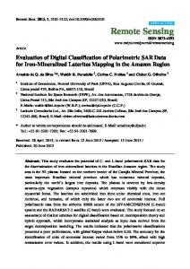
Fig. 1.—Neck as seen from left anterior view. A, Drawing shows anatomy pertinent to nodal classification. B, Drawing shows specific margins of anatomy seen in A that relate to definitions of classification levels. Note that line of separation between levels I and II is posterior margin of submandibular gland. Separation between levels II and III and level V is posterior edge of sternocleidomastoid muscle. Line of separation between levels IV and V is oblique line extending from posterior edge of sternocleidomastoid muscle to posterior edge of anterior scalene muscle. Posterior edge of internal jugular vein separates level IIA and IIB nodes. Carotid arteries separate levels III and IV from level VI. Top of manubrium separates levels VI and VII. (Reprinted with permission from [16])
838
AJR:174, March 2000
Downloaded from www.ajronline.org by 54.210.20.124 on 11/02/15 from IP address 54.210.20.124. Copyright ARRS. For personal use only; all rights reserved
Imaging-Based Nodal Classification slight variations from this approach do not change the nodal levels. The axial plane referred to in this classification is obtained with the patient’s head in a comfortable neutral position with the hard palate perpendicular to the tabletop and the shoulders down as far as possible. The scanner gantry is aligned along the inferior orbitomeatal plane and, if possible, the examination is performed with the administration of IV contrast material to allow the best possible differentiation of nodes from vessels. The recommended field of view is 16 × 18 cm. The CT examination is performed as contiguous 3-mm scans from the skull base to the manubrium or as a helical study reconstructed as contiguous 2- or 3mm slices. The helical technique uses 3-mm thick scans with a 3-mm gap and a pitch of 1:1. MR images should be no thicker than 5 mm (preferably 3– 4 mm) with a 1-mm interslice gap. If the patient has a history of thyroid or cervical esophageal cancer, the caudal margin of the studies should extend to the level of the carina to ensure inclusion of the superior mediastinum. The pertinent anatomic landmarks used for classification are depicted in the diagrams in Figure 1. The radiologist must be able to identify the essential anatomic landmarks of the classification: the skull base at the jugular fossa, the bottom of the body of the hyoid bone, the bottom of the cri-
coid arch, the top of the manubrium, the back edge of the submandibular gland, the back edge of the sternocleidomastoid muscle, the lateral posterior edge of the anterior scalene muscle, the anterior edge of the trapezius muscle, both the internal carotid and common carotid arteries, the internal jugular vein, the clavicle, the medial margin of the anterior belly of the digastric muscle, and the mylohyoid muscle. For consistency, all figure images in this article are CT scans that illustrate the use of the new classification. MR images could have been used. The final imaging-based classification is the result of continual use and refining of the classification by both radiologists and consulting clinicians during this 18-month period.
Results
The clinically and imaging-based classifications of the cervical lymphadenopathy were the same for superficial nodal disease in all 50 patients. There were five patients with retropharyngeal lymphadenopathy identified only by imaging. There were also three patients with low visceral nodes identified only by imaging. Using the imaging-based classification (Appendix), the radiologists classified all necks the same.
A
Discussion How to Use the Classification
The classification was designed to be easy to use. Each side of the neck should be evaluated separately; the lines that are used to define the boundaries of the levels should be drawn separately for each side of the neck. The lines need not actually be drawn; when one becomes familiar with the classification, one can visually approximate the lines or place a straight-line guide or ruler on the image or monitor. When a lymph node is transected by one of the lines that define the levels, the side of the line on which most of the nodal cross-sectional area lies is the level in which the lymph node should be classified. The supraclavicular fossa is defined on each axial scan when any portion of the clavicle is identified on one side of the neck; if the scan level is cranial to any portion of the clavicle, the nodes in the lower lateral neck should be classified as either level IV or level VB. Once any portion of the clavicle is seen on the scan, such nodes are classified as supraclavicular nodes. If nodes are seen below the level of the
B
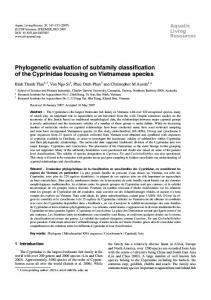
Fig. 2.—44-year-old man with lymphoma. ad = anterior belly of digastric muscle, S = submandibular gland, vn = internal jugular vein, Sc = sternocleidomastoid muscle. A, Axial contrast-enhanced CT scan of neck through floor of mouth and above level of hyoid bone. White line is drawn through back of each submandibular gland. Nodes anterior to lines are level I nodes and, because they are lateral to medial margin of each anterior belly of the digastric muscle, they can be subclassified as level IB nodes. Level II nodes are posterior to white lines, but anterior to posterior edge of sternocleidomastoid muscle. Level II nodes, either anterior to the internal jugular vein or posterior to it but touching it, are subclassified as level IIA nodes. Nodes posterior to internal jugular vein and not touching it are subclassified as level IIB nodes. B, Axial contrast-enhanced CT scan of neck through floor of mouth and at level of hyoid bone. Because level is cranial to that of bottom of body of hyoid bone, internal jugular nodes are classified as level II, not level III. On each side of neck, transverse white line (Ant) has been drawn through posterior edge of each submandibular gland. Second white line (Post) has been drawn through posterior edge of each sternocleidomastoid muscle. Nodes anterior to line (Ant) are level I nodes. Nodes between line (Ant) and line (Post) are level II nodes. Nodes posterior to line (Post) are level V nodes. Because level V nodes are cranial to level of bottom of body of cricoid cartilage arch, they are subclassified as level VA nodes.
AJR:174, March 2000
839
Downloaded from www.ajronline.org by 54.210.20.124 on 11/02/15 from IP address 54.210.20.124. Copyright ARRS. For personal use only; all rights reserved
Som et al. clavicle and lateral to the ribs, they are axillary nodes. The clinically important internal jugular nodes described by Rouvière [1] are now classified as level II, III, or IV nodes, depending on their locations with reference to the axial scan levels of the bottom of the body of the hyoid bone and the bottom of the arch (anterior rim) of the cricoid cartilage. The Imaging-Based Classification
Level I includes all nodes above the hyoid bone, below the mylohyoid muscle, and anterior to a transverse line drawn on each axial image through the posterior edge of the submandibular gland (Figs. 2 and 3). Thus, level I nodes include the previously classified submental and submandibular nodes, and they can be subclassified into levels IA and IB. Level IA represents the nodes that lie between the medial margins of the anterior bellies of the digastric muscles, above the hyoid bone, and below the mylohyoid muscle (previously classified as submental nodes) (Figs. 2 and 3). Level IB represents the nodes that lie below the mylohyoid muscle, above the hyoid bone, posterior and lateral to the medial edge of the anterior belly of the digastric muscle, and anterior to a transverse line drawn on
each axial image tangent to the posterior surface of the submandibular gland on each side of the neck (previously classified as submandibular nodes) (Figs. 2 and 3). Level II extends from the skull base, at the lower level of the bony margin of the jugular fossa, to the level of the lower body of the hyoid bone (Figs. 2–5). Level II nodes lie anterior to a transverse line drawn on each axial image through the posterior edge of the sternocleidomastoid muscle and lie posterior to a transverse line drawn on each axial scan through the posterior edge of the submandibular gland. If a node situated within 2 cm of the skull base lies anterior, lateral, or posterior to the carotid sheath, it is classified as a level II node. If the node lies medial to the internal carotid artery, it is classified as a retropharyngeal node (Fig. 5). Caudal to 2 cm below the skull base, level II nodes can lie anterior, lateral, medial, and posterior to the internal jugular vein. Level II nodes can be subclassified into levels IIA and IIB. Level IIA nodes are level II nodes that lie posterior to the internal jugular vein and are inseparable from the vein or that lie anterior, lateral, or medial to the vein (previously classified as upper internal jugular nodes) (Figs. 2–5).
Fig. 3.—36-year-old HIV-positive man. Axial contrast-enhanced CT scan of neck through level of lower mandible and above hyoid bone shows level IA node between medial margins of anterior bellies of digastric muscles (ad). Level IB nodes are lateral to level IA nodes and anterior to back of submandibular glands (S). Levels IIA and IIB nodes are seen bilaterally. v = internal jugular veins, Sc = sternocleidomastoid muscles.
840
Level IIB nodes are level II nodes (previously classified as upper spinal accessory nodes) that lie posterior to the internal jugular vein and have a fat plane separating the nodes and the vein (Figs. 2 and 3). Level III nodes lie between the level of the lower body of the hyoid bone and the level of the lower margin of the cricoid cartilage arch (Figs. 6 and 7). These nodes lie anterior to a transverse line drawn on each axial image through the posterior edge of the sternocleidomastoid muscle. Level III nodes also lie lateral to the medial margin of either the common carotid artery or the internal carotid artery. On each side of the neck, the medial margin of these arteries separates level III (lateral) nodes from level VI (medial) nodes. Level III nodes were previously known as the mid jugular nodes. Level IV nodes lie between the level of the lower margin of the cricoid cartilage arch and the level of the clavicle on each side as seen on each axial scan. These nodes lie anterior and medial to an oblique line drawn through the posterior edge of the sternocleidomastoid muscle and the posterolateral edge of the anterior scalene muscle on each axial image (Figs. 7– 10). The medial aspect of the common carotid artery is the landmark that separates level IV
Fig. 4.—Axial contrast-enhanced CT scan of neck through level of hyoid bone in 57year-old man with squamous cell carcinoma of pharynx. White line has been drawn through posterior edge of right sternocleidomastoid muscle (Sc). Note conglomerate mass of necrotic level IIA nodes in right neck posterior to submandibular gland (S). Small necrotic level VA nodes are also present on right side.
AJR:174, March 2000
Downloaded from www.ajronline.org by 54.210.20.124 on 11/02/15 from IP address 54.210.20.124. Copyright ARRS. For personal use only; all rights reserved
Imaging-Based Nodal Classification Fig. 5.—14-year-old boy (A) and 46year-old man (B) with nasopharyngeal carcinomas. A and B, Axial contrast-enhanced CT scans show neck at level just below skull base. Sagittal black line has been drawn along medial aspect of right internal carotid artery (C). Note retropharyngeal necrotic node (R) medial to this line. Also note necrotic high level II node (A), which can be subclassified as level IIA node because it is inseparable from carotid sheath.
A
B
Fig. 6.—47-year-old woman with lymphoma. Axial contrast-enhanced CT scan of neck at level of supraglottic larynx shows transverse white line drawn on each side at posterior edge of sternocleidomastoid muscle (Sc). Bilateral level III nodes and rightsided level VA node are seen. V = internal jugular vein.
A
B
Fig. 7.—46-year-old woman with lymphoma. A, Axial contrast-enhanced CT scans of neck at level of cricoid cartilage. On each side of neck, transverse white line has been drawn at posterior edge of sternocleidomastoid muscle (Sc). Because scan level is above bottom of cricoid arch, internal jugular nodes are classified as level III and not level IV. Level VA nodes are posterior to lines. B, Axial contrast-enhanced CT scans of neck through thyroid gland shows oblique white line drawn on each side of neck through posterolateral margin of anterior scalene muscle (A) and posterior margin of sternocleidomastoid muscle (Sc). Levels IV and VB nodes are seen. No portion of clavicle is present on either side so supraclavicular nodes cannot be diagnosed.
AJR:174, March 2000
841
Som et al.
Downloaded from www.ajronline.org by 54.210.20.124 on 11/02/15 from IP address 54.210.20.124. Copyright ARRS. For personal use only; all rights reserved
Fig. 8.—48-year-old man with nasopharyngeal carcinoma. Axial contrast-enhanced CT scan of neck through level of thyroid gland (T) shows oblique white line drawn on each side of neck through posterolateral margins of anterior scalene muscle (A) and posterior margin of sternocleidomastoid muscle (Sc). Necrotic nodes are seen at level IV and VB .
A
B
Fig. 9.—54-year-old woman with lymphoma. A and B, Axial contrast-enhanced CT scans of neck through level of lower thyroid gland show incidental thyroid adenoma (TA). A is slightly more cranial than B. On each side of neck oblique white line has been drawn through posterolateral margins of anterior scalene muscles (A) and posterior margin of sternocleidomastoid muscles (Sc). Level IV and VB nodes are seen. On left side, portion of clavicle (C) is seen on both images; thus, left level V nodes are classified as supraclavicular nodes (S).
nodes (lateral to this artery) from level VI nodes (medial to this artery) (Fig.10). Level IV nodes were previously known as the low jugular nodes. Level V nodes extend from the skull base, at the posterior border of the attachment of the sternocleidomastoid muscle, to the level of the clavicle as seen on each axial scan (Figs. 2, 4, 6–8, 10). Level V nodes all lie anterior to a transverse line drawn on each axial scan through the anterior edge of the trapezius muscle. Between the levels of the skull base and the bottom of the cricoid arch, these nodes are situated posterior to a transverse line drawn on each axial scan through the posterior edge of the sternocleidomastoid muscle (Figs. 4, 6, 7). Between the axial level of the bottom of the cricoid arch and the level of the clavicle, level V nodes lie posterior and lateral to an oblique line through the posterior edge of the sternocleidomastoid muscle and the posterolateral edge of the anterior scalene muscle (Figs. 7, 8–10). The
842
level V nodes can be subdivided into VA and VB nodes. Level VA (upper level V) nodes lie between the skull base and the level of the lower margin of the cricoid cartilage arch, behind the posterior edge of the sternocleidomastoid muscle (Figs. 2, 4, 6, 7). Level VB (lower level V) nodes on each side lie between the level of the lower margin of the cricoid cartilage arch and the level of the clavicle as seen on each axial scan. They are behind an oblique line through the posterior edge of the sternocleidomastoid muscle and the posterolateral edge of the anterior scalene muscle (Figs. 7–10). Level VI nodes lie inferior to the lower body of the hyoid bone, superior to the top of the manubrium, and between the medial margins of the left and right common carotid arteries or the internal carotid arteries. They are the visceral nodes (Fig.10). Level VII nodes lie caudal to the top of the
manubrium in the superior mediastinum, between the medial margins of the left and right common carotid arteries (Fig.11). These superior mediastinal nodes extend caudally to the level of the innominate vein. To be consistent with the earlier classifications, most nodal groups continue to be referred to by their anatomic names: supraclavicular, retropharyngeal, parotid, facial, occipital, postauricular, and the other superficial nodes. In conclusion, CT and MR imaging form an integral part of the assessment of most head and neck cancer patients. The imaging findings complement the physical examination, and the imaging-based classification provides the radiologist with clinically acceptable guidelines for classifying the cervical nodes and communicating these findings to clinicians. This new classification provides precision and reproducibility to nodal localization; we hope that imaging will now become a necessary component of patient classification and staging.
AJR:174, March 2000
Downloaded from www.ajronline.org by 54.210.20.124 on 11/02/15 from IP address 54.210.20.124. Copyright ARRS. For personal use only; all rights reserved
Imaging-Based Nodal Classification
A
B Fig. 10.—28-year-old woman with lymphoma. A and B, Axial contrast-enhanced CT scans through level of lower neck. A is slightly more cranial than B. On each side of neck oblique white line has been drawn through posterior and lateral margins of anterior scalene muscles (A) and posterior margins of sternocleidomastoid muscles (Sc). Sagittal white line is drawn through medial margin of each carotid artery (ca). Necrotic levels IV and VI nodes, right supraclavicular nodes (S), and level VB nodes are seen. C = clavicle.
Fig. 11.—67-year-old man with lymphoma. Axial contrast-enhanced CT scan of neck through level just below top of manubrium (M) shows medial clavicle (C) on each side. Level VII node is seen in superior mediastinum. Multiple axillary nodes (A) lie lateral to ribs on each side of upper chest.
References 1. Rouvière H. Lymphatic system of the head and neck. Ann Arbor, MI: Edwards Brothers, 1938 2. Poirer P, Charpy A. Traite d’anatomie humaine, 2nd ed., vol. 2, part IV. Paris, 1909
AJR:174, March 2000
3. Trotter HA. The surgical anatomy of the lymphatics of the head and neck. Ann Otol Rhinol Laryngol 1930;39:384–397 4. Shah JP, Strong E, Spiro RH, et al. Surgical grand rounds: neck dissection—current status and future
possibilities. Clin Bull 1981;11:25–33 5. Mancuso AA, Harnsberger HR, Muraki AS, et al. Computed tomography of cervical and retropharyngeal lymph nodes: normal anatomy, variants of normal, and application in staging head and neck cancer. II. Pathology. Radiology 1983;148:715–723 6. Spiro RH. The management of neck nodes in head and neck cancer: a surgeon's view. Bull N Y Acad Med 1985;61:629–637 7. Som PM, Norton KI, Shugar JMA, et al. Metastatic hypernephroma to the head and neck. AJNR 1987; 8:1103–1106 8. Medina JE. A rational classification of neck dissections. Otolaryngol Head Neck Surg 1989;100:169–176 9. Beahrs OH, Henson DE, Hutter RVP, et al. Manual for staging cancer, 3rd ed., Philadelphia: Lippincott, 1988 10. Robbins KT. Pocket guide to neck dissection and TNM staging of head and neck cancer. Alexandria, VA: American Academy of Otolaryngology–Head and Neck Surgery Foundation, 1991:1–31 11. Fleming ID, Cooper JS, Henson DE, et al. American Joint Committee on Cancer Staging manual, 5th ed., Philadelphia: Lippincott Raven, 1997 12. van den Brekel MWM. Assessment of lymph node metastases in the neck: a radiological and histopathological study. Utrecht, the Netherlands: University of Amsterdam, 1992:1–152 13. Curtin HD, Ishwaran H, Mancuso AA, et al. Comparison of CT and MR imaging in staging of neck metastases. Radiology 1998;207:123–130 14. Lindberg R. Distribution of cervical lymph node metastases from squamous cell carcinoma of the upper respiratory and digestive tracts. Cancer 1972;29: 1446–1449 15. Robbins KT. Classification of neck dissection: current concepts and future considerations. Otolaryngol Clin North Am 1998;31:639–655 16. Som PM, Curtin HD, Mancuso AA. An imagingbased classification for the cervical nodes designed as an adjunct to recent clinically based nodal classifications. Arch Otolaryngol–Head Neck Surg 1999; 125:388–396 17. Mancuso AA, Maceri D, Rice D, et al. CT of cervical lymph node cancer. AJR 1981;136:381–385 18. van den Brekel MW, Stel HV, Castelijns JA, et al. Cervical lymph node metastasis: assessment of radiologic criteria. Radiology 1990;177:379–384 19. Friedman M, Shelton VK, Mafee M, et al. Metastatic neck disease: evaluation by computed tomography. Arch Otolaryngol Head Neck Surg 1984;110:443–447 20. Stevens MH, Harnsberger R, Mancuso AA, et al. Computed tomography of cervical lymph nodes: staging and management of head and neck cancer. Arch Otolaryngol Head Neck Surg 1985;111:735–739 21. Close LG, Merkel M, Vuitch MF, et al. Computed tomographic evaluation of regional lymph node involvement in cancer of the oral cavity and oropharynx. Head Neck 1989;11:309–317 22. Feinmesser R, Freeman JL, Nojek AM, et al. Metastatic neck disease: a clinical/radiographic/pathologic correlative study. Arch Otolaryngol Head Neck Surg 1987;113:1307–1310 23. Som PM. Lymph nodes of the neck. Radiology 1987;165:593–600 24. Som PM. Detection of metastasis in cervical lymph nodes: CT and MR criteria and differential diagnosis. AJR 1992;158:961–969
843
Downloaded from www.ajronline.org by 54.210.20.124 on 11/02/15 from IP address 54.210.20.124. Copyright ARRS. For personal use only; all rights reserved
Som et al.
844
AJR:174, March 2000
Som et al. APPENDIX: Summary of the Imaging-Based Nodal Classification Nodes
Downloaded from www.ajronline.org by 54.210.20.124 on 11/02/15 from IP address 54.210.20.124. Copyright ARRS. For personal use only; all rights reserved
Level I:
Level IA: Level IB: Level II:
Level IIA:
Level IIB:
Definition
• Above hyoid bone • Below mylohyoid muscle • Anterior to back of submandibular gland • Previously classified as submental and submandibular nodes • Between medial margins of anterior bellies of digastric muscles • Previously classified as submental nodes • Posterolateral to level IA nodes • Previously classified as submandibular nodes • From skull base to level of lower body of hyoid bone • Posterior to back of submandibular gland • Anterior to back of sternocleidomastoid muscle • Anterior, lateral, medial, or posterior to internal jugular vein • Inseparable from internal jugular vein (if posterior to vein) • Previously classified as upper internal jugular nodes • Posterior to internal jugular vein with fat plane separating nodes and vein • Previously classified as upper spinal accessory nodes
Level III:
• From level of lower body of hyoid bone to level of lower cricoid cartilage arch • Anterior to back of sternocleidomastoid muscle • Previously known as mid jugular nodes
Level IV:
• From level of lower cricoid cartilage arch to level of clavicle • Anterior to line connecting back of sternocleidomastoid muscle and posterolateral margin of anterior scalene muscle • Lateral to carotid arteries • Previously known as low jugular nodes
Level V:
• Posterior to back of sternocleidomastoid muscle from skull base to level of lower cricoid arch • From level of lower cricoid arch to level of clavicle as seen on each axial scan • Posterior to line connecting back of sternocleidomastoid muscle and posterolateral margin of anterior scalene muscle • Anterior to anterior edge of trapezius muscle
Level VA:
• From skull base to level of bottom of cricoid cartilage arch • Posterior to back of sternocleidomastoid muscle • Previously known as upper level V nodes
Level VB:
• From level of lower cricoid arch to level of clavicle as seen on each axial scan • Posterior to line connecting back of sternocleidomastoid muscle and posterolateral margin of anterior scalene muscle • Previously known as lower level V nodes • Between carotid arteries from level of lower body of hyoid bone to level superior to top of manubrium • Previously known as visceral nodes
Level VI: Level VII:
• Between carotid arteries below level of top of manubrium • Caudal to level of innominate vein • Previously known as superior mediastinal nodes
Supraclavicular:
• At or caudal to level of clavicle as seen on each axial scan • Lateral to carotid artery on each side of neck • Above and medial to ribs
Retropharyngeal: • Within 2 cm of skull base and medial to internal carotid arteries Note.—For levels I–V, the nodes are classified for each side of the neck. The parotid nodes and other superficial nodes are referred to by their anatomic names. (Appendix modified and reprinted with permission from [16])
845
AJR:174, March 2000
Downloaded from www.ajronline.org by 54.210.20.124 on 11/02/15 from IP address 54.210.20.124. Copyright ARRS. For personal use only; all rights reserved
This article has been cited by: 1. Nancy Lee, Nadeem Riaz, Roger Ove, Marsha Laufer Reyngold, Robert L. Foote, James A. BonnerNasopharyngeal Carcinoma 629-648.e4. [CrossRef] 2. Tarita O Thomas, Tamer Refaat, Mehee Choi, Ian Bacchus, Sean Sachdev, Alfred W Rademaker, Vythialingam Sathiaseelan, Achilles Karagianis, Bharat B Mittal. 2015. Brachial plexus dose tolerance in head and neck cancer patients treated with sequential intensity modulated radiation therapy. Radiation Oncology 10. . [CrossRef] 3. Andrea Polistena, Massimo Monacelli, Roberta Lucchini, Roberta Triola, Claudia Conti, Stefano Avenia, Ivan Barillaro, Alessandro Sanguinetti, Nicola Avenia. 2015. Surgical morbidity of cervical lymphadenectomy for thyroid cancer: A retrospective cohort study over 25 years. International Journal of Surgery 21, 128-134. [CrossRef] 4. Nayana U. Patel, Kristin McKinney, Sarah M. Kreidler, Teresa M. Bieker, Paul Russ, Katherine Roberts, Deborah H. Glueck, Maria Albuja-Cruz, Joshua Klopper, Bryan R. Haugen. 2015. Ultrasound-based clinical prediction rule model for detecting papillary thyroid cancer in cervical lymph nodes: A pilot study. Journal of Clinical Ultrasound n/a-n/a. [CrossRef] 5. Hyun Jin Choi, Kyung Hee Lee, Na Hee Kim, Jun Ho Kim, In Young Hyun, Jeong-Seon Ryu. 2015. The usefulness of combined axial and coronal computed tomography for the evaluation of metastatic supraclavicular lymph nodes. Clinical Imaging 39, 608-612. [CrossRef] 6. Ho-Joon Lee, Jinna Kim, Ha Young Woo, Won Jun Kang, Jae-Hoon Lee, Yoon Woo Koh. 2015. 18 F-FDG PET-CT as a supplement to CT/MRI for detection of nodal metastasis in hypopharyngeal SCC with palpably negative neck. The Laryngoscope 125:10.1002/lary.v125.7, 1607-1612. [CrossRef] 7. Qiaojuan Guo, Jianji Pan, Jingfeng Zong, Wei Zheng, Chun Zhang, Linbo Tang, Bijuan Chen, Xiaofei Cui, Youping Xiao, Yunbin Chen, Shaojun Lin. 2015. Suggestions for Lymph Node Classification of UICC/AJCC Staging System. Medicine 94, e808. [CrossRef] 8. Na-Young Shin, Jae-Hoon Lee, Won Jun Kang, Yoon Woo Koh, Beomseok Sohn, Jinna Kim. 2015. Clinical Usefulness of [18F]FDG PET-CT and CT/MRI for Detecting Nodal Metastasis in Patients with Hypopharyngeal Squamous Cell Carcinoma. Annals of Surgical Oncology 22, 994-999. [CrossRef] 9. Andreea G. Moore, Ashok Srinivasan. 2015. Postoperative and Postradiation Head and Neck. Topics in Magnetic Resonance Imaging 24, 3-13. [CrossRef] 10. Yi-Zhuo Li, Chuan-Miao Xie, Yao-Pan Wu, Chun-Yan Cui, Zi-Lin Huang, Ci-Yong Lu, Pei-Hong Wu. 2015. Nasopharyngeal Carcinoma Patients With Retropharyngeal Lymph Node Metastases: A Minimum Axial Diameter of 6 mm Is a More Accurate Prognostic Predictor Than 5 mm. American Journal of Roentgenology 204:1, 20-23. [Abstract] [Full Text] [PDF] [PDF Plus] 11. Asari Sai, Taro Shimono, Akira Yamamoto, Toru Takeshita, Masahiko Ohsawa, Kenichi Wakasa, Yukio Miki. 2014. Incidence of abnormal retropharyngeal lymph nodes in sinonasal malignancies among adults. Neuroradiology 56, 1097-1102. [CrossRef] 12. J. Gong, W. Cao, Z. Zhang, Y. Deng, L. Kang, P. Zhu, Z. Liu, Z. Zhou. 2014. Diagnostic efficacy of whole-body diffusion-weighted imaging in the detection of tumour recurrence and metastasis by comparison with 18F-2-fluoro-2-deoxy-D-glucose positron emission tomography or computed tomography in patients with gastrointestinal cancer. Gastroenterology Report . [CrossRef] 13. Aaron K. Remenschneider, Amanda E. Dilger, Yingbing Wang, Edwin L. Palmer, James A. Scott, Kevin S. Emerick. 2014. The predictive value of single-photon emission computed tomography/computed tomography for sentinel lymph node localization in head and neck cutaneous malignancy. The Laryngoscope n/a-n/a. [CrossRef] 14. Dan Yue, Ya-Fei Xu, Fan Zhang, Li Lin, Yan-Ping Mao, Wen-Fei Li, Lei Chen, Ying Sun, Li-Zhi Liu, Ai-Hua Lin, Li Li, Jun Ma. 2014. Is replacement of the supraclavicular fossa with the lower level classification based on magnetic resonance imaging beneficial in nasopharyngeal carcinoma?. Radiotherapy and Oncology 113, 108-114. [CrossRef] 15. Hye Jin Baek, Jeong Hyun Lee, Hyun Kyung Lim, Ha Young Lee, Jung Hwan Baek. 2014. Diagnostic accuracy of the clinical and CT findings for differentiating Kikuchi’s disease and tuberculous lymphadenitis presenting with cervical lymphadenopathy. Japanese Journal of Radiology . [CrossRef] 16. Rihan Khan. 2014. Lymph Node Disease and Advanced Head and Neck Imaging: A Review of the 2013 Literature. Current Radiology Reports 2. . [CrossRef] 17. Jong-Lyel Roh, Joon Pyo Park, Jae Seung Kim, Jeong Hyun Lee, Kyung-Ja Cho, Seung-Ho Choi, Soon Yuhl Nam, Sang Yoon Kim. 2014. 18 F Fluorodeoxyglucose PET/CT in Head and Neck Squamous Cell Carcinoma with Negative Neck Palpation Findings: A Prospective Study. Radiology 271, 153-161. [CrossRef] 18. Kathryn A. Robinson, William D. Middleton. 2014. Thyroid. Ultrasound Clinics . [CrossRef]
Downloaded from www.ajronline.org by 54.210.20.124 on 11/02/15 from IP address 54.210.20.124. Copyright ARRS. For personal use only; all rights reserved
19. Yu Sung Yoon, Jong Kyu Han, Hyeong Cheol Shin, Young Tong Kim, Sang Byung Bae, Sung Shick Jou. 2014. Comparative Analysis of Tuberculous Lymphadenitis and Kikuchi Disease of the Neck. Journal of the Korean Society of Radiology 71, 6. [CrossRef] 20. Oncology 227-264. [CrossRef] 21. M. Martin, D. MacDonaldMalignant Lesions 304-340. [CrossRef] 22. Tomonori Kanda, Kazuhiro Kitajima, Yuko Suenaga, Jyunya Konishi, Ryohei Sasaki, Koichi Morimoto, Miki Saito, Naoki Otsuki, Ken-ichi Nibu, Kazuro Sugimura. 2013. Value of retrospective image fusion of 18F-FDG PET and MRI for preoperative staging of head and neck cancer: Comparison with PET/CT and contrast-enhanced neck MRI. European Journal of Radiology 82, 2005-2010. [CrossRef] 23. Chad Tang, Sirisha Komakula, Cato Chan, James D. Murphy, Wen Jiang, Christina Kong, Nancy Lee-Enriquez, Kristin C. Jensen, Nancy J. Fischbein, Quynh-Thu Le. 2013. Radiologic assessment of retropharyngeal node involvement in oropharyngeal carcinomas stratified by HPV status. Radiotherapy and Oncology 109, 293-296. [CrossRef] 24. Bulent Cetin, Tamer Atasever, Umit Ozgur Akdemir, Senem Senturk, Gulnihal Tufan, Nedim Turan, Suleyman Buyukberber, Ugur Coskun, Mustafa Benekli. 2013. The role of positron emission tomography with 18F-fluorodeoxyglucose in nodal staging of clinical and radiological N0 head and neck cancers. European Archives of Oto-Rhino-Laryngology 270, 2307-2313. [CrossRef] 25. Wen-Fei Li, Ying Sun, Yan-Ping Mao, Lei Chen, Yuan-Yuan Chen, Mo Chen, Li-Zhi Liu, Ai-Hua Lin, Li Li, Jun Ma. 2013. Proposed Lymph Node Staging System Using the International Consensus Guidelines for Lymph Node Levels Is Predictive for Nasopharyngeal Carcinoma Patients From Endemic Areas Treated With Intensity Modulated Radiation Therapy. International Journal of Radiation Oncology*Biology*Physics 86, 249-256. [CrossRef] 26. Young Jun Choi, Jeong Hyun Lee, Hyun Kyung Lim, Sang Yoon Kim, Myung Woul Han, Kyung-Ja Cho, Jung Hwan Baek. 2013. Quantitative Shear Wave Elastography in the Evaluation of Metastatic Cervical Lymph Nodes. Ultrasound in Medicine & Biology 39, 935-940. [CrossRef] 27. A SIDDIQUI, S E J CONNOR. 2013. Imaging of the pharynx and larynx. Imaging 22, 91047403. [CrossRef] 28. Ji Won Kim, Jong-Lyel Roh, Jae Seung Kim, Jeong Hyun Lee, Kyung-Ja Cho, Seung-Ho Choi, Soon Yuhl Nam, Sang Yoon Kim. 2013. Evaluation of 18F-FDG PET/CT and CT/MRI with histopathologic correlation in patients undergoing central compartment neck dissection for squamous cell carcinoma of the larynx, hypopharynx, and esophagus. Oral Oncology 49, 449-453. [CrossRef] 29. Min-Joo Kim, Jae Seung Kim, Jong-Lyel Roh, Jeong Hyun Lee, Kyung-Ja Cho, Seung-Ho Choi, Soon Yuhl Nam, Sang Yoon Kim. 2013. Utility of 18F-FDG PET/CT for Detecting Neck Metastasis in Patients with Salivary Gland Carcinomas: Preoperative Planning for Necessity and Extent of Neck Dissection. Annals of Surgical Oncology 20, 899-905. [CrossRef] 30. D.W. Lee, Y.B. Ji, E.S. Sung, J.S. Park, Y.J. Lee, D.W. Park, K. Tae. 2013. Roles of ultrasonography and computed tomography in the surgical management of cervical lymph node metastases in papillary thyroid carcinoma. European Journal of Surgical Oncology (EJSO) 39, 191-196. [CrossRef] 31. Jenny K. Hoang, Jyotsna Vanka, Benjamin J. Ludwig, Christine M. Glastonbury. 2013. Evaluation of Cervical Lymph Nodes in Head and Neck Cancer With CT and MRI: Tips, Traps, and a Systematic Approach. American Journal of Roentgenology 200:1, W17-W25. [Abstract] [Full Text] [PDF] [PDF Plus] 32. Young-Hoon Joo, Ie-Ryung Yoo, Kwang-Jae Cho, Jun-Ook Park, In-Chul Nam, Min-Sik Kim. 2013. Extracapsular spread in hypopharyngeal squamous cell carcinoma: Diagnostic value of FDG PET/CT. Head & Neck n/a-n/a. [CrossRef] 33. E. Kong, K. Chun, Y. Hong, J. Hah, I. Cho. 2013. 18F-FDG PET/CT findings in patients with Kikuchi disease. Nuklearmedizin 52, 101-106. [CrossRef] 34. Eric S. Bartlett, Thomas D. Walters, Eugene Yu. 2013. Can Axial-Based Nodal Size Criteria Be Used in Other Imaging Planes to Accurately Determine “Enlarged” Head and Neck Lymph Nodes?. ISRN Otolaryngology 2013, 1-7. [CrossRef] 35. Haruo Watanabe, Masayuki Kanematsu, Hiroki Kato, Toshihisa Kojima, Toshiharu Miyoshi, Satoshi Goshima, Hiroshi Kondo, Hiroshi Kawada, Yoshifumi Noda, Noriyuki Moriyama. 2012. Enhancement of anatomical structures and detection of metastatic cervical lymph nodes: comparison of two different contrast material doses. Japanese Journal of Radiology 30, 846-851. [CrossRef] 36. J. Matthew Debnam, Nandita Guha-Thakurta. 2012. Retropharyngeal and Prevertebral Spaces. Otolaryngologic Clinics of North America 45, 1293-1310. [CrossRef] 37. Philip Lobert, Ashok Srinivasan, Gaurang V. Shah, Suresh K. Mukherji. 2012. Postoperative and Postradiation Changes on Imaging. Otolaryngologic Clinics of North America 45, 1405-1422. [CrossRef]
Downloaded from www.ajronline.org by 54.210.20.124 on 11/02/15 from IP address 54.210.20.124. Copyright ARRS. For personal use only; all rights reserved
38. Benjamin J. Ludwig, Jimmy Wang, Rohini N. Nadgir, Naoko Saito, Ilse Castro-Aragon, Osamu Sakai. 2012. Imaging of Cervical Lymphadenopathy in Children and Young Adults. American Journal of Roentgenology 199:5, 1105-1113. [Abstract] [Full Text] [PDF] [PDF Plus] 39. Young Lan Seo, Dae Young Yoon, Sora Baek, You Jin Ku, Young-Soo Rho, Eun-Jae Chung, Sung Hye Koh. 2012. Detection of neck recurrence in patients with differentiated thyroid cancer: comparison of ultrasound, contrast-enhanced CT and 18FFDG PET/CT using surgical pathology as a reference standard: (ultrasound vs. CT vs. 18F-FDG PET/CT in recurrent thyroid cancer). European Radiology 22, 2246-2254. [CrossRef] 40. Anne W.M. Lee, W.T. Ng, L.K. Chan, Oscar S.H. Chan, W.M. Hung, C.C. Chan, Peter T.C. Cheng, Henry Sze, T.S. Lam, T.K. Yau. 2012. The strength/weakness of the AJCC/UICC staging system (7th edition) for nasopharyngeal cancer and suggestions for future improvement. Oral Oncology 48, 1007-1013. [CrossRef] 41. Ashutosh Chauhan, Pranjal Kulshrestha, Sanjay Kapoor, Harkirat Singh, M.J. Jacob, Maneel Patel, Manomoy Ganguly. 2012. Comparison of PET/CT with conventional imaging modalities (USG, CECT) in evaluation of N0 neck in head and neck squamous cell carcinoma. Medical Journal Armed Forces India 68, 322-327. [CrossRef] 42. PT de Souza Figueiredo, AF Leite, FR Barra, RF dos Anjos, AC Freitas, LA Nascimento, NS Melo, ENS Guerra. 2012. Contrastenhanced CT and MRI for detecting neck metastasis of oral cancer: comparison between analyses performed by oral and medical radiologists. Dentomaxillofacial Radiology 41, 396-404. [CrossRef] 43. Robert Ferris, David Goldenberg, Megan Rist Haymart, Ashok R. Shaha, Sheila Seth, Julie Ann Sosa, Brendan C Stack, Ralph P. Tufano. 2012. American Thyroid Association Consensus Review of the Anatomy, Terminology and Rationale for Lateral Neck Dissection in Differentiated Thyroid Cancer. Thyroid 120116130812003. [CrossRef] 44. S. Lee, J. H. Yoo, S. W. Lee. 2012. Kikuchi Disease: Differentiation from Tuberculous Lymphadenitis Based on Patterns of Nodal Necrosis on CT. American Journal of Neuroradiology 33, 135-140. [CrossRef] 45. Hiroki Kato, Masayuki Kanematsu, Zenichiro Kato, Takahide Teramoto, Naomi Kondo, Hiroaki Hoshi. 2012. Computed Tomographic Findings of Kawasaki Disease With Cervical Lymphadenopathy. Journal of Computer Assisted Tomography 36, 138-142. [CrossRef] 46. Nancy Lee, Marsha Laufer, Roger Ove, Robert L. Foote, James A. BonnerNasopharyngeal Carcinoma 619-638. [CrossRef] 47. Eun Bi Ryu, Kyung Seung Oh, Kyung Soon Jeong. 2012. Supraclavicular Lymph Node Metastasis from Various Malignancies: Assessment with 18F-Fluorodeoxyglucose Positron Emission Tomography/CT, Contrast-Enhanced CT and Ultrasound. Journal of the Korean Society of Radiology 66, 83. [CrossRef] 48. Hiroki Kato, Masayuki Kanematsu, Zenichiro Kato, Takahide Teramoto, Naomi Kondo, Yoshinobu Hirose, Hiroaki Hoshi. 2011. MR imaging findings of cervical lymphadenopathy in patients with Kikuchi disease. European Journal of Radiology 80, e576e581. [CrossRef] 49. Mohammad Ashraf, Jaydip Biswas, Jayesh Jha, Sandeep Nayak, Vikas Singh, Suparna Majumdar, Anup Bhowmick, Aniruddha Dam. 2011. Clinical utility and prospective comparison of ultrasonography and computed tomography imaging in staging of neck metastases in head and neck squamous cell cancer in an Indian setup. International Journal of Clinical Oncology 16, 686-693. [CrossRef] 50. B.M. Wensing, W.M.L.L.G. Deserno, R.B.J. de Bondt, H.A.M. Marres, M.A.W. Merkx, J.O. Barentsz, F.J.A. van den Hoogen. 2011. Diagnostic value of magnetic resonance lymphography in preoperative staging of clinically negative necks in squamous cell carcinoma of the oral cavity: A pilot study. Oral Oncology 47, 1079-1084. [CrossRef] 51. Brion Benninger, Richard Barrett. 2011. A Head and Neck Lymph Node Classification Using an Anatomical Grid System While Maintaining Clinical Relevance. Journal of Oral and Maxillofacial Surgery 69, 2670-2673. [CrossRef] 52. Sang Yoon Kim, Jae Seung Kim, Jong Sook Yi, Jeong Hyun Lee, Seung-Ho Choi, Soon Yuhl Nam, Kyung-Ja Cho, Sang-wook Lee, Sung-Bae Kim, Jong-Lyel Roh. 2011. Evaluation of 18F-FDG PET/CT and CT/MRI with Histopathologic Correlation in Patients Undergoing Salvage Surgery for Head and Neck Squamous Cell Carcinoma. Annals of Surgical Oncology 18, 2579-2584. [CrossRef] 53. Joshua A. Rubin, Jeffrey R. Wesolowski. 2011. Neck MR Imaging Anatomy. Magnetic Resonance Imaging Clinics of North America 19, 457-473. [CrossRef] 54. Allen M. Chen, D. Gregory Farwell, Quang Luu, Leon M. Chen, Srinivasan Vijayakumar, James A. Purdy. 2011. Marginal Misses After Postoperative Intensity-Modulated Radiotherapy for Head and Neck Cancer. International Journal of Radiation Oncology*Biology*Physics 80, 1423-1429. [CrossRef] 55. R. Garrel, C. Tripodi, C. Cartier, M. Makeieff, L. Crampette, B. Guerrier. 2011. Adénopathies cervicales révélatrices de microcarcinomes thyroïdiens. Étude de cas et revue de la littérature. Annales françaises d'Oto-rhino-laryngologie et de Pathologie Cervico-faciale 128, 136-140. [CrossRef]
Downloaded from www.ajronline.org by 54.210.20.124 on 11/02/15 from IP address 54.210.20.124. Copyright ARRS. For personal use only; all rights reserved
56. R. Garrel, C. Tripodi, C. Cartier, M. Makeieff, L. Crampette, B. Guerrier. 2011. Cervical lymphadenopathies signaling thyroid microcarcinoma. Case study and review of the literature. European Annals of Otorhinolaryngology, Head and Neck Diseases 128, 115-119. [CrossRef] 57. Jesus E. Medina, Alfio Ferlito, K. Thomas Robbins, Carl E. Silver, Juan P. Rodrigo, Remco de Bree, Alessandra Rinaldo, Mohamed N. Elsheikh, Randal S. Weber, Jochen A. Werner. 2011. Central compartment dissection in laryngeal cancer. Head & Neck 33:10.1002/hed.v33.5, 746-752. [CrossRef] 58. Kumar Dinesh, Sanjay Thulkar, Sameer Bakhshi, K. S. Madhusudan, Ashish Datt Upadhyay. 2011. Pediatric Hodgkin Lymphoma: CT Features at Presentation, on Treatment and its Prognostic Significance. The Indian Journal of Pediatrics 78, 549-554. [CrossRef] 59. Sang Yoon Kim, Jae Seung Kim, Hyungtak Doo, Hana Lee, Jeong Hyun Lee, Kyung-Ja Cho, Seung-Ho Choi, Soon Yuhl Nam, Jong-Lyel Roh. 2011. Combined [18F]fluorodeoxyglucose positron emission tomography and computed tomography for detecting contralateral neck metastases in patients with head and neck squamous cell carcinoma. Oral Oncology 47, 376-380. [CrossRef] 60. Shin Nakamura, Kiyoshi Okochi, Tohru Kurabayashi. 2011. Dual-Time-Point Fluorodeoxyglucose Positron Emission Tomography for Diagnosis of Cervical Lymph Node Metastases in Patients With Head and Neck Squamous Cell Carcinoma. Journal of Computer Assisted Tomography 35, 303-307. [CrossRef] 61. Rhian RhysCervical lymph nodes 920-937. [CrossRef] 62. Allen M. Chen, D. Gregory Farwell, Quang Luu, Leon M. Chen, Srinivasan Vijayakumar, James A. Purdy. 2010. Misses and near-misses after postoperative radiation therapy for head and neck cancer: Comparison of IMRT and non-IMRT techniques in the CT-simulation era. Head & Neck 32:10.1002/hed.v32:11, 1452-1459. [CrossRef] 63. N. Gopalakrishna Iyer, Jonathan R. Clark, Shahlini Singham, Jacqui Zhu. 2010. Role of pretreatment 18FDG-PET/CT in surgical decision-making for head and neck cancers. Head & Neck 32:10.1002/hed.v32:9, 1202-1208. [CrossRef] 64. P T Figueiredo, A F Leite, A C Freitas, L A Nascimento, M G Cavalcanti, N S Melo, E N Guerra. 2010. Comparison between computed tomography and clinical evaluation in tumour/node stage and follow-up of oral cavity and oropharyngeal cancer. Dentomaxillofacial Radiology 39, 140-148. [CrossRef] 65. Daniel A. Martin, Jeffery P. Hogg. 2010. Illustrated Approach to Imaging and Staging of Nodal Disease in the Neck. Current Problems in Diagnostic Radiology 39, 62-73. [CrossRef] 66. Nancy Lee, A. Dimitrios Colevas, Karen K. FuCancer of the Nasopharynx 523-545. [CrossRef] 67. Nancy J. Fischbein, Nancy LeeHead and Neck 331-361. [CrossRef] 68. Nafi Aygun, S. James ZinreichOverview of Diagnostic Imaging of the Head and Neck 130-176. [CrossRef] 69. Barton F. Branstetter,Diagnostic Imaging of the Pharynx and Esophagus 1393-1420. [CrossRef] 70. Yongnan Piao, Bayarkhuu Bold, Abulajiang Tayier, Ryuji Ishida, Ken Omura, Norihiko Okada, Hitoshi Shibuya. 2009. Evaluation of 18F-FDG PET/CT for diagnosing cervical nodal metastases in patients with oral cavity or oropharynx carcinoma. Oral Surgery, Oral Medicine, Oral Pathology, Oral Radiology, and Endodontology 108, 933-938. [CrossRef] 71. Bettina Hohlweg-Majert, Marc C. Metzger, Pit J. Voss, Frank Hölzle, Klaus-Dietrich Wolff, Dirk Schulze. 2009. Preoperative cervical lymph node size evaluation in patients with malignant head/neck tumors: comparison between ultrasound and computer tomography. Journal of Cancer Research and Clinical Oncology 135, 753-759. [CrossRef] 72. Ming-Tai Liang, Clayton Chi-Chang Chen, Ching-Ping Wang, Chen-Chi Wang, Whe-Dar Lin, Shih-An Liu. 2009. The association of lymph node volume with cervical metastatic lesions in head and neck cancer patients. European Archives of OtoRhino-Laryngology 266, 883-887. [CrossRef] 73. Jill E. Langer, Susan J. Mandel. 2009. Sonographic Imaging of Cervical Lymph Nodes in Patients with Thyroid Cancer. Ultrasound Clinics 4, 105-115. [CrossRef] 74. Dae Young Yoon, Hee Sung Hwang, Suk Ki Chang, Young-Soo Rho, Hwoe Young Ahn, Jin Hwan Kim, In Jae Lee. 2009. CT, MR, US, 18F-FDG PET/CT, and their combined use for the assessment of cervical lymph node metastases in squamous cell carcinoma of the head and neck. European Radiology 19, 634-642. [CrossRef] 75. Jong-Lyel Roh, Jae Seung Kim, Jeong Hyun Lee, Kyung-Ja Cho, Seung-Ho Choi, Soon Yuhl Nam, Sang Yoon Kim. 2009. Utility of combined 18F-fluorodeoxyglucose-positron emission tomography and computed tomography in patients with cervical metastases from unknown primary tumors. Oral Oncology 45, 218-224. [CrossRef] 76. Hye Jeong Han, Gye-Yeon Lim, Dong-Myung Yeo, Nak-Gyun Chung. 2009. Kikuchi's Disease in Children: Clinical Manifestations and Imaging Features. Journal of Korean Medical Science 24, 1105. [CrossRef]
Downloaded from www.ajronline.org by 54.210.20.124 on 11/02/15 from IP address 54.210.20.124. Copyright ARRS. For personal use only; all rights reserved
77. Xiao Shen Wang, Chao Su Hu, Hong Mei Ying, Zheng Rong Zhou, Jian Hui Ding, Yan Feng. 2009. Patterns of Retropharyngeal Node Metastasis in Nasopharyngeal Carcinoma. International Journal of Radiation Oncology*Biology*Physics 73, 194-201. [CrossRef] 78. Alexander S. Jung, Edward G. Grant. 2009. Ultrasound Interventions in the Neck with Emphasis on Postthyroidectomy Papillary Carcinoma. Ultrasound Clinics 4, 1-16. [CrossRef] 79. Nathan A. Johnson, Mitchell E. Tublin. 2008. Postoperative Surveillance of Differentiated Thyroid Carcinoma: Rationale, Techniques, and Controversies 1. Radiology 249, 429-444. [CrossRef] 80. Jill E. Langer, Susan J. Mandel. 2008. Sonographic Imaging of Cervical Lymph Nodes in Patients with Thyroid Cancer. Neuroimaging Clinics of North America 18, 479-489. [CrossRef] 81. Ji Eun Ahn, Jeong Hyun Lee, Jong Sook Yi, Young Ki Shong, Seok Joon Hong, Deok Hee Lee, Choong Gon Choi, Sang Joon Kim. 2008. Diagnostic Accuracy of CT and Ultrasonography for Evaluating Metastatic Cervical Lymph Nodes in Patients with Thyroid Cancer. World Journal of Surgery 32, 1552-1558. [CrossRef] 82. C. Ala Eddine, J.D. Piekarski, M. Benamor. 2008. Imagerie des tumeurs de l’oropharynx et de la cavité orale : IRM, TDM, TEPscan. Journal de Radiologie 89, 968-983. [CrossRef] 83. O. Monnet, F. Cohen, T. Lecorroller, V. Vidal, A. Jacquier, J.Y. Gaubert, J.M. Bartoli, G. Moulin. 2008. Adénopathies cervicales. Journal de Radiologie 89, 1020-1036. [CrossRef] 84. F. Javier García Callejo, Delfina Dualde Beltrán, Elena Benlloch Ramos, M. José Montoro Elena, Marta Hernandorena González, Jaime Marco Algarra. 2008. Empleo de patrones de imagen en la identificación de metástasis cervical mediante tomografía computarizada en tumores de cabeza y cuello. Acta Otorrinolaringológica Española 59, 257-262. [CrossRef] 85. Stephanie A. Fish, Jill E. Langer, Susan J. Mandel. 2008. Sonographic Imaging of Thyroid Nodules and Cervical Lymph Nodes. Endocrinology and Metabolism Clinics of North America 37, 401-417. [CrossRef] 86. Anamaria R. Yeung, Stanley L. Liauw, Robert J. Amdur, Anthony A. Mancuso, Russell W. Hinerman, Christopher G. Morris, Douglas B. Villaret, John W. Werning, William M. Mendenhall. 2008. Lymph node-positive head and neck cancer treated with definitive radiotherapy. Cancer 112:10.1002/cncr.v112:5, 1076-1082. [CrossRef] 87. S.Y. Kim, J.-L. Roh, J.S. Kim, C.H. Ryu, J.H. Lee, K.-J. Cho, S.-H. Choi, S.Y. Nam. 2008. Utility of FDG PET in patients with squamous cell carcinomas of the oral cavity. European Journal of Surgical Oncology (EJSO) 34, 208-215. [CrossRef] 88. Y. Kimura, M. Sumi, N. Sakihama, F. Tanaka, H. Takahashi, T. Nakamura. 2008. MR Imaging Criteria for the Prediction of Extranodal Spread of Metastatic Cancer in the Neck. American Journal of Neuroradiology 29, 1355-1359. [CrossRef] 89. Yon Mi Sung, Kyung Soo Lee, Byung-Tae Kim, Seonwoo Kim, O Jung Kwon, Joon Young Choi, Seoung-Oh Yang. 2008. Nonpalpable Supraclavicular Lymph Nodes in Lung Cancer Patients: Preoperative Characterization with 18F-FDG PET/CT. American Journal of Roentgenology 190:1, 246-252. [Abstract] [Full Text] [PDF] [PDF Plus] 90. Jong-Lyel Roh, Byoung Jae Moon, Jae Seung Kim, Jeong Hyun Lee, Kyung-Ja Cho, Seung-Ho Choi, Soon Yuhl Nam, BongJae Lee, Sang Yoon Kim. 2008. Use of 18F-Fluorodeoxyglucose Positron Emission Tomography in Patients with Rare Head and Neck Cancers. Clinical and Experimental Otorhinolaryngology 1, 103. [CrossRef] 91. Hyun Jung Kim, Jung Suk Yeom, Ji Suk Park, Eun Sil Park, Ji Hyun Seo, Jae Young Lim, Chan Hoo Park, Hyang Ok Woo, Jae Min Cho, Jeong Hee Lee, Hee Shang Youn. 2008. Analysis of disease mechanism of subacute necrotizing lymphadenitis in children. Korean Journal of Pediatrics 51, 1198. [CrossRef] 92. Gerwin P. Schmidt, Andrea Baur-Melnyk, Alexander Haug, Volker Heinemann, Ingo Bauerfeind, Maximilian F. Reiser, Stefan O. Schoenberg. 2008. Comprehensive imaging of tumor recurrence in breast cancer patients using whole-body MRI at 1.5 and 3T compared to FDG–PET–CT. European Journal of Radiology 65, 47-58. [CrossRef] 93. F. Javier García Callejo, Delfina Dualde Beltrán, Elena Benlloch Ramos, M. José Montoro Elena, Marta Hernandorena González, Jaime Marco Algarra. 2008. Use of Imaging Criteria to Identify Cervical Metastases Using CT Scans in Head and Neck Tumours. Acta Otorrinolaringologica (English Edition) 59, 257-262. [CrossRef] 94. David E. EiblingNeck Dissection 679-708. [CrossRef] 95. Barton F. Branstetter. 2007. Pertinent CT Anatomy of the Neck for Interpreting PET/CT. PET Clinics 2, 423-432. [CrossRef] 96. Jong-Lyel Roh, Nam-Kyung Yeo, Jae Seung Kim, Jeong Hyun Lee, Kyung-Ja Cho, Seung-Ho Choi, Soon Yuhl Nam, Sang Yoon Kim. 2007. Utility of 2-[18F] fluoro-2-deoxy-d-glucose positron emission tomography and positron emission tomography/ computed tomography imaging in the preoperative staging of head and neck squamous cell carcinoma. Oral Oncology 43, 887-893. [CrossRef] 97. Christine M. Glastonbury. 2007. Nasopharyngeal Carcinoma. Topics in Magnetic Resonance Imaging 18, 225-235. [CrossRef]
Downloaded from www.ajronline.org by 54.210.20.124 on 11/02/15 from IP address 54.210.20.124. Copyright ARRS. For personal use only; all rights reserved
98. Theodore S. Donta, Wendy R.K. Smoker. 2007. Head and Neck Cancer. Topics in Magnetic Resonance Imaging 18, 281-292. [CrossRef] 99. M.R. Kim, J.-L. Roh, J.S. Kim, J.H. Lee, K.-J. Cho, S.-H. Choi, S.Y. Nam, S.Y. Kim. 2007. Utility of 18F-fluorodeoxyglucose positron emission tomography in the preoperative staging of squamous cell carcinoma of the oropharynx. European Journal of Surgical Oncology (EJSO) 33, 633-638. [CrossRef] 100. Young Hen Lee, Nam Joon Lee, Jung Hyuk Kim, Jae Jun Song. 2007. US diagnosis of cervical recurrence in patients operated on thyroid cancer: Sonographic features and clinical significance. Auris Nasus Larynx 34, 213-219. [CrossRef] 101. Chunyan Cui, Xuewen Liu. 2007. Magnetic resonance imaging of retropharyngeal lymphadenopathy in nasopharyngeal carcinoma. Chinese Journal of Clinical Oncology 4, 42-47. [CrossRef] 102. Wai T. Ng, Anne W.M. Lee, Wai K. Kan, John Chan, Ellie S.Y. Pang, Tsz K. Yau, Kam Y. Lau. 2007. N-staging by magnetic resonance imaging for patients with nasopharyngeal carcinoma: Pattern of nodal involvement by radiological levels. Radiotherapy and Oncology 82, 70-75. [CrossRef] 103. C. Czerny, C. Krestan, M. Formanek. 2006. Imaging of Lymph Nodes in the Head and Neck Region. Imaging Decisions MRI 10:10.1111/mri.2006.10.issue-4, 3-8. [CrossRef] 104. Joseph M. Aulino, Megan K. Strother, Jason L. Shipman. 2006. Imaging of Oral Cavity Squamous Cell Carcinoma. Oral and Maxillofacial Surgery Clinics of North America 18, 445-463. [CrossRef] 105. Li-Zhi Liu, Guo-Yi Zhang, Chuang-Miao Xie, Xue-Wen Liu, Chun-Yan Cui, Li Li. 2006. Magnetic resonance imaging of retropharyngeal lymph node metastasis in nasopharyngeal carcinoma: Patterns of spread. International Journal of Radiation Oncology*Biology*Physics 66, 721-730. [CrossRef] 106. Billy W. Loo, Mary T. Draney, Ranjiv Sivanandan, Stefan G. Ruehm, Todd Pawlicki, Lei Xing, Robert J. Herfkens, Quynh-Thu Le. 2006. Indirect MR lymphangiography of the head and neck using conventional gadolinium contrast: A pilot study in humans. International Journal of Radiation Oncology*Biology*Physics 66, 462-468. [CrossRef] 107. S. Nangia, K.S. Chufal, V. Arivazhagan, P. Srinivas, A. Tyagi, D. Ghosh. 2006. Compensator-based Intensity-modulated Radiotherapy in Head and Neck Cancer: Our Experience in Achieving Dosimetric Parameters and their Clinical Correlation. Clinical Oncology 18, 485-492. [CrossRef] 108. Patrick Wunderbaldinger. 2006. Problems and prospects of modern lymph node imaging. European Journal of Radiology 58, 325-337. [CrossRef] 109. C. Krestan, A.M. Herneth, M. Formanek, C. Czerny. 2006. Modern imaging lymph node staging of the head and neck region. European Journal of Radiology 58, 360-366. [CrossRef] 110. Yumi Makimoto, Seiji Yamamoto, Hideyuki Takano, Ken Motoori, Takuya Ueda, Katsuhiko Kaneoya, Ryouta Shimofusa, Takashi Uno, Hisao Ito, Yoshitaka Okamoto. 2006. Lymphadenopathy in the Mesenteric Pedicle of the Free Jejunal Flap. Journal of Computer Assisted Tomography 30, 65-67. [CrossRef] 111. Marie-Madeleine Plantet, Charley Hagay, J.D. Piekarski. 2006. Larynx normal. EMC - Radiologie et imagerie médicale Cardiovasculaire - Thoracique - Cervicale 1, 1-22. [CrossRef] 112. Gerwin P. Schmidt, Andrea Baur-Melnyk, Peter Herzog, Rupert Schmid, Reinhold Tiling, Michael Schmidt, Maximilian F. Reiser, Stefan O. Schoenberg. 2005. High-Resolution Whole-Body Magnetic Resonance Image Tumor Staging With the Use of Parallel Imaging Versus Dual-Modality Positron Emission Tomography???Computed Tomography. Investigative Radiology 40, 743-753. [CrossRef] 113. Bo-Kyung Je, Min Ja Kim, Sung-Bum Kim, Dae Won Park, Taik-Kun Kim, Nam Joon Lee. 2005. Detailed Nodal Features of Cervical Tuberculous Lymphadenitis on Serial Neck Computed Tomography Before and After Chemotherapy. Journal of Computer Assisted Tomography 29, 889-894. [CrossRef] 114. D. D. Brennan, T. Gleeson, L. E. Coate, C. Cronin, D. Carney, S. J. Eustace. 2005. A Comparison of Whole-Body MRI and CT for the Staging of Lymphoma. American Journal of Roentgenology 185:3, 711-716. [Abstract] [Full Text] [PDF] [PDF Plus] 115. Dev R. Puri, William Chou, Nancy Lee. 2005. Intensity-Modulated Radiation Therapy in Head and Neck Cancers. American Journal of Clinical Oncology 28, 415-423. [CrossRef] 116. Angel I. Blanco, K.S. Clifford Chao, Issam El Naqa, Gregg E. Franklin, Konstantin Zakarian, Milos Vicic, Joseph O. Deasy. 2005. Dose–volume modeling of salivary function in patients with head-and-neck cancer receiving radiotherapy. International Journal of Radiation Oncology*Biology*Physics 62, 1055-1069. [CrossRef] 117. Florian Dammann, Marius Horger, Marcus Mueller-Berg, Heinz Schlemmer, Claus Claussen, Juergen Hoffman, Susanne Eschmann, Roland Bares. 2005. Rational Diagnosis of Squamous Cell Carcinoma of the Head and Neck Region: Comparative
Downloaded from www.ajronline.org by 54.210.20.124 on 11/02/15 from IP address 54.210.20.124. Copyright ARRS. For personal use only; all rights reserved
Evaluation of CT, MRI, and 18FDG PET. American Journal of Roentgenology 184:4, 1326-1331. [Abstract] [Full Text] [PDF] [PDF Plus] 118. A.-C. Collin, A. Girod, S. Pinet-Isaac, P. Boulétreau, I. Morelec, P. Céruse, P. Breton. 2005. Détection du ganglion sentinelle par lymphoscintigraphie dans les cancers epidermoïdes de la cavité buccale : étude prospective de 31 patients. Revue de Stomatologie et de Chirurgie Maxillo-faciale 106, 69-74. [CrossRef] 119. O. Malard, C. Toquet, F. Jegoux, P. Bordure, C. Beauvillain de Montreuil, M. Gayet-Delacroix. 2004. Computed tomography in TN stage evaluation of oral cavity and oropharyngeal cancers. Clinical Imaging 28, 360-367. [CrossRef] 120. A D King, K I Lei, A T Ahuja. 2004. MRI of neck nodes in non-Hodgkin's lymphoma of the head and neck. The British Journal of Radiology 77, 111-115. [CrossRef] 121. G. Plaza, J.M. Aparicio, G. De Los Santos, J. Ferrando. 2004. Utilidad de la nueva clasificación de la extensión regional en tc del cáncer de cabeza y cuello. Acta Otorrinolaringológica Española 55, 97-101. [CrossRef] 122. O Malard, M Gayet, C Toquet, P Bordure, C Beauvillain de Montreuil. 2003. Pretherapeutic evaluation of oropharyngeal and oral cavity squamous cell carcinomas: a clinical, radiological and histological evaluation. International Congress Series 1240, 1051-1062. [CrossRef] 123. D M Blunt. 2003. Non-laryngeal squamous cell carcinoma of the upper aerodigestive tract. Imaging 15, 95-100. [CrossRef] 124. Nancy Lee, Ping Xia, Nancy J Fischbein, Pam Akazawa, Clayton Akazawa, Jeanne M Quivey. 2003. Intensity-modulated radiation therapy for head-and-neck cancer: The UCSF experience focusing on target volume delineation. International Journal of Radiation Oncology*Biology*Physics 57, 49-60. [CrossRef] 125. Daniel T. Boll, Andrik J. Aschoff, Jonathan S. LewinLymph Node Staging in the Neck . [CrossRef] 126. Patrick J. Fultz, Amy R. Harrow, Simone P. Elvey, Richard H. Feins, John G. Strang, John C. Wandtke, David W. Johnstone, Thomas J. Watson, Ronald H. Gottlieb, Susan L. Voci, Deborah J. Rubens. 2003. Sonographically Guided Biopsy of Supraclavicular Lymph Nodes: A Simple Alternative to Lung Biopsy and Other More Invasive Procedures. American Journal of Roentgenology 180:5, 1403-1409. [Citation] [Full Text] [PDF] [PDF Plus] 127. H.-C. Kim, M. H. Han, K.-H. Do, K. H. Kim, H. J. Choi, A. Y. Kim, M. W. Sung, K.-H. Chang. 2002. Volume of cervical lymph nodes using 3D ultrasonography. Differentiation of metastatic from reactive lymphadenopathy in primary head and neck malignancy. Acta Radiologica 43, 571-574. [CrossRef] 128. Nancy Lee, Ping Xia, Jeanne M Quivey, Khalil Sultanem, Ian Poon, Clayton Akazawa, Pam Akazawa, Vivian Weinberg, Karen K Fu. 2002. Intensity-modulated radiotherapy in the treatment of nasopharyngeal carcinoma: an update of the UCSF experience. International Journal of Radiation Oncology*Biology*Physics 53, 12-22. [CrossRef] 129. Patrick J. Fultz, Richard H. Feins, John G. Strang, John C. Wandtke, David W. Johnstone, Thomas J. Watson, Ronald H. Gottlieb, Susan L. Voci, Deborah J. Rubens. 2002. Detection and Diagnosis of Nonpalpable Supraclavicular Lymph Nodes in Lung Cancer at CT and US1. Radiology 222, 245-251. [CrossRef] 130. Ichiro Ogura, Tohru Kurabayashi, Teruo Amagasa, Hiroshi Iwaki, Takehito Sasaki. 2001. Preoperative computed tomography for determining nodal status combined with histologic grading as a prognostic factor for patients with tongue carcinoma. Oral Radiology 17, 1-10. [CrossRef] 131. E. Bardet, B. Piot, M. Gayet, V. Savary. 2001. Volume tumoral macroscopique et volume–cible anatomoclinique en radiothérapie. Tumeur de la cavité buccale. Cancer/Radiothérapie 5, 606-613. [CrossRef] 132. John C. O'Brien. 2001. Letter to the editor. Head & Neck 23:10.1002/hed.v23:5, 430-430. [CrossRef] 133. Manuel Navarro, Concepción Crespo, María Carnero, María Fermina Lorente, Isabel González, Vicente Arenas. 2001. Carcinomas de lengua y suelo de boca: hallazgos en TC. Radiología 43, 19-26. [CrossRef] 134. Laura Oleaga. 2001. Resonancia magnética de la laringe. Radiología 43, 421-434. [CrossRef]