Immunobiology of DC in NOD mice Penelope A. Morel, A. Cecilia Vasquez, and Maryam Feili-Hariri Department of Medicine, University of Pittsburgh; and University of Pittsburgh Cancer Institute, Pennsylvania
Abstract: NOD mice spontaneously develop diabetes between 15 and 20 weeks of age, which is preceded by insulitis characterized by the infiltration of lymphocytes. Dendritic cells (DC) are among the first cells to infiltrate the islet and they have been implicated in the pathogenesis of the disease. Our work has been concerned with the detailed characterization of four distinct DC populations in NOD mice: two derived from bone marrow (BM) cells cultured in either granulocyte-macrophage colony-stimulating factor (GM-CSF) plus interleukin-4 (IL-4) or GM-CSF alone and two from the spleen of Flt3 ligand (Flt3L) -treated mice, isolated on the basis of CD8a expression. Phenotypic and functional differences between these DC subsets in NOD mice have been identified. In addition, we obtained a lower yield of NOD BM-derived DC and they expressed higher levels of cell-surface CD40 and IL-12 p40 mRNA than BM-derived DC from the diabetes-resistant strain, B10.BR. We have also investigated the ability of these DC populations to modulate the development and progression of diabetes in NOD mice. J. Leukoc. Biol. 66: 276–280; 1999. Key Words: autoimmunity · diabetes · antigen-presenting cells
INTRODUCTION Dendritic cells (DC) are highly potent antigen-presenting cells and are the only cells capable of initiating an immune response [1]. The recent ability to culture and propagate these cells has led to the realization that several DC subsets can be defined, both at the level of hematopoietic origin and in the maturation stage of individual lineages [2]. Thus, DC can derive from either a myeloid or a lymphoid progenitor [3], and myeloid DC at different maturational stages have been defined [4, 5]. Immature myeloid DC are characterized by high endocytic function and low major histocompatibility complex (MHC) class II/ costimulatory molecule expression [1]. Bone marrow (BM)derived DC grown in granulocyte-macrophage colony-stimulating factor (GM-CSF) alone tend to have this phenotype and have been shown to induce delay in allograft rejection [6]. As DC mature, they lose the ability to take up new antigens and markedly increase expression of MHC class II and costimulatory molecules, thus providing a strong stimulus to naive T cells [7]. The lymphoid progenitor has also been shown to give rise to a DC lineage that is characterized by the expression of the CD8 276
Journal of Leukocyte Biology
Volume 66, August 1999
aa homodimer, and low levels of the myeloid marker CD11b. This population of DC was first identified in the thymus and spleen and has been implicated in the process of negative selection and tolerance induction [8]. Recent studies have suggested that myeloid and lymphoid DC might play distinct roles in directing the immune response to either a Th1dominated or a Th2-dominated response, respectively [9, 10]. The non-obese diabetic (NOD) mouse spontaneously develops diabetes between 15 and 20 weeks of age, preceded by a period of insulitis during which the islets of Langerhans are infiltrated by autoreactive T cells and antigen-presenting cells [11]. DC and macrophages have been identified among the first cells to infiltrate the islets [12] and recently infiltrating DC have been shown to produce tumor necrosis factor a (TNF-a) before any T cell infiltration [13]. In addition, the administration of the cytokine IL-12 to NOD mice resulted in an increase in the number of infiltrating DC and an acceleration of diabetes induction [14]. The development of diabetes in NOD mice has been shown to be dependent on a Th1-dominated autoimmune response because islet cell destruction appears to be mediated by islet-specific cytotoxic T lymphocytes (CTL) [15]. In addition, manipulation of the immune response to induce a Th2-dominated response prevents the onset of diabetes in NOD mice [16]. Because few studies have been reported on the characterization of DC function and phenotype in NOD mice, we have performed a detailed analysis of the phenotype and function of four different DC populations in NOD. Two populations were derived from BM cultures with either GM-CSF 1 IL-4 or GM-CSF alone, and CD81 and CD82 DC were isolated from the spleen of Flt3-ligand (Flt3L) -treated NOD mice. Flt3L is a newly described cytokine that, when administered in vivo, results in a marked expansion of DC numbers [17]. The aim of these studies was to determine whether any differences in the function and phenotype of DC from NOD mice, compared with DC populations from diabetes-resistant strains, might contribute to the autoimmune process in NOD mice. Further studies were initiated to determine whether particular DC subsets could be used to modulate the development of diabetes in these
Abbreviations: DC, dendritic cell; BM, bone marrow; GM-CSF, granulocytemacrophage colony-stimulating factor; NOD, non-obese diabetic; CTL, cytotoxic T lymphocytes; RT-PCR, reverse-transcriptase polymerase chain reaction; MLR, mixed lymphocyte reaction; ELISA, enzyme-linked immunosorbent assay; IFN-g, interferon-g; PBS, phosphate-buffered saline. Correspondence: Penelope A. Morel, M.D., University of Pittsburgh Cancer Institute, 200 Lothrop Street, BST W1057, Pittsburgh, PA 15213. E-mail:
[email protected] Received February 10, 1999; revised March 22, 1999; accepted March 23, 1999.
http://www.jleukbio.org
mice through the use of in vivo therapeutic regimens because previous studies had suggested that DC could be used to modulate the development of diabetes in NOD mice [18].
TABLE 1.
Phenotype and Function of DC Subsets from NOD Mice Bone marrow-derived GM-CSF 1
MATERIALS AND METHODS Generation and purification of DC population The culture conditions for the generation of DC from NOD BM were optimized from published protocols [2, 19]. Briefly, BM cells were harvested from the femurs and tibias, and lineage-positive cells were removed by antibody (anti-MHC class II, anti-B220, anti-Thy1.2) and complement treatment. The remaining cells were placed in culture (1 to 1.5 3 106 cells/mL) in RPMI in the presence of either GM-CSF (1 ng/mL) alone or GM-CSF (1 ng/mL) plus IL-4 (1 ng/mL). The cells were fed with fresh medium and cytokines on day 2 and, after 4 days of incubation, the nonadherent cells were harvested and the DC purified by centrifugation over metrizamide gradients. Similar DC populations were also generated from the diabetes-resistant strain B10.BR. Lymphoid and myeloid DC populations from NOD spleen were purified from the spleen of Flt3L-treated mice. Mice were injected intraperitoneally with 10 µg/day Flt3L as previously described [17] for 10 consecutive days. The spleens were then harvested, T and B cells were removed by antibody (anti-Thy1.2 and anti-B220) and complement treatment, and DC were purified by passage over a metrizamide gradient (14.5%). CD81 and CD82 DC were further purified by sorting on a FACStar after staining of the cells with a phycoerythrin (PE)-conjugated anti-CD8a mAb.
Phenotype and function of DC subsets The four DC populations described above were analyzed for their expression of DC-specific markers (DEC-205, CD11c, CD8a), costimulator molecules (CD80, CD86, CD40), adhesion molecules (CD54, CD11a, CD11b), MHC class II, and for their level of purity (CD45, Gr1, CD3, B220, F4/80) by direct or indirect flow cytometry. The production of IL-12 p40 mRNA by BM-derived DC subsets was determined by reverse-transcriptase polymerase chain reaction (RT-PCR) as previously described [20]. The up-regulation of IL-12 p40 mRNA production and CD40 expression were also determined in DC cultured for 5 or 24 h with lipopolysaccharide (LPS; 50 ng/mL), respectively. The ability of these DC populations to stimulate a primary mixed lymphocyte reaction (MLR) was determined as previously described [5]. Cytokine production during the MLR was determined by enzyme-linked immunosorbent assays (ELISAs) for interferon-g (IFN-g) and IL-10 production in supernatants collected 96 h after initiation of culture.
IL-4a
FL-treated spleen
GM-CSF
CD81
CD82
Phenotype DEC 205 CD11b CD11c CD80 CD86 CD40 MHC II
11 111 111 111 111 111 111
111 111 111 11 1/2 1/2 1
11 1/2 11 1 1 1 11
1 111 11 1 1 1 11
Functions Primary MLR IFN-g
111 111
1 1
1/2 1/2
11 1
aThe level of marker expression (in terms of 11) is based on the phenotype shown in Figure 1 for the BM-derived GM-CSF 1 IL-4 DC populations. The other populations are represented relative to Figure 1.
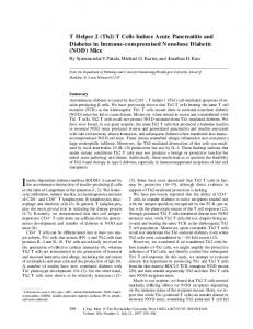
IL-4 expressed higher levels of MHC class II molecules and costimulatory molecules (Fig. 1) and were more potent in stimulating a primary MLR than DC derived from BM cultures with GM-CSF alone (Fig. 2). The CD81 and CD82 splenic DC populations expressed similar levels of MHC class II and costimulatory molecules, but expressed markers characteristic of their lineage such that the CD82 DC expressed the myeloid marker CD11b, which was not expressed on the CD81 DC. The CD81 DC were, however, less potent in stimulating a primary MLR and we are currently investigating whether this may be
In vivo therapy with DC subsets DC subsets were purified as described above and were pulsed for 2 h with a mixture of three peptides (GAD65 509-528; GAD65 524-543; hsp60 437–460) that have been implicated in the pathogenesis of diabetes in NOD mice [21–23]. The cells were then washed in phosphate-buffered saline (PBS) and injected (4 3 105/mouse) intravenously into 5-week-old NOD mice, which were then followed for the development of diabetes by the weekly monitoring of blood glucose levels (.300 mg/dL).
RESULTS AND DISCUSSION Phenotype and function of NOD DC subsets Detailed analysis of the phenotype and function of four DC populations from NOD mice revealed several distinct characteristics (summarized in Table 1) that were similar although not identical to DC populations from other, diabetes-resistant strains. The two BM-derived DC populations had phenotypic and functional characteristics of mature and immature DC, respectively, such that DC derived from cultures with GM-CSF 1
Fig. 1. Flow cytometric analysis of purified bone marrow-derived DCs that were cultured for 4 days with GM-CSF 1 IL-4. Cells were stained with the indicated mAbs specific for DC or other lineages (solid lines) and appropriate isotype controls (dotted lines). The histograms are representative of five independent experiments.
Morel et al.
Dendritic cells in murine diabetes
277
Fig. 2. Comparison of the T cell stimulatory capacity of NOD DC subsets. Purified B10.BR T cells (2 3 105 cells/well) were stimulated with the indicated DC subsets for 4 days. The cells were pulsed (0.5 µCi [3H]thymidine/well) overnight, harvested, and counted on a beta scintillation counter. The data shown are representative of five independent experiments.
related to any regulatory function of these cells. The relative abilities of these four DC populations to stimulate a primary MLR is shown in Figure 2. It is interesting that in our preliminary analysis of cytokines induced by these DC populations, we did not observe any differences in the ability of DC subsets to stimulate Th1- or Th2-dominated responses. IFN-g production was observed to be the predominant cytokine produced after stimulation with all four DC populations, with quantitative differences presumably related to the overall stimulatory capacity of these cells. Further studies are required to determine whether this represents a difference between NOD DC and DC from diabetes-resistant strains.
Comparison of DC populations between NOD and B10.BR mice When we compared the two BM-derived NOD DC populations with similar populations from the diabetes-resistant strain B10.BR we observed some interesting differences, particularly in the BM-derived DC from GM-CSF 1 IL-4 cultures (Table 2). The yield of DC from NOD mice was consistently lower from these cultures compared to side-by-side B10.BR DC cultures. This is consistent with previous reports from human diabetics who have been shown to generate fewer monocyte-derived DC after culture in GM-CSF 1 IL-4 [24, 25]. Analysis of the phenotype revealed that the NOD DC expressed higher levels of CD40 than B10.BR DC (Table 2). This is illustrated in Figure 3 and was consistent throughout a 6-day culture period (data not shown), suggesting that control of constitutive CD40 expression might be different in NOD than in B10.BR mice. Similarly, unstimulated NOD DC were found to express higher levels of IL-12 p40 mRNA than similar B10.BR DC. When NOD and B10.BR DC were stimulated with LPS for 24 h, marked up-regulation of CD40 expression was observed in both 278
Journal of Leukocyte Biology
Volume 66, August 1999
strains of mice (Table 2). LPS stimulation of the DC populations also resulted in up-regulation of IL-12 p40 mRNA to equivalent levels in both NOD and B10.BR mice (Table 2). These data suggest that myeloid DC, derived from GM-CSF 1 IL-4 BM cultures, in NOD mice might have a more activated phenotype than similar populations from other non-autoimmune strains of mice. Signaling through CD40 has been implicated in the up-regulation of IL-12 production by DC [26], and IL-12 is a necessary cytokine for the induction of Th1 responses [27]. Recent studies have demonstrated that the treatment of NOD mice with anti-CD40L mAb was effective in preventing diabetes and inhibiting the development of isletspecific Th1 responses [28]. The fact that NOD DC might express more CD40 and IL-12 could be responsible for a bias toward the development of Th1 responses in these mice, which could be important in the development of the destructive anti-islet response. Further work is required to determine whether this difference is observed with a larger panel of strains, and whether the production of IL-12 p40 mRNA is correlated with the production of bioactive IL-12. Comparative studies of the lymphoid DC populations are currently underway.
In vivo therapy with NOD DC subsets The ability of DC subsets to modulate the development of diabetes in young prediabetic NOD mice was determined. In these experiments, 5-week-old NOD mice were given a single injection (intravenous) of BM-derived DC (from either GM-CSF 1 IL-4 or GM-CSF alone cultures). Before injection the DC had been either pulsed for 2 h with a mixture of three peptides from two antigens that have been implicated in the autoimmune response or had been pulsed in medium alone. The animals were then followed for the development of diabetes by the weekly monitoring of blood glucose levels. Injection of BM DC from GM-CSF cultures reduced the incidence of diabetes development from 70% in PBS-injected mice to 33% in mice given peptide-pulsed DC, and 40% in mice given DC without peptide (Fig. 4). It is interesting that DC derived from BM cultures in GM-CSF 1 IL-4 were more effective in the prevention of diabetes in NOD mice [Feili-Hariri et al., unpublished results]. This suggests that the more mature DC might be capable of stimulating a regulatory T cell response, and preliminary data suggest that peptide-specific Th2 responses may be stimulated after GM 1 IL-4 DC administration. We performed in vivo trafficking experiments to determine whether these two populations maintained their phenotypic differences after in vivo injection and we found that CD40 TABLE 2. Differences Between BM-Derived Mature DC (GM-CSF 1 IL-4) from NOD and B10.BR Mice Feature
NOD DC
B10.BR DC
0.59 6 0.15
1.29 6 0.52
Phenotype CD40 (unstimulated) CD40 (after LPS)
111 1111
1 111
Function IL-12 p40 mRNA (unstimulated) IL-12 p40 mRNA (after LPS)
111 1111
1 1111
Yield
(3106
cells/107
BM cells)
http://www.jleukbio.org
Fig. 3. NOD DC derived from bone marrow cultures with GM-CSF 1 IL-4 express higher levels of CD40 than a similar population from the diabetes-resistant strain, B10.BR. Cells were stained with the indicated mAbs specific for CD40, other DC markers (solid lines), and appropriate isotype controls (dotted lines). The histograms are representative of three independent experiments.
expression was low in both populations, whereas the difference in B7.1 expression was maintained [Feili-Hariri et al., unpublished results]. The fact that peptide pulsing did not appear to make a major difference to the results suggests that the choice of peptides as an antigen to induce regulatory cells might not be optimal. Potential reasons for this include the recent demonstration that the NOD MHC class II molecule (I-Ag7) forms unstable complexes and does not bind peptide efficiently [29]. Thus it is possible that peptide pulsing according to the protocol we used did not result in a significant number of peptide/MHC complexes on the surface of the DC. Therefore, we are in the process of exploring other ways to deliver antigen to the DC.
Conclusions The results presented here represent a comprehensive assessment of four distinct DC populations in a murine model of autoimmune disease, namely NOD mice, which has not yet
been reported. We identify some potentially interesting differences between one of the myeloid DC populations derived from the BM of NOD mice and a similar population of DC from B10.BR mice. These differences, in CD40 and IL-12 p40 expression, may play a role in the initiation of an autoimmune Th1 response, an area that is actively under investigation in our lab. The ability of the DC populations to modulate the development of diabetes in this model is modest, and may reflect the difficulty in influencing an ongoing autoimmune response as compared to affecting a primary response, as is the case in the induction of tolerance to an allograft [30]. Finally, the mechanism by which any of these DC populations influence the development of diabetes is actively under investigation. The recent data all suggest that DC may be the most important APC in determining the outcome of an immune response, and in particular whether this leads to stimulation or tolerance induction. The detailed understanding of the phenotype and function of DC subsets, particularly in the scenario of autoimmune predisposition, will lead to the development of therapeutic strategies to induce tolerance to autoantigens and prevent disease development.
ACKNOWLEDGMENTS The authors would like to acknowledge the technical assistance of Marylin Frantz, Dewayne Falkner, and Martha Campana, and the University of Pittsburgh Cancer Institute Flow Cytometry Facility. We would also like to thank Immunex for the kind gift of Flt3 ligand. The work presented here was supported by NIH Grant CA73743 and AI25151.
REFERENCES Fig. 4. Bone marrow-derived DC can modulate the development of diabetes in NOD mice. NOD mice (5 weeks of age) were given DC (4 3 105/mouse, intravenously) derived from BM cultures in GM-CSF alone that were either pulsed with peptides (mix. pept; nine mice) or in medium alone (Med; five mice), and were monitored for the development of diabetes. Control mice were given an intravenous injection of PBS (PBS; 10 mice). Statistical analysis revealed no significant difference between the groups (P . 0.05).
1. Banchereau, J., Steinman, R. M. (1998) Dendritic cells and the control of immunity. Nature 392, 245–252. 2. Inaba, K., Inaba, M., Romani, N., Aya, H., Deguchi, M., Ikehara, S., Muramatsu, S., Steinman, R. M. (1992) Generation of large numbers of dendritic cells from mouse bone marow cultures supplemented with granulocyte/macrophage colony-stimulating factor. J. Exp. Med. 176, 1693–1702. 3. Wu, L., Vremee, D., Ardavin, C., Winkel, K., Suss, G., Georgiou, H., Maraskovsky, E., Cook, W., Shortman, K. (1995) Mouse thymus dendritic
Morel et al.
Dendritic cells in murine diabetes
279
4.
5.
6.
7. 8.
9.
10. 11. 12.
13. 14. 15.
16.
cells: kinetics of development and changes in surface markers during maturation. Eur. J. Immunol. 25, 418–425. Lu, L., Woo, J., Rao, A. S., Li, Y., Watkins, S. C., Qian, S., Starzl, T. E., Demetris, A. J., Thomson, A. W. (1994) Propagation of dendritic cell progenitors from normal mouse liver using GM-CSF and their maturational development in the presence of type-1 collagen. J. Exp. Med. 179, 1823–1833. Lu, L., Hsieh, M., Oriss, T. B., Morel, P. A., Starzl, T. E., Rao, A. S., Thomson, A. W. (1995) Generation of DC from mouse spleen cell cultures in response to GM-CSF: immunophenotypic and functional analyses. Immunol. 84, 127–134. Fu, F., Li, Y., Qian, S., Lu, L., Chambers, F., Starzl, T. E., Fung, J. J., Thomson, A. W. (1996) Costimulatory molecule-deficient dendritic cell progenitors (MHC class II1, CD80dim, CD862) prolong cardiac allograft survival in nonimmunosuppressed recipients. Transplant. 62, 659–665. Cella, M., Sallusto, F., Lanzavecchia, A. (1997) Origin, maturation and antigen presenting function of dendritic cell. Curr. Opin. Immunol. 9, 10–16. Vremec, D., Zorbas, M., Scollay, R., Saunders, D. J., Ardavin, C. F., Wu, L., Shortman, K. (1992) The surface phenotype of dendritic cells purified from mouse thymus and spleen: investigation of the CD8 expression by a subpopulation of dendritic cells. J. Exp. Med. 176, 47–58. Maldonado-Lopez, R., de Smedt, T., Michel, P., Godfroid, J., Pajak, B., Heirman, C., Thielemans, K., Leo, O., Urbain, J., Moser, M. (1999) CD8a1 and CD8a2 subclasses of dendritic cells direct the development of distinct T helper cells in vivo. J. Exp. Med. 189, 587–592. Rissoan, M.-C., Soumelis, V., Kadowaki, N., Grouard, G., Briere, F., de Waal Malefyt, R., Liu, Y.-J. (1999) Reciprocal control of T helper cell and dendritic cell differentiation. Science 283, 1183–1186. Castano, L., Eisenbarth, G. S. (1990) Type-I diabetes: a chronic autoimmune disease of human, mouse and rat. Annu. Rev. Immunol. 8, 647–679. Jansen, A., Homo-Delarche, F., Hooijkaas, H., Leenen, P. J., Dardenne, M., Drexhage, H. A. (1994) Immunohistochemical characterization of monocytes-macrophages and dendritic cells involved in the initiation of the insulitis and b-cell destruction in NOD mice. Diabetes 43, 667–675. Dahlen, E., Dawe, K., Ohlsson, L., Hedlund, G. (1998) Dendritic cells and macrophages are the first and major producers of TNF-a in pancreatic islets in the nonobese diabetic mouse. J. Immunol. 160, 3585–3593. Trembleau, S., Penna, G., Bosi, E., Mortara, A., Gately, M. K., Adorini, L. (1995) Interleukin 12 administration induces T helper type 1 cells and accelerates autoimmune diabetes. J. Exp. Med. 181, 817–821. Nagata, M., Santamaria, P., Kawamura, T., Utsugi, T., Yoon, J.-W. (1994) Evidence for the role of CD81 cytotoxic T cells in the destruction of pancreatic b-cells in nonobese diabetic mice. J. Immunol. 152, 2042– 2050. Cameron, M. J., Arreaza, G. A., Zucker, P., Chensue, S. W., Strieter, R. M., Chakrabarti, S., Delovitch, T. L. (1997) IL-4 prevents insulitis and insulin-dependent diabetes mellitus in nonobese diabetic mice by potentiation of regulatory T helper-2 cell function. J. Immunol. 159, 4686–4692.
280
Journal of Leukocyte Biology
Volume 66, August 1999
17. Maraskovsky, E., Brasel, K., Teepe, M., Roux, E. R., Lyman, S. D., Shortman, K., McKenna, H. J. (1996) Dramatic increase in the numbers of functionally mature dendritic cells in Flt3 ligand-treated mice: multiple dendritic cell subpopulations identified. J. Exp. Med. 184, 1953–1962. 18. Clare-Salzler, M. J., Brooks, J., Chai, A., van Herle, K., Anderson, C. (1992) Prevention of diabetes in nonobese diabetic mice by dendritic cell transfer. J. Clin. Invest. 90, 741–748. 19. Lu, L., Rudert, W. A., Qian, S., McCaslin, D., Fu, F., Rao, A. S., Trucco, M., Fung, J. J., Starzl, T. E., Thomson, A. W. (1995) Growth of donor-derived dendritic cells from the bone marrow of murine liver allograft recipients in response to granulocyte/macrophage colony-stimulating factor. J. Exp. Med. 182, 379–387. 20. Wang, S. C., Jordan, M. L., Morel, P. A., Simmons, R. L., Tweardy, D. J. (1993) A dual mechanism of immunosuppression by FK-506: FK-506 markedly suppresses IL-4 but not IL-10 mRNA levels in T helper 2 clones. Transplant. 56, 978–985. 21. Atkinson, M. A., Maclaren, N. K. (1993) Islet cell autoantigens in insulin-dependent diabetes. J. Clin. Invest. 92, 1608–1616. 22. Elias, D., Reshef, T., Birk, O., van der Zee, R., Walker, M. D., Cohen, I. R. (1991) Vaccination against autoimmune mouse diabetes with a T-cell epitope of the human 65-kDa heat shock protein. Proc. Natl. Acad. Sci. USA 88, 3088–3091. 23. Tisch, R., Yang, X. D., Singer, S. M., Liblau, R. S., Fugger, L., McDevitt, H. O. (1993) Immune response to glutamic acid decarboxylase correlates with insulitis in non-obese diabetic mice. Nature 366, 72–75. 24. Takahashi, K., Honeyman, M. C., Harrison, L. C. (1998) Impaired yield, phenotype, and function of monocyte-derived dendritic cells in humans at risk for insulin-dependent diabetes. J. Immunol. 161, 2629–2635. 25. Jansen, A., van Hagen, M., Drexhage, H. A. (1995) Defective maturation and function of antigen-presenting cells in type 1 diabetes. Lancet 345, 491–492. 26. Cella, M., Scheidegger, D., Palmer-Lehmann, K., Lane, P., Lanzavecchia, A., Alber, G. (1996) Ligation of CD40 on dendritic cells triggers production of high levels of interleukin-12 and enhances T cell stimulatory capacity: T-T help via APC function. J. Exp. Med. 184, 747–752. 27. Macatonia, S. E., Hosken, N. A., Litton, M., Vieira, P., Hsieh, C. S., Culpepper, J. A., Wysocka, M., Trinchieri, G., Murphy, K. M., O’Garra, A. (1995) Dendritic cells produce IL-12 and direct the development of Th1 cells from naive CD41 T cells. J. Immunol. 154, 5071–5079. 28. Balasa, B., Krahl, T., Patstone, G., Lee, J., Tisch, R., McDevitt, H. O., Sarvetnick, N. (1997) CD40 ligand-CD40 interactions are necessary for the initiation of insulitis and diabetes in nonobese diabetic mice. J. Immunol. 159, 4620–4627. 29. Carrasco-Marin, E., Shimizu, J., Kanagawa, O., Unanue, E. R. (1996) The class II MHC I-Ag7 molecules from non-obese diabetic mice are poor peptide binders. J. Immunol. 156, 450–458. 30. Steptoe, R. J., Thomson, A. W. (1996) Dendritic cells and tolerance induction. Clin. Exp. Immunol. 105, 397–402.
http://www.jleukbio.org