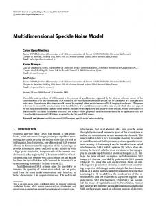
Implementation of wavelet filters for speckle noise reduction in ultrasound medical Images: a comparative study R. Sivakumar and D. Nedumaran Central Instrumentation and Service Laboratory, University of Madras, Guindy campus, Chennai-600025, Tamil Nadu, INDIA, E-mail:
[email protected],
[email protected] Abstract Speckle is an inherent noise in ultrasound images and
generally tends to reduce the image resolution and contrast thereby reducing the diagnostic value of ultrasound imaging modality. Removing noises from biomedical image is still a challenging problem for the biomedical researchers and lots of denoising techniques have been developed over a period. Wavelet transform has been gaining popularity due to its sparsity and multiresolution properties in the area of biomedical image denoising. In this paper, we have implemented the multiresolution wavelet denoising technique to remove the speckle noise in biomedical ultrasound images. Several wavelets such as Haar, Daubechies, Symlet, Discrete Meyer and Coiflets have been employed and compared the performance of each wavelet by calculating the Root Mean Square Error (RMSE) and Peak signal to noise ratio (PSNR) of the denoised image. This study will provide the knowledge of selecting a particular wavelet for denoising the speckle noise with optimum efficiency. Keywords: Ultrasound Image, Speckle Noise, RMSE, PSNR
1. Introduction Ultrasound is a commonly used diagnostic imaging modality for heart, liver, spleen, and lungs, etc [1]. The main advantage of the ultrasound imaging is that it is quick, economic, relatively safe, noninvasive and the machinery is highly portable and versatile. However, the main disadvantage of the ultrasound image is poor quality of images [2], due to speckle noise. Ultrasound images are usually affected with an intrinsic artifact called speckle, which is the result of the constructive and destructive coherent summation of ultrasound echoes. The presence of speckle is undesirable since it degrades image quality and affects the tasks of human interpretation and diagnosis. As a result, speckle filtering is a critical method for feature extraction, analysis, and recognition in medical imagery measurements. Several techniques for removing the speckle noise [3] have been developed over a decade. In this study we have demonstrated the important wavelet filtering techniques to remove the speckle noise effectively and preserve most of the diagnostic details. We have attempted denoising of
speckle with several wavelet basis functions [4] and compared their peak signal to noise ratio (PSNR) in order to select the best optimum wavelet basis function for effective denoising.
2. Wavelet technique Wavelets have been employed for denoising of images more than a decade. Wavelet transformation is a multiresolution representation of signal and image in two dependant domains, which decompose the signal and image into multiscale resolution. The localization of the wavelet basis functions in both time and frequency domain leads to multiresolution analysis and effective filter designs for specific applications. Wavelet decomposition preserved and depicted the sharp transition in images, which results in very accurate edge detection in images. These properties of the wavelet transform make it a very effective alternative to the classical Short-Time Fourier Transform (STFT). The continuous wavelet transform (CWT) is expressed as ∞
1 ⎛ t −τ ⎞ X (s,τ ) = ψ⎜ ⎟x(t)dt , for s>0 ∫ s −∞ ⎝ s ⎠
(1)
where s is the scale factor and τ is the time shift. Discretized continuous wavelet transform [5] is simply a sampled version of the CWT, which enables the computation of the continuous wavelet transform effectively in PCs; it is not a true discrete transform. The information it provides is highly redundant and requires a significant amount of computation time and resources. The discrete wavelet transform (DWT), on the other hand, provides sufficient information both for analysis and synthesis of the original signal, with a significant reduction in the computation time. The continuous wavelet transform was computed by changing the scale of the analysis window, shifting the window in time, multiplying by the signal, and integrating over all times. In the discrete case, filters of different cutoff frequencies are used to analyze the signal at different scales. The resolution of the signal, which is a measure of the amount of detail information in the signal, is changed by the filtering operations, and the scale is changed by upsampling and downsampling (subsampling) operations. DWT employs two sets of functions, called scaling
functions and wavelet functions, which are associated with low pass and high pass filters, respectively. The decomposition of the signal into different frequency bands is simply obtained by successive high pass and low pass filtering of the time domain signal.
3. Wavelet based denoising Donoho and Johnstone [6, 7] have done lot of pioneering work on wavelet based noise removal employing thresholding of the Discrete Wavelet Transform (DWT) coefficients. Fig 1 shows the scheme we have employed for denoising speckle noise in biomedical ultrasound images in the Matlab 7.1 environment [8].
Patient-1
Patient-2
Patient-3
Fig. 2a Original biomedical ultrasound Image
Fig. 2b Denoised with Haar wavelet
Ultrasound image with speckle Noise Application of Discrete Wavelet Transform and testing with different basis functions Calculation of Wavelet Coefficients after soft thresholding
Fig. 2c Denoised with Db2 wavelet
Fig. 2d Denoised with Db4 wavelet
Inverse Discrete Wavelet Transform
Denoised Ultrasound image Fig. 2e Denoised with Db8 wavelet Fig1. Block diagram of the Wavelet Based Denoising Scheme
The 2-D data of the ultrasound biomedical images were obtained from the GE healthcare machine (Model: VIVID7) available at Sri Ramachandra Medical College Hospital, Porur, Chennai. The data were subjected to DWT with important wavelet basis functions. The wavelet coefficients calculated from the DWT were subjected to soft thresholding [9]. After soft thresholding of the wavelet coefficients, the denoised image is reconstructed using Inverse Discrete Wavelet Transform. The quality of the denoised image was measured by calculating the traditional PSNR (peak signal-to-noise ratio) of the image [10]. Various Wavelet basis functions such as haar, daubechies, symlet, coiflets, and discrete Meyer are demonstrated in this study to estimate the performance of the wavelet basis function in denoising the speckle noise from biomedical ultrasound images. The raw ultrasound biomedical images and the denoised images using various wavelet filters are shown in fig 2a to 2n.
Fig. 2f Denoised with Db10 wavelet
Fig. 2g Denoised with Symlet2 wavelet
Fig. 2h Denoised with Symlet4 wavelet
Fig. 2i Denoised with Symlet6 wavelet
of gray scale resolution (8-bit) of the image, MN is the number of pixels of the ultrasound image, Fi and Di are the threshold values of the original and the denoised image respectively. The table 1 shows the calculated RMSE and PSNR values for different wavelet filters employed in this study.
Fig. 2j Denoised with Symlet10 wavelet
Wavelet filters
Fig. 2l Denoised with Coiflet1 wavelet
Fig. 2m Denoised with Coiflet 2 wavelet
Fig. 2n Denoised with Coiflet 3 wavelet
4. Experimental analysis The performance of noise reduction of the wavelet filter is measured using quantitative performance measures such as Peak Signal-to-Noise Ratio (PSNR) and in terms of visual quality of the ultrasound images [11]. The PSNR is the ratio between the maximum possible power of a signal and the power of corrupting noise that affects the fidelity of its representation. The PSNR is most commonly used as a measure of quality of reconstruction in image compression, denoising, etc. The PSNR and RMSE are given by equation 2 and 3. ⎛ 255 ⎞ S = 20 log10 ⎜ ⎟ ⎝ RMSE ⎠
PSNR
RMSE
PSNR
RMSE
PSNR
26.593
11.393
26.993
10.906
27.372
Db2
9.982
28.141
9.345
28.714
8.728
29.307
Db4
8.177
29.873
8.139
29.913
7.858
30.224
Db8
8.074
29.983
8.109
29.946
7.869
30.206
Db10
8.070
29.987
8.083
29.973
7.807
30.275
Sym2
9.213
28.837
9.045
28.997
9.002
29.038
Sym4
8.138
29.914
8.993
29.047
7.857
30.220
Sym6
8.108
29.947
8.138
29.914
7.968
30.098
Sym10
8.062
29.996
8.015
30.047
7.878
30.197
D.M
8.076
29.981
8.118
29.936
7.972
30.093
Coif1
9.067
28.976
9.025
29.016
8.677
29.358
Coif2
8.121
29.933
8.078
29.979
7.812
30.270
Coif3
8.138
29.914
8.124
29.930
7.960
30.107
Table 1 Calculated RMSE and PSNR values of different wavelet filters tested in three ultrasound biomedical images From the experimental values, optimum wavelet filter is chosen for denoising the speckle based on the criteria that the RMSE value is low and PSNR value is large. It is clearly indicated in the performance analysis chart as shown in fig 3a to 3c Patient-1 35 30 25 20
RMSE
15
PSNR
10 5
oi f2 C
-6
.m ey is
-2
D
Sy m
D
Sy m
MN
b8
0
(3)
b4
i
2
D
∑ (F − D ) i
RMSE
aa r
RMSE =
Patient-3
11.930
H
Where
(2)
Patient-2
Haar
RMSE and PSNR
Fig. 2k Denoised with Discrete meyer
Patient-1
Wavelet Filters
Here S is the Peak Signal-to-Noise Ratio (PSNR) in dB, RMSE is the Root Mean Square Error, 255 is the number
Fig 3a Performance analysis chart of wavelet filters for the fetus image of patient-1
6. Acknowledgement
Patient-2
RMSE and PSNR
35 30 25 20
RMSE
15
PSNR
10 5
We acknowledged the help rendered by Prof. Dr. S. Thanigasalam, Chairman, Cardiac Care Centre, Sri Ramachandra Medical College Hospital, Chennai for providing the ultrasound echo images used in this study. We also thank the Tamilnadu State Council for Science and Technology for the financial assistance extended to conduct this study
0
H aa r D b4 D b8 Sy m -2 Sy m D -6 is .m ey C oi f2
7. References
Wavelet Filters
Fig 3b Performance analysis chart of wavelet filter for the cardiac image of patient-2 Patient-3
RMSE and PSNR
35 30 25 20
RMSE
15
PSNR
10 5
H aa r D b4 D b8 Sy m -2 Sy m D -6 is .m ey C oi f2
0
Wavelet filters
Fig 3c Performance analysis chart of wavelet filter for the cardiac image of patient-3
5. Results and discussion In this work, various wavelets filters have been attempted to test the efficiency of denoising the speckle in ultrasound biomedical images. The performance of denoising the speckle noise using the wavelet basis functions such as, Haar, Daubechies, Symlet, Coiflets, and Discrete Meyer have been demonstrated for three different patient images with two levels of DWT decomposition and soft thresholding. The RMSE and PSNR of individual wavelet filters were calculated and are used to estimate the performance of each wavelet filter. From the experimental values, it is observed that higher value of the PSNR and lower value of RMSE results in better performance in denoising the speckle noise. Experimental results showed that Symlet10 and Daubechies10 wavelet filters exhibit much better performance in PSNR, RMSE and visual effect..
[1] Gjenna Stippel, Wilfried Philips, Ignace Lemahieu, Paul Govart, “A New Medical feature Enhancing speckle suppression method for ultrasound images of Neonatal Brain,” Proceeding of (359) Signal and Image Processing – 2002. [2] Alin Achim, Anastasios Bezerianos panagiotis Tsakalides, “An Alpha-Stable based Bayesian Algorithm for speckle noise removal in the wavelet domain”, NSIP-01, June0306, Baltimore, Maryland USA, 2001. [3] Ivana Duskunovic, Aleksandra Pizurica, Gjenna Stippel, Wilfried Philips and Ignace Lemahieu, “Wavelet Based denoising techniques for ultrasound images”, Proceedings of 22nd Annual International conference of the IEEE, Volume 4 issue, pp. 2662 – 2665. [4] Su Cheol Kang, and Seung Hong Hong, “Experimental and Theoretical Analysis of Wavelet based denoising filter for Echocardiographic images”, MEDINFO 2001. [5] Robi Polikar, “The Wavelet Tutorial”, Part I-IV, Second Edition. [6] David L. Donoho and Iain M. Johnstone, “Ideal spatial adaptation via wavelet shrinkage”, Biometrika, Vol.81, pp.425-455, 1994. [7] David L. Donoho and Iain M. Johnstone, “Adapting to unknown smoothness via wavelet shrinkage”, Journal of the American Statistical Association, Vol.90, No.432, pp. 1200-1224, 1995. [8] Rafael C. Gonzalez, Richard E. Woods, and Steven L. Eddins, “Digital Image Processing Using MATLAB®,” Pearson Education, Inc, 2004. [9] S.Arivazhagan, S. Deivalakshmi, K. Kannan, B. N. Gajbhiye, C. Muralidhar, Sijo Lukose, and M P. Subramanian “Performance Analysis of Wavelet filters for image denoising”, Advance in Computational Sciences and Technology Vol. 1, pp1-4. [10] Antonio Fernandez-Caballero and Juhan L. Mateo, Methodological Approach to Reducing Speckle Noise in Ultrasound images”, International Conference on Biomedical Engineering and Informatics, Computer Society, 978-0-7695-3118-2/08, 2008. [11] Kil Dong Hong Young-Gn.Maeng, “Wavelet- based image denoising with optimal filter”, ISSN 17388899,KIPS, 2005.