The Role of Amygdala in Emotional and Social Functions: Implications for Temporal Lobe Epilepsy* Chiara Cristinzio1,2 and Patrik Vuilleumier1,2 1 Laboratory for Behavioural Neurology & Imaging of Cognition, Department of Neuroscience & Clinic of Neurology, University of Geneva 2 Swiss Center for Affective Sciences, University of Geneva
Summary Temporal lobe epilepsy is among the most frequent causes of chronic and drug-resistant seizure disorders. It is typically associated with lesions involving critical limbic structures within the anterior medial temporal lobe, such as the amygdala and hippocampus. While the role of the hippocampus and adjacent cortical regions in memory function is now well established, the role of the amygdala and related brain circuits is still poorly known. The amygdala is a complex neural structure implicated in several aspects of emotional and social behaviour, but the varieties and the consequences of amygdala dysfunction in patients with temporal lobe epilepsy remain unclear, and insufficiently examined in standard neuropsychological assessments. Here we review data from recent research in humans indicating that amygdala lesions may impair selective domains of affect and cognition, all related to the appraisal of emotional and social significance of sensory events. We describe neurophysiological and behavioural evidence to illustrate how the amygdala may contribute to a wide range of affective functions, including recognition of facial expressions, perception of gaze direction, modulation of attention and memory, perception of musical emotions, theory of mind, plus mood and psychiatric disorders. We argue that a more systematic assessment of affective functions mediated by the amygdala and related circuits might provide useful information about temporal lobe pathology and neuropsychological outcome after surgery.
Epileptologie 2007; 24: 78 – 89 Key words: Facial expression recognition, temporal lobe epilepsy, eye gaze, perception, emotional memory
critiques au sein du lobe temporal antérieur médial telles que l'amygdale ou l'hippocampe. Tandis que le rôle de l'hippocampe et des régions corticales adjacentes dans le fonctionnement de la mémoire a fait l'objet d'études étendues, celui de l'amygdale et des régions du cerveau y liées reste encore largement inconnu. L'amygdale est une structure neurale implexe qui participe à de nombreux aspects du comportement émotionnel et social. Cependant, nous ne possédons pas une vision très claire des différentes dysfonctions possibles de l'amygdale et de leurs conséquences pour les patients atteints d'une épilepsie du lobe temporal et elles sont insuffisamment saisies lors d'examens neuropsychologiques standard. Nous rapportons ici les résultats d'études récentes sur l'homme suggérant que les lésions de l'amygdale pourraient affecter des zones déterminées liées à l'affect et à la cognition, affects et perceptions qui seraient tous associés à l'appréciation d'impressions sensorielles importantes pour l'interaction émotionnelle et sociale. Nous décrivons des signes neurophysiologiques et comportementaux pour montrer en quoi l'amygdale peut jouer un rôle dans un vaste éventail de fonctions affectives, y compris la reconnaissance d'expressions faciales, la perception d'orientations du regard, les altérations de l'attention et de la mémoire, la perception de sentiments musicaux, la capacité de se mettre en pensée à la place d'autres personnes (theory of mind), les humeurs et les troubles psychiatriques. Nous estimons qu'une étude plus systématique des fonctions affectives de l'amygdale et de ses zones d'influence pourrait fournir des informations importantes sur la pathologie du lobe temporal et sur les conséquences neuropsychologiques d'un traitement chirurgical de l'épilepsie. Mots clés : reconnaissance d'expressions faciales, orientation du regard, perception, mémoire émotionale
Le rôle de l'amygdale dans les fonctions émotionnelles et sociales : conclusions pour l'épilepsie du lobe temporal * Acknowledgements
L'épilepsie du lobe temporal est une des causes les plus fréquentes de maladies chroniques et réfractaires aux traitements se révélant par des crises. Elle est typiquement associée à des lésions de structures limbiques
78
Epileptologie 2007
This work was supported by a grant of the SNF (No 105311-108187) to David Sander and Patrik Vuilleumier, and by the Swiss National Center for Affective Sciences. We thank David Sander, Gilles Pourtois, and Margitta Seeck for their collaboration and insightful discussions during this research.
The Role of Amygdala in Emotional and Social Functions | Chiara Cristinzio and Patrik Vuilleumier
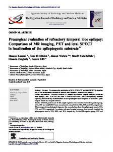
B A
C amygdala hippocampus
Figure 1: (A) Illustration of the anatomy of the medial temporal lobe and location of the amygdala. (B) Activation of amygdala in a healthy subject to fearful relative to neutral face expression. (C) Example of unilateral temporal lobectomy in a patient with refractory epilepsy.
Die Rolle der Amygdala bei emotionalen und sozialen Funktionen: Folgerungen für die Temporallappenepilepsie Die Temporallappenepilepsie ist eine der häufigsten Ursachen von chronischen und therapieresistenten Anfallskrankheiten. Typischerweise ist sie verknüpft mit Läsionen von kritischen limbischen Strukturen innerhalb des anterioren medialen Temporallappens wie der Amygdala und des Hippokampus. Während die Rolle des Hippokampus und der anliegenden kortikalen Regionen für die Gedächtnisfunktionen heute gut erforscht ist, ist diejenige der Amygdala und der mit ihr in Verbindung stehenden Hirnregionen immer noch weitgehend unbekannt. Die Amygdala ist eine implexe neurale Struktur, die an vielen Aspekten des emotionalen und sozialen Verhaltens beteiligt ist. Die verschiedenen Möglichkeiten von Amygdaladysfunktionen und deren Konsequenzen bei Patienten mit Temporallappenepilepsie sind aber unklar und werden bei neuropsychologischen Standarduntersuchungen unzureichend erfasst. Hier berichten wir über Resultate von kürzlichen Untersuchungen beim Menschen, welche darauf hinweisen, dass Amygdala-Läsionen bestimmte Zonen für Affekte und Kognition beeinträchti-
gen können, wobei diese Affekte und Wahrnehmungen alle mit der Beurteilung von emotionalen und sozial bedeutsamen Sinneseindrücken verbunden sind. Wir beschreiben neurophysiologische und Verhaltens-Zeichen, um zu zeigen, wie die Amygdala bei einer weiten Reihe von affektiven Funktionen inkl. Wiedererkennen von Gesichtsausdrücken, Wahrnehmung von Blickrichtungen, Veränderungen der Aufmerksamkeit und des Gedächtnisses, Wahrnehmung von musikalischen Gefühlen, die Fähigkeit, sich in das Denken anderer Menschen hineinzuversetzen (theory of mind), Stimmungen und psychiatrischen Störungen eine Rolle spielen kann. Wir meinen, dass eine systematischere Untersuchung von affektiven Funktionen der Amygdala und ihrer Einflussbereiche wichtige Informationen über die Pathologie des Temporallappens und über die neuropsychologischen Folgen einer Epilepsieoperation liefern könnten. Schlüsselwörter: Wiedererkennen des Gesichtsausdrucks, Temporallappenepilepsie, Blickrichtung, Wahrnehmung, emotionales Gedächtnis
The Role of Amygdala in Emotional and Social Functions | Chiara Cristinzio and Patrik Vuilleumier
Epileptologie 2007
79
Introduction The epilepsies are a complex group of disorders characterized by repeated seizures due to paroxysmal changes in electrical brain activity. According to the International Classification of Epilepsies, four main localization-related types of epilepsy can be distinguished: temporal lobe epilepsies, frontal lobe epilepsies, parietal lobe epilepsies, and occipital lobe epilepsies [1]. Temporal lobe epilepsy (TLE) is the most frequent type of focal epilepsy, and the form associated with hippocampal sclerosis is the most prevalent. Aetiology can be genetic, with frequent onset during childhood, or secondary to brain lesion, with variable onset in adult age. Mesial temporal epilepsy is characterized by focal discharges, often with partial complex seizures, associated with an epileptogenic lesion in anterior medial temporal lobe (typically hippocampal sclerosis), and a strong potential for drug resistance [2]. The anterior temporal lobe is composed of several important neuronal structures, including the amygdala, hippocampus, and surrounding cortex (Figure 1), all intimately connected with the limbic system [3]. Although sclerosis of the hippocampus is clearly established in patients with TLE, similar damage to the amygdala is also frequent. Just as hippocampus lesions, amygdala damage in TLE may be either unilateral or bilateral, but it can occur independent of hippocampus lesion [4]. Thus, a recent MRI study clearly demonstrated that significant amygdala damage may sometimes be found even when no evidence of hippocampal sclerosis or atrophy is detected [4]. However, in the majority of patients, amygdala sclerosis appears together with hippocampal damage, and both lesion sites may therefore contribute to the clinical manifestations. The amygdala is a complex neural structure, composed of many subdivisions with different cytoarchitectonic and connectional characteristics, which plays a major role in emotional and social processes. Such a role in the affective domain has been supported by a large amount of neurobiology research in animals as well as functional neuroimaging in healthy humans [5]. However, neuropsychological deficits due to amygdala damage are still poorly known, despite several studies that reported selective impairments in emotional and social functions in patients suffering from focal lesions or destruction of the amygdala. Because these studies were frequently conducted in rare cases with rare structural lesions, still little is known about the nature and prevalence of neuropsychological consequences of amygdala damage in TLE. In epilepsy patients, amygdala damage may result from idiopathic sclerosis; but also from various tumoral or pseudo-tumoral diseases arising in the temporal lobe; from progressive calcification associated with Urbach-Wiethe syndrome; or from surgical removal or lobectomy to cure pharmacoresistant seizures (Figure 1). Yet, deficits in emotional and social processes have rarely been investigated in a
80
Epileptologie 2007
systematic manner. Most of the neuropsychological research and clinical assessment in TLE has traditionally focused on memory function, associated with hippocampus and temporal neocortex. However, as we review here, amygdala lesions can cause a wide range of distinctive manifestations in emotion and social cognition. We believe that a more systematic exploration of such manifestations might be warranted to improve both the management of TLE and the understanding of human amygdala functions. Seizure affecting the amygdala and interconnected regions (such as anterior temporal cortex, ventromedial cortex, or insula) may also produce acute disturbances in emotional experience and behaviour, such as ictal fear, grimacing, or screaming [6, 7]. However, in the present review we will not address this issue, but rather focus on the selective neuropsychological deficits associated with amygdala lesions, which may arise either prior or after surgery for epilepsy, and concern a wide range of emotional and social processes. Our goal is not to provide an exhaustive review of amygdala functions or TLE manifestations, but to illustrate the importance of medial temporal lobe structures in affective processing, and to emphasize the need for the development of appropriate tests in the neuropsychological assessment of these patients.
The role of the amygdala in emotions A crucial role of the amygdala in emotional and social processing has long been proposed on the basis of focal lesion studies in primate as well as other animal species. A first classic observation was reported by Klüver and Bucy [8] who found that extensive, bilateral removal of the anterior temporal lobes led to a highly distinctive pattern of severe behavioural changes, characterized by a loss of emotional reactivity and lack of fear responses (tameness), as well as hypersexuality, orality, hypermetamorphosis, and visual agnosia. Subsequent work by Weiskrantz [9] revealed that the behavioural and emotional abnormalities associated with the Klüver-Bucy syndrome could be produced by lesions of the amygdala region alone. This finding has then been replicated many times in monkeys [10, 11] following complete destruction of the amygdala and its connections in both hemispheres. However, it is now thought that the full symptoms of Klüver-Bucy syndrome, including some aspects of hypo-emotionality, may reflect the extent of the lesions to adjacent structures in the rhinal and polar temporal cortices, as well as in the white matter connecting temporal regions with orbitofrontal cortex [12, 13], rather than damage to the amygdala itself. Focal lesions produced by local infusion of cytotoxic agents produce a more selective destruction of amygdala neurons, sparing neighbouring axonal tracts, and thus lead to less dramatic disturbances than amygdala destruction
The Role of Amygdala in Emotional and Social Functions | Chiara Cristinzio and Patrik Vuilleumier
by surgical removal or aspiration. Moreover, in humans, a complete form of the Klüver-Bucy syndrome is rarely, if ever, observed after damage to the amygdala, unless such damage extends to other limbic regions within temporal, insular, and orbitofrontal areas [14, 15]. This is best exemplified by the famous patient HM who suffered from severe amnesia after bilateral temporal lobectomy, which included both the amygdala and anterior hippocampus. HM is reported to show some signs of emotional indifference but no other striking disorders in affective and social behaviour. The rarity of Klüver-Bucy syndrome in humans may reflect the fact that other regions in temporal and frontal cortex play a greater role in the control of emotional and social functions, as compared with other primates, such that lesions to one single area within this neural network may not be sufficient to produce similar disorders. Alternatively, amygdala functions may have changed in humans over the course of evolution, perhaps due to the greater complexity and influences of cortical interactions which could have resulted in a somewhat different processing role. Several patients with bilateral amygdala damage have now been reported in the literature and extensively studied in the past two decades, but their deficits are relatively subtle as compared with emotional and social disturbances previously associated with the Klüver-Bucy syndrome. Moreover, the deficits are quite variable across cases [16]. Thus, in humans, the exact role of the amygdala in emotion and social cognition, and the exact deficits caused by selective amygdala lesions, still remain unresolved. Nevertheless, neuropsychological studies of the rare patients have clearly established the existence of selective impairments in a variety of affective and social domains, as we will review in details below. Taken together, these studies clearly indicate that the human amygdala play a major role in assigning affective values and recognizing emotional meaning of environmental stimuli, including faces, voices, or other events. Accordingly, patients with amygdala dysfunction show deficits of various degrees in processing and learning the affective values of stimuli, particularly those associated with a negative or aversive content (see below). A major role of the amygdala in aversive or threatrelated processing is further suggested by studies of fear-conditioning in animals [17]. A large body of work in neurophysiology and molecular biology has now clearly established that the amygdala is involved in pavlovian associative learning situations, where an innocuous stimulus (e.g. a tone) is paired with a noxious event (e.g. a electric shock): after a few pairings, the initially neutral stimulus (conditioned stimulus, CS+) will elicit a set of behavioural and physiological responses that are indicative of fear (e.g. freezing, tachycardia, increased blood pressure, sweating, papillary dilatation) even in the absence of the threatening event (unconditioned stimulus, US). Such fear-conditioning has been extensively studied in a variety of animal
species, and shown to depend on amygdala integrity, including in humans [5]. Furthermore, different aspects of the acquisition and expression of fear responses have been shown to depend on different subregions within the amygdala, which can be separately disrupted by different types of lesions or neuropharmacological interventions. In epilepsy patients, amygdala lesions arising due to sclerosis, calcifications, tumoral diseases, or surgery are typically unilateral. As a consequence, any emotional and social deficits after unilateral lesion can potentially be alleviated by the intact amygdala on the opposite side. However, as for memory disturbances, the impact of unilateral damage or surgery might depend on the presence of subclinical anomalies in the other hemisphere, and therefore vary across patients. Moreover, the severity of lesion (e.g. sclerosis) or the extent of surgical resection (cortical and subcortical) may also differ across patients, and thus produce distinct patterns of deficits, rather than a complete loss of amygdala functions. Furthermore, epileptic activity and/or underlying pathology may induce slowly progressive changes in the functional organization of the medial temporal lobe, such that removal of neuronal tissue on the affected side may not produce similar deficits after surgery as would be expected if such removal was made in a healthy brain. Likewise, amygdala dysfunction arising in early childhood, or later during life, is likely to result in distinct degree of neuronal plasticity and reorganization, and perhaps to differentially affect the development of emotional and social functions (see below). Accordingly, the type and severity of emotional deficits associated with amygdala damage in temporal lobe epilepsy will certainly depend not only on the nature and site of the underlying disease but also on the age of onset, its time-course, and duration. Moreover, some studies have suggested that amygdala function may be influenced by laterality as well as by gender, with slightly different roles in the right vs left hemisphere [18], and different degrees of hemispheric asymmetry in men vs women [19]. These factors may therefore also influence the nature of emotional symptoms caused by right or left amygdala dysfunction. Finally, it is important to bear in mind that epilepsy patients are usually treated by one or several drugs that can have direct effects on neural activity within the amygdala and other limbic brain regions. Indeed, many anti-epileptic drugs also have psychotropic effects on mood, arousal, and learning. These effects might potentially interact with some emotional processes normally associated with amygdala function, and hence contribute to the pattern of anomalies seen in temporal lobe epilepsy patients. However, the exact interplay between these different factors also remains poorly known.
The Role of Amygdala in Emotional and Social Functions | Chiara Cristinzio and Patrik Vuilleumier
Epileptologie 2007
81
Anatomy and physiology of the amygdala A great deal of knowledge has accumulated over the last years concerning the precise anatomical organization of the amygdala, particularly through the study of fear conditioning in mice and rats. However, less is known about the amygdala in primates, although the general structure and function appear remarkably shared across the different species. Comparative studies suggest that the relative size of the amygdala has increased rather than decreased in primates and humans, as compared with smaller animals, especially for the more lateral and basal subnuclei that are strongly connected to cortical brain regions. Thus, much of what is known about amygdala functions in rodents is thought to also apply to primates, but presumably with a much richer repertoire of interactions between amygdala and neocortex in humans. Anatomically, the amygdala is not a homogenous structure. It contains more than 15 different nuclei that are interconnected together to form different processing circuits, each with distinct specialized functions [20, 21]. It is therefore often preferable to refer to this structure as the “amygdaloid complex”, particularly because it is often difficult to distinguish between different subnuclei with conventional imaging techniques (including MRI or fMRI). These nuclei can be schematically grouped into a few major functional units. The lateral nucleus receives sensory inputs from neocortical areas in all modalities, and thus constitutes the main site of convergence for afferent information about the association between stimuli during fear-conditioning. However, olfactory inputs enter the amygdala by a different route, via corticomedial nuclei and periamygdaloid cortex. The lateral nucleus then projects to all other nuclei, in particular the basal, accessory basal, and central nuclei. The central nucleus provides the main output system projecting to various brain areas that are involved in the behavioural expression of emotional responses (e.g. fear), such as the hypothalamus (for sympathetic autonomic responses and stress hormone release), brainstem (for freezing or startle), cholinergic nuclei in basal forebrain (for arousal and attention), etc. The basal nucleus receives information from both the lateral nucleus and other brain regions such as orbitofrontal cortex and hippocampus, and in turn projects to the central nucleus, as well as to several cortical regions and hippocampus. This pattern of connectivity suggests that the basal nucleus may serve to integrate contextual information with incoming sensory information in order to modulate the emotional responses orchestrated by central nucleus and other target regions [22]. For example, studies of fear conditioning have shown that inputs from the hippocampus to the amygdala are involved in the association of fear responses with a specific place or context [23]; whereas inputs from ventromedial frontal areas play a crucial role in the inhibition of fear responses during the extinction of
82
Epileptologie 2007
conditioning [24]. Furthermore, neural processing within amygdala circuits can also be finely controlled by various other modulatory afferents, including dopamine, hormones, or neuropeptides such as oxytocine and orexin. This complex functional architecture testifies that the amygdala complex occupies a key position at the interface between the appraisal of external environmental events and adjustment of internal motivational processes. This privileged position makes it capable of eliciting and learning adaptive responses to various stimuli with intrinsic or acquired affective values, including social cues, as we will describe in more details in the following sections of this review. However, in humans, relatively little is known about the role of different subregions of amygdala in different aspects of emotional behaviour. Recent functional brain imaging studies [25, 26] converge with neurophysiology data in monkeys to point to the same general principles of organization. Nevertheless, it is conceivable that distinct patterns of emotional or social disorders might be associated with different types of lesion or dysfunction within amygdala circuits. Elucidating these differences is clearly a challenge for future research.
Recognition of facial emotional expressions A large body of neuropsychological and neuroimaging studies has demonstrated an implication of the amygdala in the recognition of facial expression of emotions (for reviews see [27, 28]). In particular, many observations have supported the hypothesis of a specialisation of amygdala responsiveness to negative emotions, especially fear, but also anger and sadness. The most famous case reported in the literature is that of SM, a 40-year-old woman who has complete bilateral amygdala destruction resulting from Urbach-Wiethe disease [29]. Her rare pathology has allowed a detailed study of the role of amygdala across a range of face processing tasks. Overall, she shows a disproportionate impairment in recognizing fear in facial expressions, and only a much milder impairment in recognizing the intensity of related emotions such as surprise and anger [29]. She is also impaired at making judgements of other subtle aspects of facial appearance that have particular threat or social meaning, such as perceived trustworthiness. Likewise, in patients with temporal epilepsy, several recent studies have now also reported significant deficits in the recognition of facial emotions, presumably caused by amygdala pathology. Such deficits have been observed in patients with mesial sclerosis lesions [30, 31] or after anterior lobectomy for surgical treatment [32]. Some reports have emphasized more frequent deficits with right than left hemisphere disease, and with an early age at epilepsy onset, consistent with a righthemisphere dominance in emotion and face processing.
The Role of Amygdala in Emotional and Social Functions | Chiara Cristinzio and Patrik Vuilleumier
This selective loss for fear recognition is paralleled with a disruption of fear conditioning in some cases with unilateral temporal lobe damage [33]. Remarkably, however, these patients may still be able to produce appropriate fear expressions on their own face when verbally instructed to do so, even though they fail to recognize the same expression presented visually in pictures or movies. According to some authors, patients with bilateral temporal lobe lesions might show more extensive deficits, affecting not only the recognition of fear, but also disgust, anger, sadness, and even happiness in faces (and perhaps in other modalities, e.g. voices) [34] – unlike the selective deficit for fear in patient SM who has bilateral lesions restricted to the amygdala. These findings suggest that some other cortical areas within the temporal pole, more extensively damaged after epilepsy surgery, might also contribute to expression recognition deficits. In addition, some patients with amygdala damage due to UrbachWiethe disease do not show deficits for a single emotion category (e.g. fear), but make more confusions between categories and report ”blends” of emotion more often than normal controls do [35]. Selective impairments in the visual recognition of fearful expressions may arise without any corresponding deficits in the subjective experience of fear and anxiety in everyday life. For instance, Anderson and Phelps [36] investigated self-evaluation of emotion states in amygdala-damaged patients and healthy control subjects. They observed that patients described their emotional life not differently than controls, and concluded that the amygdala is not necessarily implicated in the generation of affective states. But different results were reported by Tranel and colleagues [37]. They investigated personal feelings of emotion in SM (the same patient with bilateral amygdala destruction as described before; see [29]). These authors observed that when SM reports events of her life, the content of her reports misses negative emotions or subjective threat experiences: even when she talks about negative events or illnesses, no negative emotional state is verbally conveyed, making her appear as “brave” and “unusually courageous” in the face of her medical disease for other naive observers. Thus, in this case, the deficit in recognizing negative facial emotions was accompanied by a parallel defect in negative experiential aspects of emotional life. Finally, recent observations in patient SM suggest that her deficit in recognizing fearful expressions might result at least in part from an abnormal scanning of faces, with a lack of exploration of the eye region in faces, where the critical “diagnostic” features of fear expression are present. Thus, when instructed to look at the eyes in faces, SM can significantly improve her recognition of fearful expressions. This finding points to an important role of eye information in driving amygdala responses, and of active visual exploration for accurate recognition of expressions.
Eye gaze perception While facial expressions of emotion are an important source of information in normal communicative and social life, concomitant information conveyed by eye gaze is also crucial to appropriate interpretation and interaction between individuals. Several studies have emphasized an implication of the amygdala in processing both facial emotion and eye gaze. It has been reported that eye cues are sufficient to produce a threat superiority [38] in healthy subjects. Direction of eye gaze also informs about the emotional state and intention of other individuals. In the context of some expressions of emotion, the direction of gaze can modulate the interpretation of emotional state perceived from the other. Thus, Adams et al. [39] reported that perception of anger expression is enhanced for faces with straight/direct eye-gaze, whereas perception of fear expression is enhanced in faces with averted/deviated gaze. Recently, Sander et al. (in press) also investigate the perceived intensity of emotion in faces when gaze was direct or averted. They used dynamic faces expressing anger, fear, or happiness. They observed that angry faces with direct gaze were perceived as expressing more intense anger than with averted gaze. Inversely, fearful faces with averted gaze were perceived as expressing more intense fear than with direct gaze. These results support appraisal theories of emotion [40] according to which the interaction between both facial expression and gaze direction can modulate the personal relevance of stimuli, and hence the resulting emotion. However, two fMRI studies investigated whether such effects were mediated by amygdala and reported contradictory results. A first study [41] confirmed this interaction between gaze direction and expression, with greater amygdala activation to fearful faces with averted gaze and to angry faces with straight gaze. But conversely, a second study [42] found that angry faces with averted gaze and fearful faces with straight gaze elicited stronger left amygdala responses, as compared with angry faces with direct gaze and fearful faces with averted gaze, which was attributed to the fact that threat signals were more ambiguous in the former than the later situation. Other imaging results suggest that the amygdala may be particularly sensitive to the perception of eye-gaze contact, a facial cue with particular affective relevance in interpersonal interactions [43, 44]. Neuropsychological findings in a patient with a large temporal lobe lesion including the amygdala also indicate a deficit in discriminating between different eye gaze direction in faces [45]. However, to our knowledge, no study has yet investigated whether such tasks might reveal deficits in temporal lobe epilepsy. Gaze direction can also provide important signals to orient attention in space. Seeing a face with gaze deviated can induce a reflexive shift in attention towards the gazed-at location [46, 47]. A recent study has investiga-
The Role of Amygdala in Emotional and Social Functions | Chiara Cristinzio and Patrik Vuilleumier
Epileptologie 2007
83
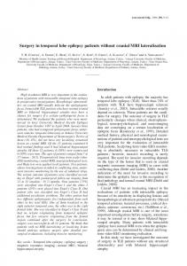
Figure 2: Consequences of amygdala lesion on neural activity in distant cortical areas. The upper left panel shows a coronal brain section on T2-FLAIR MRI, demonstrating bilateral sclerosis in the amygdala-hippocampal region in a patient with temporal lobe epilepsy. All other panels show fMRI activations in brain areas responding less to fearful face expression in patients with amygdala sclerosis, relative to patients with hippocampal sclerosis only. These areas include (from top to bottom, left to right): visual cortex, retrosplenial cortex, anterior cingulate, fusiform gyrus, bilateral posterior superior temporal sulcus, hypothalamus, somatosensory cortex, and peri-hippocampal regions. (Adapted from Vuilleumier et al., 2003).
ted these effects in patients with amygdala damage subsequent to lobectomy for epilepsy surgery, and found a selective deficit in attentional orienting by perceived gaze direction. In this study, attention was manipulated in a simple visual detection task by using two kinds of stimuli to indicate the position of an upcoming target: a photograph of a face with gaze averted either rightward or leftward; and a schematic arrow pointing either rightward or leftward (all presented centrally on a computer screen). Results revealed that healthy subjects show a congruency effect (i.e. faster reaction times) when the target appears in the position indicated by gaze as well by the arrow. By contrast, patients with amygdala lesions showed a specific deficit in orienting attention to the direction indicated by gaze, but normal orienting when direction was indicated by the arrow. Taken together, these findings indicate that some anomalies in the perception of, and reaction to, eye gaze direction in faces may provide a useful measure of amygdala integrity.
84
Epileptologie 2007
Emotional influences on perception and attention The amygdala and related limbic areas are not only responsible for appraising the affective value of external stimuli or events, but also critically involved in orchestrating adaptive responses to emotionally relevant information, such as modulating perceptual analyis and attention. Anatomical studies have shown strong bidirectional connections between amygdala and sensory cortical regions, with important feedback projections from the amygdala to sensory areas which might serve to modulate perceptual processing taking place in these regions [22]. Such modulatory influences might be responsible for the enhanced activation of sensory areas in response to emotional stimuli, as compared with neutral stimuli, a typical finding in many functional brain imaging studies in humans [48, 49] that may facilitate detection and subsequent memory for emotional stimuli [50].
The Role of Amygdala in Emotional and Social Functions | Chiara Cristinzio and Patrik Vuilleumier
Damage to the amygdala in temporal lobe epilepsy can therefore also impair this modulatory feedback of emotional processes on cortical sensory function and perception. Such a deficit was demonstrated in a recent fMRI study of medial temporal lobe sclerosis that compared patients in whom sclerotic lesions involved the hippocampus alone, but not the amygdala, with patients in whom sclerosis involved both the hippocampus and the amygdala [51]. During this fMRI study, patients had to perform a same/different judgment task on pictures of faces or houses, while faces could have either a neutral or fearful expression. Severity of medial temporal lobe sclerosis was determined independently by the intensity of T2 signal on FLAIR MRI scans. Results from the fMRI task showed normal activation in fusiform cortex when the task required face judgments, and normal activation in parahippocampal cortex when the task required house judgments, just like expected from healthy subjects. By contrast, the enhancement of fusiform activation to fearful vs neutral faces was seen in patients with sclerosis affecting the hippocampus alone, but not in those with sclerosis affecting the amygdala in addition to hippocampus (Figure 2). This difference was found even though the two groups of patients did not differ otherwise, had the same clinical epilepsy history and drug treatment, and performed at the same level on standard cognitive tasks. Moreover, amygdala lesions had a predominant impact on visual activation in the same (ipsilateral) hemisphere, such that the greater the sclerosis in left amygdala, the weaker the emotional response to fearful face expressions in left fusiform cortex; and conversely, the greater the sclerosis in right amygdala, the weaker the response to fearful expressions in right fusiform. In addition, this fMRI study [51] also showed that amygdala lesion (right or left) altered the normal pattern of activations to fearful face expressions in a variety of other intact brain regions, suggesting that activity in these regions may normally be influenced by amygdala processing, but be impaired as a consequence of amygdala sclerosis. Regions showing such a loss of emotional effect due to amygdala lesion included the rostral anterior cingulate cortex (rACC), retrosplenial cortex, superior temporal sulcus (STS), secondary somatosensory cortex (SII), and hypothalamus (Figure 2). All these regions have previously been associated with some aspects of affective or social processing. For instance, rACC is involved in emotion regulation and depression [52]; STS is involved in theory of mind and eye gaze perception [53], whereas SII is implicated in somatic markers and facial mimicry during extinction recognition [54]. These fMRI results therefore indicate that abnormal amygdala function due to sclerosis and epilepsy may alter a large cortico-subcortical network of regions normally engaged by emotional face expression and social cognition. However, the exact behavioural correlates of such distant consequences of temporal
lobe sclerosis still remain unknown. It is generally thought that a likely function of the increased activation of sensory areas in response to emotional stimuli [48, 55] is to enhance perceptual analysis, and perhaps subsequent memory traces, for emotionally salient relative to neutral stimuli [50]. In support of this, many studies in healthy subjects have shown an advantage for detecting and/or orienting attention towards emotional stimuli, in conditions where neutral stimuli are typically difficult to detect (e.g. visual search, attentional blink). Remarkably, such facilitation is abolished in patients with amygdala lesion or anterior temporal lobectomy [56]. This surprising finding that amygdala disease can impair visual perception in detection tasks underscores the major role of amygdala in modulating cortical visual pathways, and the important interactions between emotion and attention processes.
Emotional memory Beyond modulating perceptual analysis, the amygdala has also been shown to be critical for emotional memory, by enhancing the storage (and perhaps retrieval) of affectively salient events. Thus, recall is typically better for emotional relative to neutral material. Several studies have found deficits of emotional memory in epilepsy patients with temporal lobectomy and amygdala damage. In a recent work [34], lobectomy patients were initially presented with a series of sentences containing neutral and emotional target words, and then tested one hour later on a recognition task where these target words were presented using a forced choice paradigm. Results showed that memory for emotional items was impaired after bilateral temporal lobe damage, while the performance of unilateral patients was comparable to that of healthy controls. Emotional memory therefore appears to be more adversely affected when lesions to the amygdala are bilateral. Another study [57] on verbal emotional memory reported a loss of the normal enhancement for emotional aspects of a story in patients with unilateral amygdala and hippocampus lesions, relative to controls. Recent work in lobectomy patients suggests that such emotional memory deficits may concern the gist more than the details of emotional events [58]. In the latter study, epilepsy patients with unilateral lobectomy were first shown neutral target pictures in the context of either a neutral or an emotional story (determined by the content of other pictures); and then they were tested on the next day by asking them to recall these target stimuli, in response to a fixed set of questions about the gist and details of each story. Results showed a specific impairment in emotional memory associated with unilateral damage to medial temporal lobe including the amygdala. However, in contrast with previous findings, Phelps
The Role of Amygdala in Emotional and Social Functions | Chiara Cristinzio and Patrik Vuilleumier
Epileptologie 2007
85
et al. [59] reported a study where amygdala-damaged patients did not show any significant deficit on some emotional memory tasks. This study investigated emotional memory in patients with unilateral damage to the medial temporal lobe including both the hippocampus and amygdala. The authors examined memory for emotional words and memory for neutral words embedded in emotional sentences. They found that all groups showed superior recall for positive and negative words in comparison to neutral words; and positive words were recalled significantly more often than negative words. However, no difference was found between unilateral damage patients and healthy controls. The authors concluded that unilateral temporal lobectomy may preserve a normal pattern of performance when recalling affective words. Yet, such effects might partly be mediated by semantic associations rather than by purely emotional influences. Therefore, although some results are still partly contradictory and controversial, it now seems clearly established that the amygdala is crucially involved in processing emotional memories. But more work remains to be done to better characterize the nature of emotional effects on memory, and the possible hemispheric asymmetries for different types of information.
Emotion in music perception A more recent focus of research is the role of amygdala in the perception of emotion in music. Gosselin et al. [60] have explored the ability to recognize the emotional character of different musical excerpts in a series of patients with unilateral left or right medial temporal lesion, including amygdala, after surgery for epilepsy. Because the amygdala involvement in recognizing fear in faces is well known, these authors were particularly interested in the perception of threat in music. They used musical excerpts composed to induce fear, peacefulness, happiness and sadness; and then asked patients to judge how much of each labelled emotion (threat, peacefulness, happiness and sadness) was present in the music. They found that unilateral lobectomy patients showed a specific deficit in recognition of “scary” music, and this impairment was more pronounced in right hemisphere-damaged patients. It is important to notice that the amygdala was completely removed in all patients, but the lesion involved several other regions, so it is difficult to know if the deficit was due to amygdala damage alone or to some disconnection with other brain structure. To ascertain the specific contribution of amygdala in recognition of scary music, the same experimental design was administered to patient SM, who has selective bilateral amygdala atrophy due to Urbach-Wiethe disease. This patient has been studied in detail and her selective lesion has already established the role of amygdala in the perception of fear in facial expressions
86
Epileptologie 2007
(see above) [29, 61]. In the music paradigm, Gosselin et al. [62] confirmed that her deficit in the recognition of fear extended to musical stimuli. Recognition of joyful music was not impaired. This result emphasizes the specificity of amygdala function in the identification of emotion from music.
Theory of mind An important aspect of social cognition is the ability to make inferences about others' mental states, an ability that is necessary to interpret people's behaviour, beliefs and intentions. This important aspect of social cognition is named “theory of mind” (ToM) [63, 64], and its reliance on amygdala function is still partly unresolved. Many tasks have been designed to evaluate the ToM. A classic task is the detection of “faux-pas”, i.e., socially inappropriate actions. For instance, a “faux-pas” occurs when a person involuntary makes a remark that he should not have made and that was perceived as hurtful, or insulting. Many studies have demonstrated that this ability, acquired during early childhood, is impaired in some developmental disorders such as autism [64]. Recently, some authors have proposed an implication of amygdala function in ToM, and tested this hypothesis in epileptic patient with lesion involving the amygdala, using various tasks such as the detection of “faux-pas”. Shaw and coworkers [65] compared the effects of early and late developmental damage to the amygdala on ToM abilities. They divided epileptic patients in two groups: “early” and “late” damage. The first included patient who had amygdala dysfunction since the age of their first seizure; the second group consisted of patients in whom amygdala damage occurred at the time of surgical resection, but who had a normal amygdala function before operation. Theory of mind abilities were examined by a “faux-pas” task and other tests. The authors found that only patients with amygdala lesion occurring during the first two decades of life showed deficits in complex abilities of reasoning about the mental states of others. In contrast, subjects with acquired lesions of the amygdala showed no significant impairment in ToM tasks, as compared with a control group of patients. Different results were provided by a study of Stone et al. [66]. The latter researchers investigated the social abilities of patients with acquired bilateral amygdala lesion due to focal brain injuries, using both the recognition of “faux-pas“ and another classic task, the test of “reading the mind in the eyes”. This latter task was created by Baron-Cohen et al. [67] to study autistic subjects and consists of photographs showing only the eye region of 25 different faces, from which the participant is asked to decide what the depicted person is feeling or thinking (i.e. envy, seduction, etc.). Stone et al. [66] observed that amygdala-damaged patients per-
The Role of Amygdala in Emotional and Social Functions | Chiara Cristinzio and Patrik Vuilleumier
formed worse than control subjects in both of these tasks probing theory of mind. These results therefore suggest that theory of mind can be impaired by amygdala lesion even if the lesion occurs in adult age. Similarly, Schacher et al. [68] have investigated the ability to detect „faux-pas“ in patients with mesial temporal lobe epilepsy compared to patients suffering from epilepsy originating outside of the mesial temporal lobe. They found that only patients with mesial temporal epilepsy (tested either preoperatively or postoperatively) showed an impairment in the recognition of “faux-pas“, but not those with extra-mesiotemporal epilepsy. No correlation was found with the age of seizure onset. Taken together, these data corroborate the idea that, in humans, the amygdala plays a crucial role in a wide range of complex social abilities, in addition to just fear perception.
Mood and psychiatric disorders Besides deficits in processing stimuli or events with emotional significance, as reviewed above, temporal lobe epilepsy is frequently associated with mood or psychiatric disorders. The risk of psychiatric comorbidity is 20-40 % in TLE patients, and it is greater in those with a form of drug-resistant epilepsy [69]. Affective disorders, such as depression and anxiety, are the most common disturbances with a prevalence as high as 50 % in patients with drug-resistant epilepsy [70]. Clinical symptoms are often different from the classic endogenous forms of depression, and this may cause some difficulty for diagnosis. The causal relationship between epilepsy and depression is not really clear. Many researchers have proposed that depression is a consequence or a correlate of the chronic illness, but it was shown that in some patients the affective disorder may appear before the onset of epilepsy. Moreover, in many cases, epileptic patients with depression present a family history for depression or other psychiatric disorders. As some abnormalities in the size and function of the amygdala are commonly observed in depression, similar anomalies might also be present in patients at risk of depression. In keeping with this, Richardson et al. [71] reported bilateral increases in amygdala volume for temporal epileptic patients with self-reported depressive symptoms. A particular form of psychosis can also be associated with temporal lobe epilepsy. This has been recognized since the early 1950s, when the term psychosis of epilepsy (POE) was introduced to define a sample of psychiatric symptoms related to seizure disorder. However, POE can arise in different situations with different clinical characteristics. Psychotic symptoms can appear during a seizure (ictal), with a high prevalence of confusion and hallucination; or following seizures (postictal), with variable delays [72]. Finally, other psychotic signs can alternate with seizures (inter-
ictal), characterized by hallucinations, delusions, aggression, and disorganized behaviour. The relation to some dysfunction within the limbic system is still poorly known, but anomalies in both amygdala and prefrontal projections have long been suspected in schizophrenia [73] and may also play an important role in POE. A few studies have evaluated the effect of surgical intervention on psychiatric disorders in epileptic patients. Devinsky et al. [74] tested a large sample of patients with temporal or extratemporal epilepsy before and after surgery, using self-reported and structural interview to evaluate various psychiatric disorders. The observed prevalence of depression was approximately 25% in their population before surgical intervention. At 3, 12, and 24 months of postoperative follow-up, there was a significant reduction of depression symptoms. Similarly, other longitudinal studies [75, 76] observed an improvement of depression after surgery in patients with temporal epilepsy who presented clinically relevant affective disorders before surgery. By contrast, other authors have noted the emergence of mood disturbances after surgery. Kanemoto et al. [77] tested a sample of patients before and after temporal lobectomy including amygdala and hippocampus. They observed that patients with a history of psychiatric disease before surgery also present a higher risk of manifesting new mood deficits after surgery, relative to those with no previous history of psychiatric disease. Future studies may usefully employ new neuroimaging measures, both prior and after surgery, in order to monitor changes in brain activity that may correlate or predict subsequent changes in mood states in these patients.
Conclusion While the importance of medial temporal lobe structures has long been established for the hippocampus and memory function, the role of the amygdala and of its dense projections to widespread brain areas is still largely underappreciated in temporal lobe epilepsy. Damage to the amygdala may cause a wide range of deficits in the appraisal of emotional and social significance of sensory events, although these deficits are often variable and still poorly understood. These deficits may include the recognition of facial expressions, perception of gaze direction, attention, memory, musical emotions, theory of mind, as well as mood and other neuropsychiatric disorders. Some of these deficits might result from the loss of distant modulatory inputs from the amygdala on other intact regions, as can now be demonstrated by functional neuroimaging methods. We believe that a more systematic assessment of the rich repertoire of affective functions mediated by the amygdala might provide useful information about temporal lobe pathology and
The Role of Amygdala in Emotional and Social Functions | Chiara Cristinzio and Patrik Vuilleumier
Epileptologie 2007
87
neuropsychological outcome after surgery in TLE patients. In the future, new tests probing emotional and social processing should be highly desirable for clinical applications in these patients, to improve clinical management and to shed new lights on amygdala functions in humans.
20. Pitkanen A, Savander V, LeDoux JE. Organization of intra-amygdaloid circuitries in the rat: an emerging framework for understanding functions of the amygdala. Trends Neurosci 1997; 20: 517-523 21. Amaral DG. The primate amygdala and the neurobiology of social behavior: implications for understanding social anxiety. Biol Psychiatry 2002; 51: 11-17 22. Amaral DG, Behniea H, Kelly JL. Topographic organization of projections from the amygdala to the visual cortex in the macaque monkey. Neuro-
References
science 2003; 118: 1099-1120 23. Phillips RG, LeDoux JE. Differential contribution of amygdala and hippo-
1. Jokeit H, Schacher M. Neuropsychological aspects of type of epilepsy and etiological factors in adults. Epilepsy Behav 2004; 5(Suppl 1): S14-20 2. Elger CE, Helmstaedter C, Kurthen M. Chronic epilepsy and cognition. Lancet neurology 2004; 3: 663-672
106: 274-285 24. Quirk GJ, Beer JS. Prefrontal involvement in the regulation of emotion: convergence of rat and human studies. Curr Opin Neurobiol 2006; 16: 723-
3. Van Hoesen GW. Anatomy of the medial temporal lobe. Magn Reson Imaging 1995; 13: 1047-1055
727 25. Morris JS, Buchel C, Dolan RJ. Parallel neural responses in amygdala sub-
4. Cendes F, Leproux F, Melanson D et al. MRI of amygdala and hippocampus in temporal lobe epilepsy. Journal of computer assisted tomography 1993; 17: 206-210
regions and sensory cortex during implicit fear conditioning. Neuroimage 2001; 13: 1044-1052 26. Ball T, Rahm B, Eickhoff SB et al. Response properties of human amygdala
5. Phelps EA, LeDoux JE. Contributions of the amygdala to emotion proces-
subregions: evidence based on functional MRI combined with probabilistic
sing: from animal models to human behavior. Neuron 2005; 48: 175-187
anatomical maps. PLoS ONE 2007; 2: e307 doi 10.1371/journal.po-
6. Cendes F, Andermann F, Gloor P et al. Relationship between atrophy of the amygdala and ictal fear in temporal lobe epilepsy. Brain 1994; 117: 739746
ne.0000307 27. Adolphs R. Neural systems for recognizing emotion. Curr Opin Neurobiol 2002; 12: 169-177
7. Lanteaume L, Khalfa S, Regis J et al. Emotion induction after direct intra-
28. Vuilleumier P, Armony J, Dolan R. Reciprocal links between emotion and
cerebral stimulations of human amygdala. Cereb Cortex 2006; doi:
attenton. In: Frackowiak R et al. (eds): Human Brain Function, 2nd ed. San
10.1093/cercor/bhl041
Diego: Academic Press, 2003: 419-444
8. Klüver H, Bucy PC. Preliminary analysis of functions of the temporal lobes in monkeys. Archives of Neurology and Psychiatry 1939; 42: 979-1000 9. Weiskrantz L. Behavioral changes associated with ablation of the amygdaloid complex in monkeys. J Comp Physiol Psychol 1956; 49: 381-391 10. Horel JA, Keating EG. Recovery from a partial Kluver-Bucy syndrome in the monkey produced by disconnection. J Comp Physiol Psychol 1972; 79: 105-114
29. Adolphs R, Tranel D, Damasio H, Damasio A. Impaired recognition of emotion in facial expressions following bilateral damage to the human amygdala. Nature 1994; 372: 669-672 30. Fowler HL, Baker GA, Tipples J et al. Recognition of emotion with temporal lobe epilepsy and asymmetrical amygdala damage. Epilepsy Behav 2006; 9: 164-172 31. Meletti S, Benuzzi F, Rubboli G et al. Impaired facial emotion recognition
11. Aggleton JP, Passingham RE. Syndrome produced by lesions of the amygdala in monkeys (Macaca mulatta). J Comp Physiol Psychol 1981; 95: 961-977
in early-onset right mesial temporal lobe epilepsy. Neurology 2003; 60: 426-431 32. Anderson AK, Phelps EA. Expression without recognition: contributions of
12. Kling AS, Tachiki K, Lloyd R. Neurochemical correlates of the Kluver-Bucy syndrome by in vivo microdialysis in monkey. Behav Brain Res 1993; 56: 161-170
the human amygdala to emotional communication. Psychol Sci 2000; 11: 106-111 33. LaBar KS, LeDoux JE, Spencer DD, Phelps EA. Impaired fear conditioning
13. Meunier M, Hadfield W, Bachevalier J, Murray EA. Effects of rhinal cortex lesions combined with hippocampectomy on visual recognition memory in rhesus monkeys. J Neurophysiol 1996; 75: 1190-1205 14. Marlowe WB, Mancall EL, Thomas JJ. Complete Kluver-Bucy syndrome in man. Cortex 1975; 11: 53-59
following unilateral temporal lobectomy in humans. J Neurosci 1995; 15: 6846-6855 34. Brierley B, Medford N, Shaw P, David AS. Emotional memory and perception in temporal lobectomy patients with amygdala damage. Journal of neurology, neurosurgery, and psychiatry 2004; 75: 593-599
15. Kapur N, Barker S, Burrows EH et al. Herpes simplex encephalitis: long term
35. Siebert M, Markowitsch HJ, Bartel P. Amygdala, affect and cognition:
magnetic resonance imaging and neuropsychological profile. J Neurol
evidence from 10 patients with Urbach-Wiethe disease. Brain 2003;
Neurosurg Psychiatry 1994; 57: 1334-1342
126: 2627-2637
16. Adolphs R. Social cognition and the human brain. Trends Cogn Sci 1999; 3: 469-479
36. Anderson AK, Phelps EA. Is the human amygdala critical for the subjective experience of emotion? Evidence of intact dispositional affect in patients
17. LeDoux JE. Emotion circuits in the brain. Annu Rev Neurosci 2000; 23: 155184
with amygdala lesions. Journal of cognitive neuroscience 2002; 14: 709720
18. Baas D, Aleman A, Kahn RS. Lateralization of amygdala activation: a
37. Tranel D, Gullickson G, Koch M, Adolphs R. Altered experience of emotion
systematic review of functional neuroimaging studies. Brain Res Brain Res
following bilateral amygdala damage. Cognitive neuropsychiatry 2006;
Rev 2004; 45: 96-103
11: 219-232
19. Cahill L, Uncapher M, Kilpatrick L et al. Sex-related hemispheric lateralization of amygdala function in emotionally influenced memory: an FMRI investigation. Learn Mem 2004; 11: 261-266
88
campus to cued and contextual fear conditioning. Behav Neurosci 1992;
Epileptologie 2007
38. Fox E, Damjanovic L. The eyes are sufficient to produce a threat superiority effect. Emotion 2006; 6: 534-539 39. Adams RB, Jr., Kleck RE. Perceived gaze direction and the processing of
The Role of Amygdala in Emotional and Social Functions | Chiara Cristinzio and Patrik Vuilleumier
facial displays of emotion. Psychol Sci 2003; 14: 644-647 40. Scherem KR, Schorr A, Johnstone T. Appraisal processes in emotion: Theory, methods, research. New York: Oxford University Press, 2001 41. Sato W, Yoshikawa S, Kochiyama T, Matsumura M. The amygdala processes the emotional significance of facial expressions: an fMRI investigation using the interaction between expression and face direction. Neuroimage 2004; 22: 1006-1013 42. Adams RB, Jr., Gordon HL, Baird AA et al. Effects of gaze on amygdala sensitivity to anger and fear faces. Science 2003; 300: 1536
62. Gosselin N, Peretz I, Johnsen E, Adolphs R. Amygdala damage impairs emotion recognition from music. Neuropsychologia 2007; 45: 236-244 63. Baron-Cohen S, Ring HA, Bullmore ET et al. The amygdala theory of autism. Neuroscience and biobehavioral reviews 2000; 24: 355-364 64. Baron-Cohen S, Leslie AM, Frith U. Does the autistic child have a "theory of mind"? Cognition 1985; 21: 37-46 65. Shaw P, Lawrence EJ, Radbourne C et al. The impact of early and late damage to the human amygdala on 'theory of mind' reasoning. Brain 2004; 127: 1535-1548
43. Kawashima R, Sugiura M, Kato T et al. The human amygdala plays an
66. Stone VE, Baron-Cohen S, Calder A et al. Acquired theory of mind impair-
important role in gaze monitoring: a PET study. Brain 1999; 122: 779-783
ments in individuals with bilateral amygdala lesions. Neuropsychologia
44. George N, Driver J, Dolan RJ. Seen gaze-direction modulates fusiform activity and its coupling with other brain areas during face processing. Neuroimage 2001; 13: 1102-1112 45. Young AW, Aggleton JP, Hellawell DJ et al. Face processing impairments after amygdalotomy. Brain 1995; 118 ( Pt 1): 15-24 46. Friesen CK, Kingstone A. The eyes have it! Reflexive orienting is triggered by nonpredictive gaze. Psychonomic bulletin & review 1998;5:490-495 47. Vuilleumier P. Perceived gaze direction in faces and spatial attention: a study in patients with parietal damage and unilateral neglect. Neuropsychologia 2002; 40: 1013-1026 48. Vuilleumier P, Armony JL, Driver J, Dolan RJ. Effects of attention and emotion on face processing in the human brain: an event-related fMRI study. Neuron 2001; 30: 829-841 49. Sabatinelli D, Bradley MM, Fitzsimmons JR, Lang PJ. Parallel amygdala and inferotemporal activation reflect emotional intensity and fear relevance. Neuroimage 2005; 24: 1265-1270 50. Vuilleumier P. How brains beware: neural mechanisms of emotional attention. Trends Cogn Sci 2005; 9: 585-594 51. Vuilleumier P, Richardson MP, Armony JL et al. Distant influences of amygdala lesion on visual cortical activation during emotional face processing. Nat Neurosci 2004; 7: 1271-1278
2003; 41: 209-220 67. Baron-Cohen S, Jolliffe T, Mortimore C, Robertson M. Another advanced test of theory of mind: evidence from very high functioning adults with autism or asperger syndrome. Journal of child psychology and psychiatry, and allied disciplines 1997; 38: 813-822 68. Schacher M, Winkler R, Grunwald T et al. Mesial temporal lobe epilepsy impairs advanced social cognition. Epilepsia 2006; 47: 2141-2146 69. Devinsky O. Psychiatric comorbidity in patients with epilepsy: implications for diagnosis and treatment. Epilepsy Behav 2003; 4(Suppl 4): S2-10 70. Schmitz B. Depression and mania in patients with epilepsy. Epilepsia 2005; 46(Suppl 4): 45-49 71. Richardson EJ, Griffith HR, Martin RC et al. Structural and functional neuroimaging correlates of depression in temporal lobe epilepsy. Epilepsy Behav 2007 72. Kanemoto K, Kawasaki J, Kawai I. Postictal psychosis: a comparison with acute interictal and chronic psychoses. Epilepsia 1996; 37: 551-556 73. Aleman A, Kahn RS. Strange feelings: do amygdala abnormalities dysregulate the emotional brain in schizophrenia? Prog Neurobiol 2005; 77: 283-298 74. Devinsky O, Barr WB, Vickrey BG et al. Changes in depression and anxiety after resective surgery for epilepsy. Neurology 2005; 65: 1744-1749
52. Seminowicz DA, Mayberg HS, McIntosh AR et al. Limbic-frontal circuitry in
75. Pintor L, Bailles E, Fernandez-Egea E et al. Psychiatric disorders in tempo-
major depression: a path modeling metanalysis. Neuroimage 2004; 22:
ral lobe epilepsy patients over the first year after surgical treatment.
409-418 53. Allison T, Puce A, McCarthy G. Social perception from visual cues: role of the STS region. Trends Cogn Sci 2000; 4: 267-278
Seizure 2007; 16: 218-225 76. Inoue Y, Mihara T. Psychiatric disorders before and after surgery for epilepsy. Epilepsia 2001; 42(Suppl 6): 13-18
54. Adolphs R, Damasio H, Tranel D, Damasio AR. Cortical systems for the
77. Kanemoto K, Kawasaki J, Mori E. Postictal psychosis as a risk factor for
recognition of emotion in facial expressions. J Neurosci 1996; 16: 7678-
mood disorders after temporal lobe surgery. Journal of neurology, neuro-
7687
surgery, and psychiatry 1998; 65: 587-589
55. Grandjean D, Sander D, Pourtois G et al. The voices of wrath: brain responses to angry prosody in meaningless speech. Nat Neurosci 2005; 8: 145-146 56. Anderson AK, Phelps EA. Lesions of the human amygdala impair enhanced perception of emotionally salient events. Nature 2001; 411: 305-309 57. Edith Frank J, Tomaz C. Lateralized impairment of the emotional enhancement of verbal memory in patients with amygdala-hippocampus lesion. Brain and cognition 2003; 52: 223-230 58. Adolphs R, Tranel D, Buchanan TW. Amygdala damage impairs emotional memory for gist but not details of complex stimuli. Nature neuroscience 2005; 8: 512-518 59. Phelps EA, LaBar KS, Spencer DD. Memory for emotional words following unilateral temporal lobectomy. Brain and cognition 1997; 35: 85-109 60. Gosselin N, Peretz I, Noulhiane M et al. Impaired recognition of scary
Address for correspondence: Dr. Chiara Cristinzio Lab NIC, Dept of Neurosciences & Clinic of Neurology Centre Médical Universitaire (CMU) 1 rue Michel Servet CH 1211 Geneva Tel. 0041 22 3795 382 Fax 0041 22 3795 402
[email protected] or
music following unilateral temporal lobe excision. Brain 2005; 128: 628640 61. Adolphs R, Tranel D, Damasio H, Damasio AR. Fear and the human amygdala. J Neurosci 1995; 15: 5879-5891
The Role of Amygdala in Emotional and Social Functions | Chiara Cristinzio and Patrik Vuilleumier
Epileptologie 2007
89