Hindawi Publishing Corporation Epilepsy Research and Treatment Volume 2012, Article ID 103160, 15 pages doi:10.1155/2012/103160
Review Article Neocortical Temporal Lobe Epilepsy Eduard Bercovici,1 Balagobal Santosh Kumar,2 and Seyed M. Mirsattari2, 3, 4, 5, 6 1 Division
of Neurology, University of Toronto, Toronto, ON, Canada of Clinical Neurological Sciences, University of Western Ontario, London, ON, Canada 3 Department of Medical Imaging, University of Western Ontario, London, ON, Canada 4 Department of Medical Biophysics, University of Western Ontario, London, ON, Canada 5 Department of Psychology, University of Western Ontario, London, ON, Canada 6 London Health Sciences Centre, B10-110, London, ON, Canada N6A 5A5 2 Department
Correspondence should be addressed to Seyed M. Mirsattari,
[email protected] Received 20 May 2011; Revised 4 January 2012; Accepted 22 May 2012 Academic Editor: Warren T. Blume Copyright © 2012 Eduard Bercovici et al. This is an open access article distributed under the Creative Commons Attribution License, which permits unrestricted use, distribution, and reproduction in any medium, provided the original work is properly cited. Complex partial seizures (CPSs) can present with various semiologies, while mesial temporal lobe epilepsy (mTLE) is a wellrecognized cause of CPS, neocortical temporal lobe epilepsy (nTLE) albeit being less common is increasingly recognized as separate disease entity. Differentiating the two remains a challenge for epileptologists as many symptoms overlap due to reciprocal connections between the neocortical and the mesial temporal regions. Various studies have attempted to correctly localize the seizure focus in nTLE as patients with this disorder may benefit from surgery. While earlier work predicted poor outcomes in this population, recent work challenges those ideas yielding good outcomes in part due to better localization using improved anatomical and functional techniques. This paper provides a comprehensive review of the diagnostic workup, particularly the application of recent advances in electroencephalography and functional brain imaging, in neocortical temporal lobe epilepsy.
1. Introduction Neocortical temporal lobe epilepsy (nTLE) is a rather newly recognized entity that is different than the wellknown entity of mesial temporal lobe epilepsy (mTLE) although not as well characterized [1]. The documented cases of patients with nonlesional neocortical temporal lobe seizure origin are not as rare as previously reported. In one study, out of 31 patients seizure-free more than 18 months after temporal lobectomy, only 3 patients (9.6%) were found to have NTLE [2]. More recently, Schramm et al. [1] demonstrated 62/581 of the temporal lobe epilepsy (TLE) cases as being neocortical. With better structuralfunctional imaging modalities as well as invasive monitoring, more of these cases are being described. Unfortunately, the nomenclature is inconsistent in the literature, often being dubbed as nonlesional, extrahippocampal, or lateral neocortical. For the purpose of this review, we will use the term nTLE. Recognition of nTLE is important because these patients may either be considered non surgical candidates or
undergo extensive surgeries due to the poor localization of their seizure focus. Lesional nTLE cases are often not reported in the literature as compared to the nonlesional cases because they may be less likely to be admitted to an epilepsy monitoring unit (EMU) for video-electroencephalography (EEG) telemetry unless the lesion is closely associated with eloquent cortex, thereby limiting surgical resection or may be unamenable to surgery. Thus, the reported prevalence of nTLE is low. Therefore, it is difficult to know the prognosis of nTLE because lesional nTLE cases typically have better outcomes than nonlesional nTLE cases [2] although classically they were dubbed as having poorer outcomes compared to MTLE [3–9].
2. Historical Background Although nonconvulsive seizures and seizures manifesting with complex behaviours have been recognized since antiquity [10], their relationship to temporal lobe origin was first
2 described in the late 1800s by Jackson [11]. The psychic and motor characteristics of these seizures prompted the term of psychomotor seizures [12]. With the first application of EEG to human by Berger in 1929 [13] and the increased interest in surgical treatment of epilepsy, the anatomical significance of these seizures led them to be labeled temporal lobe seizures [14]. The prominent role of mesial temporal structures in the genesis of temporal lobe seizures was first suspected by Falconer et al. [15] and was confirmed and widely recognized thereafter [15–17]. The majority of temporal lobe seizures originate in the mesial structures, primarily in the hippocampus, with the rest beginning in temporal neocortical regions. Mesial temporal lobe seizures are far more common than lateral neocortical seizures [18]. Wieser [19] was the first to propose 5 subtypes of temporal lobe seizures depending on electroclinical features. They included temporal-basal limbic, temporal polar, posterior temporal neocortical, opercular, and frontobasal cingulate. The classification was revised to simplify the nomenclature in 1989, and only 2 of the subtypes remained. Thus temporal lobe epilepsy is now categorized into mesial and lateral [20]. Whether or not these 2 types can reliably be separated based on noninvasive evaluation was disputed [1, 21, 22]. Differentiation between mTLE and nTLE remains a challenge even for epileptologists, as many symptoms overlap. This may be due to extensive reciprocal connections between the mesial and lateral temporal structures, allowing spread of ictal discharge in either direction [23–25]. The clinical profile of patients with nTLE is different from mTLE. The average age of onset in nTLE is approximately 5–10 years more than in mTLE [6, 26]. There is no known gender, cultural, or racial risk factors for nTLE. Patients with nTLE usually do not have a history of the typical risk factors associated with mTLE such as febrile seizures, head injury, perinatal insults, or previous central nervous system (CNS) infections as compared to mTLE [3, 7, 26, 27]. Many of the clinical characteristics of the seizures described in autosomal dominant lateral temporal lobe epilepsy (ADLTE) are similar to those seen in patients with nTLE. ADLTE is a well-defined, albeit rare, condition characterized by onset in adolescence or early adulthood of lateral temporal seizures with prominent auditory auras sometimes triggered by external noises, normal conventional magnetic resonance imaging (MRI), good response to antiepileptic treatment, and overall benign outcome. The same phenotype is shared by sporadic and familial cases with complex inheritance. Mutations in the LGI1 gene in the 10-cM region on chromosome 10 q24 are found in about 50% of ADLTE families and 2% of sporadic cases. LGI1 shows no homology with known ion channel genes. Recent findings suggest that LGI1 may exert multiple functions, but it is not known which of them is actually related to lateral temporal epilepsy [28–30].
Epilepsy Research and Treatment
3. Clinical Semiology Ictal manifestations common in mTLE (ipsilateral limb automatisms, contralateral dystonic posturing, and oroalimentary automatisms) are significantly less frequent in nTLE [1, 5, 31]. These differences are summarized in Table 1. Dupont et al. [32] compared the ictal semiology of 45 mTLE patients with 13 nTLE patients and found that contralateral dystonic posturing with ipsilateral automatisms occurred in a third of the mTLE group but was never seen in those with nTLE. Auditory and vertiginous auras have been associated with the temporal neocortex, and visceral sensations and fear with the mesial temporal lobe [23, 33]. One study analyzing ictal semiology between nTLE and mTLE reported that seizures in the nTLE group were of shorter duration (46 seconds) as compared to the mesial group (67.5 seconds) [8]. Patients with mTLE were more likely to display manual or oroalimentary automatisms, dystonic posturing, hyperventilation, or postictal cough [7, 34]. nTLE patients had experienced only experiential auras, whereas mTLE patients had epigastric or olfactory/gustatory sensations or fear as their auras. Comparison of clinical semiology of 28 mTLE patients and 12 nTLE patients [35] showed that epigastric sensations, fear, olfactory auras, and dystonic posturing were typical of mTLE, whereas auditory auras, cephalic/indescribable sensations, vocalizations, ictal speech, whole-body movements, rapid onset of version, and secondary generalization were significantly more common in patients with nTLE. A recent study [26] where 55 patients with TLE (same as above) were classified into 3 distinct groups (mTLE, nTLE, and mixed and characterized them based on semiology and spatiotemporal pattern of discharges. At seizure onset, patients with nTLE were less likely to describe rising epigastric sensation, fear) or dreamy state but more likely to describe any type of hallucination or illusion. As the seizures progressed, mesial seizures produced oroalimentary, verbal and upper limb automatisms. In general, nTLE were shorter but more frequently generalized. In another study of 21 patients with nTLE, 71% of them had auras, with the experiential auras being the most common [4]. The most common initial behavioural change was motionless stare in 48% of patients. Only 2/21 patients had hippocampal atrophy (HA). A lateralized memory deficit was observed in 62% [4].
4. Ictal Semiology in nTLE Several studies have attempted to localize the semiology based on the anterior-posterior axis of the neocortical temporal lobe. One study in particular separated the groups based on interictal temporal lobe discharges (anterior, posterior, or diffuse) and correlated those to subjective ictal phenomena [36]. Olfactory and gustatory phenomena and d´ej`a vu were present exclusively in patients with anterior foci, whereas visual auras were more common in the posterior temporal group. Complex automatisms were more common in the anterior group, and neurological abnormalities were more common in the posterior group.
Epilepsy Research and Treatment Studying patients with posterior TLE (n = 14), the authors found that behavioural arrest was the first manifestation and was followed by motor signs as the seizure activity spread to the frontoparietal convexity. They also observed that behavioural automatisms (oro-alimentary and gestural), although present in half the patients, were never the first or most prominent ictal manifestations [37]. Another study attempted to analyze semiologic differences between mTLE and different anatomic subgroups of nTLE included 1-year postsurgical followup. Total of 107 seizures in 13 patients with anterior TLE, 8 patients with posterior TLE, and 21 patients with mTLE were reviewed. Frequent behavioural arrest, absence of initial oroalimentary automatisms, and early generalization were characteristic findings of posterior TLE, although they were insufficient to differentiate from anterior TLE or mTLE.
5. Diagnostic Workup As described in other articles of this special issue of the journal, patients with mTLE have typical characteristic seizure semiology and may demonstrate MTS on MRI. Their prognosis after surgical resection is said to be good if the lesion is definable. However, occasionally HS is not evident as in the nonlesional cases (i.e., symptomatic epilepsy) or the semiology and EEG findings do not fully localize to the mesial temporal lobes. Some of these patients undergo more invasive monitoring to consider the possibility of nTLE. The following includes a summary of literature depicting how EEG, structural and functional imaging can help to differentiate nTLE. Later, other advanced techniques are discussed for their putative roles. 5.1. Electroencephalography 5.1.1. Interictal Scalp EEG. A prospective study [38] on 132 consecutive patients (mTLE = 86 and nTLE = 36) with epilepsy showed that a history of febrile seizures, abdominal auras, contralateral dystonic posturing, and predominance of mesial temporal spikes point to mTLE (positive predictive value 81% and negative predictive value 70%). They concluded that analysing the clinical and EEG features, particularly the distribution of interictal epileptiform discharges (IEDs), helps to differentiate between mTLE and nTLE. There is little evidence to support the use of interictal scalp EEG in differentiating nTLE from mTLE. In one study [2], the utility of the interictal EEG was examined in patients with neocortical symptomatic epilepsies. It was useful in 9/17 of patients (52%) with nTLE. In another study, 22 patients admitted to an EMU were enrolled, and the findings were correlated the results from PET scans [39]. They found that the interictal rhythmic slow activity was highly correlated to nTLE. In contrast, no significant difference was found among 14 patients with nTLE and those with mTLE in another study when using standard intracranial EEG as comparison [40].
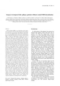
3 5.1.2. Ictal Scalp EEG. Ictal scalp EEG has the potential to localize seizures better than interictal especially in a longterm video-EEG monitoring unit. Recording a unilateral IED cannot always distinguish between mesial or lateral temporal or extratemporal foci [41]. In one case series, the sensitivity of localizing IEDs to the temporal neocortex increased from 52% (interictal) to 76% (ictal) [2]. The most commonly observed scalp ictal pattern is ipsilateral temporal rhythmic theta activity, seen in both nTLE and mTLE [8] although slower in frequency in nTLE. In NTLE, it may be preceded by an irregular 2 to 5 Hz polymorphic slowing that may or may not be lateralized [8, 42]. The characteristic ictal EEG in patients with mTLE is rhythmic theta activity of 5 to 7 Hz [43, 44]. In a clinicopathologic study comparing 46 patients with MTS or neocortical lesions by EEG, those with mTLE had significantly more fast rhythmic activity (>4 Hz). Patients with nTLE tended to develop bilateral ictal EEG changes, occurring significantly more often and faster onset of bilaterality [27]. Similar results were obtained studying ictal scalp EEG in 93 patients with seizure origin verified by intracranial EEG, showing an association between irregular polymorphic, slow (2 to 5 Hz) seizure onsets and nTLE, and regular (5 to 9 Hz) ictal onsets in mTLE patients [44]. The nTLE pattern was either preceded by periodic sharp waves or followed by theta rhythms. Unfortunately, a subsequent study by the same authors using simultaneous scalp and intracranial EEG study revealed that these associated patterns were not established on the scalp at seizure onset but resulted from differences in the development, propagation, and synchrony of cortical discharges as seizures progressed in mTLE and nTLE patients [45]. While some studies have demonstrated good localization, others have not been able to differentiate mTLE from nTLE based on ictal EEG [7]. A relatively novel and underrecognized EEG analysis involves transitional sharp waves. In one study involving 52 ictal discharges from 13 patients, the authors were able to determine with certainty that the pattern localized to the nTLE, versus mTLE. Interestingly, none of the 61 discharges in 15 patients with mTLE had the transitional sharp waves [46]. Further studies are warranted to determine if this is an easily demonstrable and reproducible scalp EEG finding Although at this time scalp EEG is not highly sensitive or specific to differentiate mTLE from nTLE practically, it has been used to assess outcomes in patients with known nTLE. In a study of 29 patients with nTLE a localized or even lateralized EEG pattern was associated with good outcomes [47]. Similarly, in larger study comparing 80 patients with nTLE to other neocortical epilepsies localized EEG rhythms highly predicted seizure freedom after resection [48]. Therefore, ictal rhythm cannot be used in isolation to definite localization (Figures 1(a)–1(f)). However, even among patients whose scalp data are complex to require invasive recording, lateralization of temporal scalp IEDs, and ictal activity should be included when assessing the side of temporal epileptogenesis [49]. 5.1.3. Intracranial EEG. Neocortical foci are seen in up to 65% of patients with TLE in some series [5, 50]. Differentiating nTLE from mTLE often requires intracranial EEG
4
Epilepsy Research and Treatment
Fp1-F3 F3-C3 C3-P3 P3-O1 Fp2-F4 F4-C4 C4-P4 P4-O2 Fp1-F7 F7-T3 T3-T5 T5-O1 Fp2-F8 F8-T4 140 µV
T4-T6 T6-O2
1s (a) Interictal scalp showing right anterior temporal spikes during sleep
Fp1-F3 F3-C3 C3-P3 P3-O1 Fp2-F4 F4-C4 C4-P4 P4-O2 Fp1-F7 F7-T3 T3-T5 T5-O1 Fp2-F8 F8-T4 140 µV
T4-T6
1s
T6-O2 (b) Ictal scalp EEG in the same patient showing possible bitemporal onset of seizure
(c) Sagital T2 FLAIR MRI brain showing left posterior temporal neoplasm (pathologically proven ganglioglioma)
Figure 1: Continued.
Epilepsy Research and Treatment
5
013 014 015 031 032 034 052 053 054 113 114 021 022 023 043 044
1000 µV 1s
(d) Interictal subdural EEG showing spikes arising independently from left mesial and neocortical temporal lobe (electrodes 10s, 30s, and 50s) as well as right anterior mesial and neocortical regions (electrodes 20s and 40s)
011 012 032 033 053 057 058 074 111 112 113 114 131 133 042
1000 µV 1s
043
(e) Ictal subdural EEG showing seizure onsetting electrode 114 (left posterior temporal region, coinciding with the lesion)
(f) Subdural electrode insertion with MRI coregistration. Cortical stimulation produced speech arrest over left anterior inferolateral temporal region (electrode 52), anterior to the lesion (electrode 114). FMRI
Figure 1: EEG of a 37-year-old right-handed male with complex partial seizures since 16 years of age characterized by speech arrest, transient impaired consciousness, automatisms with right hand, and secondary generalization, with no contributory antecedent history.
6
Epilepsy Research and Treatment
Table 1: Comparison of ictal semiology between mTLE and nTLE based on data from references (see text). If 1 minute No More likely Less likely Less likely More likely More likely Yes Yes Less likely More likely Yes Yes Yes More likely More likely
nTLE 50%) [124]. MEG predicted the outcome following surgery for medically intractable epilepsy in children with normal or nonfocal MRI findings [125]. Systematic review on the use of MEG in the presurgical evaluation of localization-related epilepsy (between 1987 and 2006) reported insufficient evidence to support the use of MEG in surgical planning [126]. MEG’s effects on outcomes have also been recently examined demonstrating good surgical outcomes in 22 children with intractable seizures and nonlocalizing or normal MRI [125]. Similarly, in the presurgical evaluation of 67 adults using EEG and MEG the authors found almost similar sensitivities between the two in detecting IED but MEG could correctly identify a source in 1/3 of patients that were EEG negative. They predicted that MEG would be useful in patients with neocortical epilepsy or those with focal cortical dysplasia [117]. 5.3.5. Treatment and Outcome. Pharmacotherapy for focal epilepsy does not depend on anatomical diagnosis, and there is no difference in response to antiepileptic drugs (AEDs) based on anatomical localization of a neocortical epileptogenic region [127]. In patients with medically refractory seizures, surgical treatment depends on precise localization and delineation of the epileptogenic region rather than anatomical diagnosis, and outcome reflects the accuracy of this process and the ability to resect the abnormal tissue. Total resection may be limited by the involvement of the adjacent eloquent areas, or by the failure to correctly map the epileptogenic substrate, as can often occur in nonlesional localization-related epilepsy. If an epileptogenic region involves eloquent cortex, seizures may be relieved by the removal of a structural lesion alone (i.e., lesionectomy) or by multiple subpial transections [128, 129]. There have been many studies reporting less favourable postsurgical outcome in patients with lesional/nonlesional nTLE [3–9]. In one study, improved outcomes were apparent by resecting the combined neocortical and mesial temporal areas [5]. Recent research has challenged this view and suggests the poor outcomes is due to poor patient selection, poor localization, and incomplete resection of the seizure focus. While this current paper highlights the challenges in distinguishing mTLE from nTLE, there have recent studies demonstrating good-to-excellent outcomes in nTLE patients as compared to mTLE [1, 26, 40] or compared to other focal cortical epilepsies [2, 130]. These outcomes are reported using tailored neocorticectomy or multiple subpial transections in addition to a standard ATL, after intracranial ictal EEG recording and cortical mapping [4, 131] even in patients with nonlesional nTLE [1, 132]. Factors affecting outcomes have been assessed by several studies. In a study comparing all neocortical focal epilepsies, the authors found that FDG-PET and interictal EEG localization predicted good outcome in nTLE [2]. The extent of neocortical resection is seen as a positive predictor [131], especially when the underlying area demonstrates pathological delta waves on intracranial monitoring [133].
Epilepsy Research and Treatment In a large prospective-retrospective series in patients with TLE, 10% of the cases had nTLE. Pathological analysis demonstrated only 5% being nonlesional, whereas 57% were neoplastic (ganglioglioma predominating) and 38% nonneoplastic (e.g., MCD). Outcomes seemed to be best for those with neoplastic lesions [1]. This is echoed by another clinicpathological study showing left hemispheric lesions and focal MCD associated with poor outcomes as compared to tumours [47]. However, it should be noted that dual pathology can exist even in patients with presumed nTLE. In a recent analysis of 243 samples from patients with TLE, 86% had HS in addition to other pathologies (33% having tumours and 45% having MCD). Nonetheless, they still demonstrated excellent surgical outcomes (Engel class I) in 87% of those with tumours and 79% with MCD [64].
6. Conclusions nTLE is less common compared to mTLE and accounts for about 10% of TLE. The risk factors for nTLE are quite different from mTLE. Seizure freedom for lesional nTLE is approximately 70%. During the past decade, additional converging evidence has been provided that there are some clinical and electrophysiological characteristics that can help to differentiate nTLE from mTLE. Advances in brain imaging with currently available high-resolution structural MRI can reveal previously covert epileptic lesions, with quantitative and voxel-based MRI analysis increasing the diagnostic yield. HFOs in intracranial EEGs increase the detection rate of ictal onset zone that further helps surgical planning in nonlesional cases. MEG provides complementary electrophysiological information to the EEG but can also determine epileptogenic focus in EEG negative patients, possibly raising surgery as a viable option in previously nonsurgical candidates. The data from MEG and EEG-fMRI can assist in the placement of intracranial electrodes to further define the seizure onset zone. PET and SPECT have also provided some data in localization of neocortical epileptic focus. Thus, reliable integration of all the structural and functional data will help to establish the neocortical origin of the seizures in patients with nonlesional nTLE which is crucial to achieve a good surgical outcome.
Acknowledgment The authors wish to thank Dr. Jean Gotman, Montreal Neurological Institute, Canada, for the illustration on HFOs (Figure 2).
References [1] J. Schramm, T. Kral, T. Grunwald, and I. Bl¨umcke, “Surgical treatment for neocortical temporal lobe epilepsy: clinical and surgical aspects and seizure outcome,” Journal of Neurosurgery, vol. 94, no. 1, pp. 33–42, 2001. [2] S. K. Lee, S. Y. Lee, K. K. Kim, K. S. Hong, D. S. Lee, and C. K. Chung, “Surgical outcome and prognostic factors of cryptogenic neocortical epilepsy,” Annals of Neurology, vol. 58, no. 4, pp. 525–532, 2005.
11 [3] S. Saygi, S. S. Spencer, R. Scheyer, A. Katz, R. Mattson, and D. D. Spencer, “Differentiation of temporal lobe ictal behavior associated with hippocampal sclerosis and tumors of temporal lobe,” Epilepsia, vol. 35, no. 4, pp. 737–742, 1994. [4] P. Kotagal, H. O. Luders, G. Williams, T. R. Nichols, and J. McPherson, “Psychomotor seizures of temporal lobe onset: analysis of symptom clusters and sequences,” Epilepsy Research, vol. 20, no. 1, pp. 49–67, 1995. [5] S. S. Spencer, D. D. Spencer, P. D. Williamson, and R. Mattson, “Combined depth and subdural electrode investigation in uncontrolled epilepsy,” Neurology, vol. 40, no. 1, pp. 74–79, 1990. [6] S. V. Pacia, O. Devinsky, K. Perrine et al., “Clinical features of neocortical temporal lobe epilepsy,” Annals of Neurology, vol. 40, no. 5, pp. 724–730, 1996. [7] A. Gil-Nagel and M. W. Risinger, “Ictal semiology in hippocampal versus extrahippocampal temporal lobe epilepsy,” Brain, vol. 120, no. 1, pp. 183–192, 1997. [8] N. R. Foldvary, N. Lee, G. Thwaites et al., “Clinical and electrographic manifestations of lesional neocortical temporal lobe epilepsy,” Neurology, vol. 49, no. 3, pp. 757–768, 1997. [9] M. Hajek, A. Antonini, K. L. Leenders, and H. G. Wieser, “Mesiobasal versus lateral temporal lobe epilepsy: metabolic differences in the temporal lobe shown by interictal 18F-FDG positron emission tomography,” Neurology, vol. 43, no. 1, pp. 79–86, 1993. [10] O. Temkin, The Falling Sickness, The Johns Hopkins University Press, Baltimore, Md, USA, 1945. [11] J. H. Jackson, Selected Writings of John Hughlings Jackson, vol. 1, 2, Edited by J. Taylor, Staples Press, London, UK, 1958. [12] E. L. Gibbs, F. A. Gibbs, and B. Fuster, “Psychomotor epilepsy,” Archives of Neurology and Psychiatry, vol. 60, no. 4, pp. 331–339, 1948. ¨ [13] H. Berger, “Uber das Elektrenkephalogramm des Menschen,” Archiv f¨ur Psychiatrie und Nervenkrankheiten, vol. 87, no. 1, pp. 527–570, 1929. [14] L. N. Sutton, R. J. Packer, R. A. Zimmerman, D. A. Bruce, and L. Schut, “Cerebral gangliogliomas of childhood,” Progress in Experimental Tumor Research, vol. 30, pp. 239–246, 1987. [15] M. A. Falconer, E. A. Serafetinides, and J. A. Corsellis, “Etiology and pathogenesis of temporal lobe epilepsy,” Archives of Neurology, vol. 10, pp. 233–248, 1964. [16] P. D. Williamson, “Frontal lobe seizures: problems of diagnosis and classification,” in Frontal Lobe Seizures and Epilepsies: Advances in Neurology, P. Chauvel, A. Delgado-Escueta, E. Halgren, and J. Bancaud, Eds., pp. 289–309, Raven Press, New York, NY, USA, 1992. [17] J. A. French, P. D. Williamson, V. M. Thadani et al., “Characteristics of medial temporal lobe epilepsy: I. Results of history and physical examination,” Annals of Neurology, vol. 34, no. 6, pp. 774–780, 1993. [18] P. D. Williamson, J. J. Engel, and C. Munari, “Anatomic classification of localization-related epilepsies,” in Epilepsy: A Comprehensive Textbook, J. J. Engel and T. A. Pedley, Eds., pp. 2405–2416, Lippincott-Raven, Philadelphia, Pa, USA, 1997. [19] H. G. Wieser, Electroclinical Features of Psychomotor Seizure, Butterworths, London, UK, 1983. [20] J. Roger, F. E. Dreifuss, M. Martinez-Lage et al., “Proposal for revised classification of epilepsies and epileptic syndromes,” Epilepsia, vol. 30, no. 4, pp. 389–399, 1989. [21] S. S. Spencer, N. K. So, J. J. Engel et al., “Depth electrodes,” in Surgical Treatment of the Epilepsies, J. J. Engel, Ed., pp. 359– 376, Raven Press, New York, NY, USA, 1993.
12 [22] T. S. Walczak, “Neocortical temporal lobe epilepsy: characterizing the syndrome,” Epilepsia, vol. 36, no. 7, pp. 633–635, 1995. [23] P. Gloor, A. Olivier, and L. F. Quesney, “The role of the limbic system in experimental phenomena of temporal lobe epilepsy,” Annals of Neurology, vol. 12, no. 2, pp. 129–144, 1982. [24] W. T. Blume, J. P. Girvin, and P. Stenerson, “Temporal neocortical role in ictal experiential phenomena,” Annals of Neurology, vol. 33, no. 1, pp. 105–107, 1993. [25] P. Kotagal, H. O. Luders, G. Williams, T. R. Nichols, and J. McPherson, “Psychomotor seizures of temporal lobe onset: analysis of symptom clusters and sequences,” Epilepsy Research, vol. 20, no. 1, pp. 49–67, 1995. [26] L. Maillard, J. P. Vignal, M. Gavaret et al., “Semiologic and electrophysiologic correlations in temporal lobe seizure subtypes,” Epilepsia, vol. 45, no. 12, pp. 1590–1599, 2004. [27] T. J. O’Brien, C. Kilpatrick, V. Murrie, S. Vogrin, K. Morris, and M. J. Cook, “Temporal lobe epilepsy caused by mesial temporal sclerosis and temporal neocortical lesions: a clinical and electroencephalographic study of 46 pathologically proven cases,” Brain, vol. 119, no. 6, pp. 2133–2143, 1996. [28] J. J. Poza, A. Saenz, A. Martinez-Gil et al., “Autosomal dominant lateral temporal lobe epilepsy: clinical and genetic study of a large basque pedigree linked to chromosome 10q,” Annals of Neurology, vol. 45, pp. 182–188, 1999. [29] R. Michelucci, J. J. Poza, V. Sofia et al., “Autosomal dominant lateral temporal epilepsy: clinical spectrum, new epitempin mutations, and genetic heterogeneity in seven european families,” Epilepsia, vol. 44, no. 10, pp. 1289–1297, 2003. [30] P. Hedera, B. Abou-Khalil, A. E. Crunk, K. A. Taylor, J. L. Haines, and J. S. Sutcliffe, “Autosomal dominant lateral temporal epilepsy: two families with novel mutations in the LGI1 gene,” Epilepsia, vol. 45, no. 3, pp. 218–222, 2004. [31] S. Y. Lee, S. K. Lee, C. H. Yun et al., “Clinico-electrical characteristics of lateral temporal lobe epilepsy, anterior and posterior lateral temproal lobe epilepsy,” Journal of Clinical Neurology, vol. 2, pp. 118–125, 2006. [32] S. Dupont, F. Semah, P. Boon et al., “Association of ipsilateral motor automatisms and contralateral dystonic posturing: a clinical feature differentiating medial from neocortical temporal lobe epilepsy,” Archives of Neurology, vol. 56, no. 8, pp. 927–932, 1999. [33] W. Penfield and H. Jasper, Epilepsy and Functional Anatomy of the Human Brain, Little Brown and Company, Boston, Mass, USA, 1954. [34] T. Mihara, Y. Inoue, T. Hiyoshi et al., “Localizing value of seizure manifestations of temporal lobe epilepsies and the consequence of analyzing their sequential appearance,” Japanese Journal of Psychiatry and Neurology, vol. 47, no. 2, pp. 175–182, 1993. [35] I. Anand, P. Kotagal, J. Hammel et al., “Seizure semiology of lateral versus mesial temporal lobe epilepsy using statistical analysis,” Neurology, vol. 48, pp. A240–A241, 1997. [36] D. W. King and C. A. Marson, “Clinical features and epileptic patterns with EEG temporal lobe foci,” Annals of Neurology, vol. 2, pp. 138–147, 1977. [37] M. Duchowny, P. Jayakar, T. Resnick, B. Levin, and L. Alvarez, “Posterior temporal epilepsy: electroclinical features,” Annals of Neurology, vol. 35, no. 4, pp. 427–431, 1994. [38] M. Pf¨ander, S. Arnold, A. Henkel et al., “Clinical features and EEG findings differentiating mesial from neocortical temporal lobe epilepsy,” Epileptic Disorders, vol. 4, no. 3, pp. 189–195, 2002.
Epilepsy Research and Treatment [39] M. Koutroumanidis, C. D. Binnie, R. D. C. Elwes et al., “Interictal regional slow activity in temporal lobe epilepsy correlates with lateral temporal hypometabolism as imaged with 18FDG PET: neurophysiological and metabolic implications,” Journal of Neurology Neurosurgery and Psychiatry, vol. 65, no. 2, pp. 170–176, 1998. [40] R. S. Burgerman, M. R. Sperling, J. A. French, A. J. Saykin, and M. J. O’Connor, “Comparison of mesial versus neocortical onset temporal lobe seizures: neurodiagnostic findings and surgical outcome,” Epilepsia, vol. 36, no. 7, pp. 662–670, 1995. [41] N. So, P. Gloor, L. F. Quesney, M. Jones-Gotman, A. Olivier, and F. Andermann, “Depth electrode investigations in patients with bitemporal epileptiform abnormalities,” Annals of Neurology, vol. 25, no. 5, pp. 423–431, 1989. [42] T. Walczak, C. Bazil, N. Lee et al., “Scalp ictal EEG differs in temporal neocortical and hippocampal seizures,” Epilepsia, vol. 35, article 134, 1994. [43] M. W. Risinger, J. Engel, P. C. van Ness, T. R. Henry, and P. H. Crandall, “Ictal localization of temporal lobe seizures with scalp/sphenoidal recordings,” Neurology, vol. 39, no. 10, pp. 1288–1293, 1989. [44] J. S. Ebersole and S. V. Pacia, “Localization of temporal lobe foci by ictal EEG patterns,” Epilepsia, vol. 37, no. 4, pp. 386– 399, 1996. [45] S. V. Pacia and J. S. Ebersole, “Intracranial EEG substrates of scalp ictal patterns from temporal lobe foci,” Epilepsia, vol. 38, no. 6, pp. 642–654, 1997. [46] N. J. Azar, A. H. Lagrange, and B. W. Abou-Khalil, “Transitional sharp waves at ictal onset—a neocortical ictal pattern,” Clinical Neurophysiology, vol. 120, no. 4, pp. 665–672, 2009. [47] J. Janszky, H. W. Pannek, A. Fogarasi et al., “Prognostic factors for surgery of neocortical temporal lobe epilepsy,” Seizure, vol. 15, no. 2, pp. 125–132, 2006. [48] C. H. Yun, S. K. Lee, S. Y. Lee, K. K. Kim, S. W. Jeong, and C. K. Chung, “Prognostic factors in neocortical epilepsy surgery: multivariate analysis,” Epilepsia, vol. 47, no. 3, pp. 574–579, 2006. [49] W. T. Blume, G. M. Holloway, and S. Wiebe, “Temporal epileptogenesis: localizing value of scalp and subdural interictal and ictal EEG data,” Epilepsia, vol. 42, no. 4, pp. 508–514, 2001. [50] N. K. So, “Depth electrode studies in mesial temporal epilepsy,” in Epilepsy Surgery, H. O. Luders, Ed., pp. 371–384, Raven Press, New York, NY, USA, 1991. [51] M. Carreno and H. O. Luders, “General principles of presurgical evaluation,” in Epilepsy Surgery, H. O. Luders and U. G. Comair, Eds., pp. 185–190, Lippincott Williams & Wilkins, Philadelphia, Pa, USA, 2nd edition, 2001. [52] I. I. Goncharova, H. P. Zaveri, R. B. Duckrow, E. J. Novotny, and S. S. Spencer, “Spatial distribution of intracranially recorded spikes in medial and lateral temporal epilepsies,” Epilepsia, vol. 50, no. 12, pp. 2575–2585, 2009. [53] J. Gotman and D. J. Koffler, “Interictal spiking increases after seizures but does not after decrease in medication,” Electroencephalography and Clinical Neurophysiology, vol. 72, no. 1, pp. 7–15, 1989. [54] A. Hufnagel, M. D¨umpelmann, J. Zentner, O. Schijns, and C. E. Elger, “Clinical relevance of quantified intracranial interictal spike activity in presurgical evaluation of epilepsy,” Epilepsia, vol. 41, no. 4, pp. 467–478, 2000. [55] K. Perrine, O. Devinsky, S. Uysal, D. J. Luciano, and M. Dogali, “Left temporal neocortex mediation of verbal memory: evidence from functional mapping with cortical stimulation,” Neurology, vol. 44, no. 10, pp. 1845–1850, 1994.
Epilepsy Research and Treatment [56] J. X. Tao, X. J. Chen, M. Baldwin et al., “Interictal regional delta slowing is an EEG marker of epileptic network in temporal lobe epilepsy,” Epilepsia, vol. 52, no. 3, pp. 467–476, 2011. [57] S. S. Spencer, P. Guimaraes, A. Katz, J. Kim, and D. Spencer, “Morphological patterns of seizures recorded intracranially,” Epilepsia, vol. 33, no. 3, pp. 537–545, 1992. [58] J. P. Lieb, J. Engel, and T. L. Babb, “Interhemispheric propagation time of human hippocampal seizures: I. Relationship to surgical outcome,” Epilepsia, vol. 27, no. 3, pp. 286–293, 1986. [59] R. L. Kutsy, D. F. Farrell, and G. A. Ojemann, “Ictal patterns of neocortical seizures monitored with intracranial electrodes: correlation with surgical outcome,” Epilepsia, vol. 40, no. 3, pp. 257–266, 1999. [60] J. P. Lieb, J. Engel, and W. J. Brown, “Neuropathological findings following temporal lobectomy related to surface and deep EEG patterns,” Epilepsia, vol. 22, no. 5, pp. 539–549, 1981. [61] W. Y. Jung, S. V. Pacia, and O. Devinsky, “Neocortical temporal lobe epilepsy: intracranial EEG features and surgical outcome,” Journal of Clinical Neurophysiology, vol. 16, no. 5, pp. 419–425, 1999. [62] D. L. A. Camacho and M. Castillo, “MR Imaging of temporal lobe epilepsy,” Seminars in Ultrasound, CT and MRI, vol. 28, no. 6, pp. 424–436, 2007. [63] M. R. Q. Pascual, “Temporal lobe epilepsy: clinical semiology and neurophysiological studies,” Seminars in Ultrasound, CT and MRI, vol. 28, no. 6, pp. 416–423, 2007. [64] L. Tassi, A. Meroni, F. Deleo et al., “Temporal lobe epilepsy: neuropathological and clinical correlations in 243 surgically treated patients,” Epileptic Disorders, vol. 11, no. 4, pp. 281– 292, 2009. [65] S. F. Berkovic, J. S. Duancan, A. Barxovich et al., “ILAE neuroimaging commission recommendations for neuroimaging of persons with refractory epilepsy,” Epilepsia, vol. 39, pp. 1375–1376, 1998. [66] N. K. Focke, M. R. Symms, J. L. Burdett, and J. S. Duncan, “Voxel-based analysis of whole brain FLAIR at 3T detects focal cortical dysplasia,” Epilepsia, vol. 49, no. 5, pp. 786–793, 2008. [67] N. K. Focke, S. B. Bonelli, M. Yogarajah, C. Scott, M. R. Symms, and J. S. Duncan, “Automated normalized FLAIR imaging in MRI-negative patients with refractory focal epilepsy,” Epilepsia, vol. 50, no. 6, pp. 1484–1490, 2009. [68] R. Kuba, I. Tyrl´ıkov´a, J. Chrastina et al., “‘MRI-negative PET-positive’ temporal lobe epilepsy: invasive EEG findings, histopathology, and postoperative outcomes,” Epilepsy & Behavior, vol. 22, pp. 537–541, 2011. [69] Y. K. Kim, D. S. Lee, S. K. Lee et al., “Differential features of metabolic abnormalities between medial and lateral temporal lobe epilepsy: quantitative analysis of 18F-FDG PET using SPM,” Journal of Nuclear Medicine, vol. 44, no. 7, pp. 1006–1012, 2003. [70] M. Hajek, A. Antonini, K. L. Leenders, and H. G. Wieser, “Mesiobasal versus lateral temporal lobe epilepsy: metabolic differences in the temporal lobe shown by interictal 18F-FDG positron emission tomography,” Neurology, vol. 43, no. 1, pp. 79–86, 1993. [71] P. Dupont, W. van Paesschen, A. Palmini et al., “Ictal perfusion patterns associated with single MRI-visible focal dysplastic lesions: implications for the noninvasive delineation of the epileptogenic zone,” Epilepsia, vol. 47, no. 9, pp. 1550–1557, 2006.
13 [72] S. S. Ho, S. F. Berkovic, W. J. McKay, R. M. Kalnins, and P. F. Bladin, “Temporal lobe epilepsy subtypes: differential patterns of cerebral perfusion on ictal SPECT,” Epilepsia, vol. 37, no. 8, pp. 788–795, 1996. [73] N. J. Kazemi, G. A. Worrell, S. M. Stead et al., “Ictal SPECT statistical parametric mapping in temporal lobe epilepsy surgery,” Neurology, vol. 74, no. 1, pp. 70–76, 2010. [74] J. R. Binder, S. J. Swanson, T. A. Hammeke et al., “Determination of language dominance using functional MRI: a comparison with the Wada test,” Neurology, vol. 46, no. 4, pp. 978–984, 1996. [75] F. G. Woermann, H. Jokeit, R. Luerding et al., “Language lateralization by Wada test and fMRI in 100 patients with epilepsy,” Neurology, vol. 61, no. 5, pp. 699–701, 2003. [76] J. E. Adcock, R. G. Wise, J. M. Oxbury, S. M. Oxbury, and P. M. Matthews, “Quantitative fMRI assessment of the differences in lateralization of language-related brain activation in patients with temporal lobe epilepsy,” NeuroImage, vol. 18, no. 2, pp. 423–438, 2003. [77] M. M. Berl, L. M. Balsamo, B. Xu et al., “Seizure focus affects regional language networks assessed by fMRI,” Neurology, vol. 65, no. 10, pp. 1604–1611, 2005. [78] J. Janszky, M. Mertens, I. Janszky, A. Ebner, and F. G. Woermann, “Left-sided interictal epileptic activity induces shift of language lateralization in temporal lobe epilepsy: an fMRI study,” Epilepsia, vol. 47, no. 5, pp. 921–927, 2006. [79] K. G. Davies, B. D. Bell, A. J. Bush, B. P. Hermann, F. C. Dohan, and A. S. Jaap, “Naming decline after left anterior temporal lobectomy correlates with pathological status of resected hippocampus,” Epilepsia, vol. 39, no. 4, pp. 407–419, 1998. [80] W. D. Gaillard, M. M. Berl, E. N. Moore et al., “Atypical language in lesional and nonlesional complex partial epilepsy,” Neurology, vol. 69, no. 18, pp. 1761–1771, 2007. [81] Z. Wang, J. R. Ives, and S. M. Mirsattari, “Simultaneous electroencephalogram-functional magnetic resonance imaging in neocortical epilepsies,” in Advances in NeurologyIntractable Epilepsies, W. T. Blume, Ed., vol. 97, chapter 15, pp. 129–139, Lippincott Williams & Wilkins, New York, NY, USA, 2006. [82] E. Formaggio, S. F. Storti, A. Bertoldo, P. Manganotti, A. Fiaschi, and G. M. Toffolo, “Integrating EEG and fMRI in epilepsy,” NeuroImage, vol. 54, no. 4, pp. 2719–2731, 2011. [83] S. Ogawa, D. W. Tank, R. Menon et al., “Intrinsic signal changes accompanying sensory stimulation: functional brain mapping with magnetic resonance imaging,” Proceedings of the National Academy of Sciences of the United States of America, vol. 89, no. 13, pp. 5951–5955, 1992. [84] A. Salek-Haddadi, B. Diehl, K. Hamandi et al., “Hemodynamic correlates of epileptiform discharges: an EEG-fMRI study of 63 patients with focal epilepsy,” Brain Research, vol. 1088, no. 1, pp. 148–166, 2006. [85] J. Gotman, C. G. B´enar, and F. Dubeau, “Combining EEG and fMRI in epilepsy: methodological challenges and clinical results,” Journal of Clinical Neurophysiology, vol. 21, no. 4, pp. 229–240, 2004. [86] A. Al-Asmi, C. G. B´enar, D. W. Gross et al., “fMRI activation in continuous and spike-triggered EEG-fMRI studies of epileptic spikes,” Epilepsia, vol. 44, no. 10, pp. 1328–1339, 2003. [87] P. Federico, J. S. Archer, D. F. Abbott, and G. D. Jackson, “Cortical/subcortical BOLD changes associated with epileptic discharges: an EEG-fMRI study at 3 T,” Neurology, vol. 64, no. 7, pp. 1125–1130, 2005.
14 [88] Y. Aghakhani, E. Kobayashi, A. P. Bagshaw et al., “Cortical and thalamic fMRI responses in partial epilepsy with focal and bilateral synchronous spikes,” Clinical Neurophysiology, vol. 117, no. 1, pp. 177–191, 2006. [89] E. Kobayashi, A. P. Bagshaw, C. Grova, F. Dubeau, and J. Gotman, “Negative BOLD responses to epileptic spikes,” Human Brain Mapping, vol. 27, no. 6, pp. 488–497, 2006. [90] J. S. Archer, R. S. Briellmann, A. Syngeniotis, D. F. Abbott, and G. D. Jackson, “Spike-triggered fMRI in reading epilepsy: involvement of left frontal cortex working memory area,” Neurology, vol. 60, no. 3, pp. 415–421, 2003. [91] C. S. Hawco, A. P. Bagshaw, Y. Lu, F. Dubeau, and J. Gotman, “BOLD changes occur prior to epileptic spikes seen on scalp EEG,” NeuroImage, vol. 35, no. 4, pp. 1450–1458, 2007. [92] M. Zijlmans, G. Huiskamp, M. Hersevoort, J. H. Seppenwoolde, A. C. van Huffelen, and F. S. S. Leijten, “EEG-fMRI in the preoperative work-up for epilepsy surgery,” Brain, vol. 130, part 9, pp. 2343–2353, 2007. [93] C. Grova, J. Daunizeau, E. Kobayashi et al., “Concordance between distributed EEG source localization and simultaneous EEG-fMRI studies of epileptic spikes,” NeuroImage, vol. 39, no. 2, pp. 755–774, 2008. [94] F. Lazeyras, O. Blanke, S. Perrig et al., “EEG-triggered functional MRI in patients with pharmacoresistant epilepsy,” Journal of Magnetic Resonance Imaging, vol. 12, pp. 177–185, 2000. [95] R. Thornton, H. Laufs, and R. Rodionov, EEG-Correlated fMRI and Post-Operative Outcome in Focal Epilepsy, European Epilepsy Congress, Berlin, Germany, 2008. [96] R. J. Staba, C. L. Wilson, A. Bragin, and I. Fried, “Quantitative analysis of high-frequency oscillations (80–500 Hz) recorded in human epileptic hippocampus and entorhinal cortex,” Journal of Neurophysiology, vol. 88, no. 4, pp. 1743–1752, 2002. [97] A. Bragin, C. L. Wilson, J. Almajano, I. Mody, and J. Engel, “High-frequency oscillations after status epilepticus: epileptogenesis and seizure genesis,” Epilepsia, vol. 45, no. 9, pp. 1017–1023, 2004. [98] J. D. Jirsch, E. Urrestarazu, P. LeVan, A. Olivier, F. Dubeau, and J. Gotman, “High-frequency oscillations during human focal seizures,” Brain, vol. 129, no. 6, pp. 1593–1608, 2006. [99] M. L´evesque, A. Bortel, J. Gotman, and M. Avoli, “Highfrequency (80–500 Hz) oscillations and epileptogenesis in temporal lobe epilepsy,” Neurobiology of Disease, vol. 42, no. 3, pp. 231–241, 2011. [100] E. Urrestarazu, R. Chander, F. Dubeau, and J. Gotman, “Interictal high-frequency oscillations (10–500 Hz) in the intracerebral EEG of epileptic patients,” Brain, vol. 130, no. 9, pp. 2354–2366, 2007. [101] J. Jacobs, P. LeVan, R. Chander, J. Hall, F. Dubeau, and J. Gotman, “Interictal high-frequency oscillations (80–500 Hz) are an indicator of seizure onset areas independent of spikes in the human epileptic brain,” Epilepsia, vol. 49, no. 11, pp. 1893–1907, 2008. [102] H. Khosravani, N. Mehrotra, M. Rigby et al., “Spatial localization and time-dependant changes of electrographic high frequency oscillations in human temporal lobe epilepsy,” Epilepsia, vol. 50, no. 4, pp. 605–616, 2009. [103] A. Bragin, J. Engel Jr., and R. J. Staba, “High-frequency oscillations in epileptic brain,” Current Opinion in Neurology, vol. 23, no. 2, pp. 151–156, 2010. [104] M. Zijlmans, J. Jacobs, Y. U. Kahn, R. Zelmann, F. Dubeau, and J. Gotman, “Ictal and interictal high frequency oscillations in patients with focal epilepsy,” Clinical Neurophysiology, vol. 122, no. 4, pp. 664–671, 2011.
Epilepsy Research and Treatment [105] J. Jacobs, P. Levan, C. D. Chatillon, A. Olivier, F. Dubeau, and J. Gotman, “High frequency oscillations in intracranial EEGs mark epileptogenicity rather than lesion type,” Brain, vol. 132, no. 4, pp. 1022–1037, 2009. [106] M. Zijlmans, J. Jacobs, R. Zelmann, F. Dubeau, and J. Gotman, “High-frequency oscillations mirror disease activity in patients with epilepsy,” Neurology, vol. 72, no. 11, pp. 979– 986, 2009. [107] J. Jacobs, K. Kobayashi, and J. Gotman, “High-frequency changes during interictal spikes detected by time-frequency analysis,” Clinical Neurophysiology, vol. 122, no. 1, pp. 32–42, 2011. [108] G. A. Worrell, L. Parish, S. D. Cranstoun, R. Jonas, G. Baltuch, and B. Litt, “High-frequency oscillations and seizure generation in neocortical epilepsy,” Brain, vol. 127, no. 7, pp. 1496–1506, 2004. [109] J. Jacobs, M. Zijlmans, R. Zelmann et al., “High-frequency electroencephalographic oscillations correlate with outcome of epilepsy surgery,” Annals of Neurology, vol. 67, no. 2, pp. 209–220, 2010. [110] H. Stefan, S. Rampp, and R. C. Knowlton, “Magnetoencephalography adds to the surgical evaluation process,” Epilepsy and Behavior, vol. 20, no. 2, pp. 172–177, 2011. [111] W. W. Sutherling, A. N. Mamelak, D. Thyerlei et al., “Influence of magnetic source imaging for planning intracranial EEG in epilepsy,” Neurology, vol. 71, no. 13, pp. 990–996, 2008. [112] H. Shibasaki, A. Ikeda, and T. Nagamine, “Use of magnetoencephalography in the presurgical evaluation of epilepsy patients,” Clinical Neurophysiology, vol. 118, no. 7, pp. 1438– 1448, 2007. [113] R. C. Knowlton, K. D. Laxer, M. J. Aminoff, T. P. L. Roberts, S. T. C. Wong, and H. A. Rowley, “Magnetoencephalography in partial epilepsy: clinical yield and localization accuracy,” Annals of Neurology, vol. 42, no. 4, pp. 622–631, 1997. [114] R. C. Knowlton and J. Shih, “Magnetoencephalography in epilepsy,” Epilepsia, vol. 45, no. 4, pp. 61–71, 2004. [115] C. Baumgartner, E. Pataraia, G. Lindinger, and L. Deecke, “Neuromagnetic recordings in temporal lobe epilepsy,” Journal of Clinical Neurophysiology, vol. 17, no. 2, pp. 177–189, 2000. [116] M. Iwasaki, N. Nakasato, H. Shamoto et al., “Surgical implications of neuromagnetic spike localization in temporal lobe epilepsy,” Epilepsia, vol. 43, no. 4, pp. 415–424, 2002. [117] S. Knake, E. Halgren, H. Shiraishi et al., “The value of multichannel MEG and EEG in the presurgical evaluation of 70 epilepsy patients,” Epilepsy Research, vol. 69, no. 1, pp. 80– 86, 2006. [118] H. Park, N. Nakasato, M. Iwasaki et al., “Detectability of convexity spikes by conventional EEG and helmet MEG,” in Biomag 2002: Proceedings of the Thirteenth International Conference on Biomagnetism, H. Nowak, J. Haueisen, F. Giessler, and R. Huonker, Eds., pp. 260–262, VDE Verlag GMBH, Berlin, Germany, 2002. [119] C. Tilz, C. Hummel, B. Kettenmann, and H. Stefan, “Ictal onset localization of epileptic seizures by magnetoencephalography,” Acta Neurologica Scandinavica, vol. 106, no. 4, pp. 190–195, 2002. [120] D. S. Eliashiv, S. M. Elsas, K. Squires, I. Fried, and J. Engel, “Ictal magnetic source imaging as a localizing tool in partial epilepsy,” Neurology, vol. 59, no. 10, pp. 1600–1610, 2002. [121] J. Xiang, Y. Liu, Y. Wang et al., “Frequency and spatial characteristics of high-frequency neuromagnetic signals in childhood epilepsy,” Epileptic Disorders, vol. 11, no. 2, pp. 113–125, 2009.
Epilepsy Research and Treatment [122] C. C. Gallen, D. F. Sobel, T. Waltz et al., “Noninvasive presurgical neuromagnetic mapping of somatosensory cortex,” Neurosurgery, vol. 33, no. 2, pp. 260–268, 1993. [123] S. M. Bowyer, T. Fleming, M. L. Greenwald et al., “Magnetoencephalographic localization of the basal temporal language area,” Epilepsy and Behavior, vol. 6, no. 2, pp. 229– 234, 2005. [124] A. Paulini, M. Fischer, S. Rampp et al., “Lobar localization information in epilepsy patients: MEG-A useful tool in routine presurgical diagnosis,” Epilepsy Research, vol. 76, no. 2-3, pp. 124–130, 2007. [125] R. Ramachandrannair, H. Otsubo, M. M. Shroff et al., “MEG predicts outcome following surgery for intractable epilepsy in children with normal or nonfocal MRI findings,” Epilepsia, vol. 48, no. 1, pp. 149–157, 2007. [126] M. Lau, D. Yam, and J. G. Burneo, “A systematic review on MEG and its use in the presurgical evaluation of localizationrelated epilepsy,” Epilepsy Research, vol. 79, no. 2, pp. 97–104, 2008. [127] J. Engel Jr. and T. A. Pedley, Epilepsy: A Comprehensive Text Book, vol. 3, Lippincott Williams & Wilkins, Philadelphia, Pa, USA, 2nd edition, 2007. [128] F. Morrell, W. W. Whisler, and T. P. Bleck, “Multiple subpial transection: a new approach to the surgical treatment of focal epilepsy,” Journal of Neurosurgery, vol. 70, no. 2, pp. 231–239, 1989. [129] G. D. Cascino, P. J. Kelly, K. A. Hirschorn, W. R. Marsh, and F. W. Sharbrough, “Stereotactic resection of intra-axial cerebral lesions in partial epilepsy,” Mayo Clinic Proceedings, vol. 65, no. 8, pp. 1053–1060, 1990. [130] P. C. van Ness, I. A. Awad, H. O. Luders et al., “The relationship of epileptogenic zone resection, lesion resection and outcome in 27 cases with neocortical epilepsy,” Annals of Neurology, vol. 28, article 236, 1991. [131] M. Keogan, M. S. Peng, P. T. Burke et al., “Temporal neocorticectomy in management of intractable epilepsy: long-term outcome and predictive factors,” Epilepsia, vol. 33, no. 5, pp. 852–861, 1992. [132] J. Engel Jr., S. Wiebe, J. French et al., “Practice parameter: temporal lobe and localized neocortical resections for epilepsy: report of the quality standards subcommittee of the american academy of neurology, in association with the american epilepsy society and the american association of neurological surgeons,” Neurology, vol. 60, no. 4, pp. 538– 547, 2003. [133] D. W. Kim, H. K. Kim, S. K. Lee, K. Chu, and C. K. Chung, “Extent of neocortical resection and surgical outcome of epilepsy: intracranial EEG analysis,” Epilepsia, vol. 51, no. 6, pp. 1010–1017, 2010.
15
MEDIATORS of
INFLAMMATION
The Scientific World Journal Hindawi Publishing Corporation http://www.hindawi.com
Volume 2014
Gastroenterology Research and Practice Hindawi Publishing Corporation http://www.hindawi.com
Volume 2014
Journal of
Hindawi Publishing Corporation http://www.hindawi.com
Diabetes Research Volume 2014
Hindawi Publishing Corporation http://www.hindawi.com
Volume 2014
Hindawi Publishing Corporation http://www.hindawi.com
Volume 2014
International Journal of
Journal of
Endocrinology
Immunology Research Hindawi Publishing Corporation http://www.hindawi.com
Disease Markers
Hindawi Publishing Corporation http://www.hindawi.com
Volume 2014
Volume 2014
Submit your manuscripts at http://www.hindawi.com BioMed Research International
PPAR Research Hindawi Publishing Corporation http://www.hindawi.com
Hindawi Publishing Corporation http://www.hindawi.com
Volume 2014
Volume 2014
Journal of
Obesity
Journal of
Ophthalmology Hindawi Publishing Corporation http://www.hindawi.com
Volume 2014
Evidence-Based Complementary and Alternative Medicine
Stem Cells International Hindawi Publishing Corporation http://www.hindawi.com
Volume 2014
Hindawi Publishing Corporation http://www.hindawi.com
Volume 2014
Journal of
Oncology Hindawi Publishing Corporation http://www.hindawi.com
Volume 2014
Hindawi Publishing Corporation http://www.hindawi.com
Volume 2014
Parkinson’s Disease
Computational and Mathematical Methods in Medicine Hindawi Publishing Corporation http://www.hindawi.com
Volume 2014
AIDS
Behavioural Neurology Hindawi Publishing Corporation http://www.hindawi.com
Research and Treatment Volume 2014
Hindawi Publishing Corporation http://www.hindawi.com
Volume 2014
Hindawi Publishing Corporation http://www.hindawi.com
Volume 2014
Oxidative Medicine and Cellular Longevity Hindawi Publishing Corporation http://www.hindawi.com
Volume 2014