Indian Journal of Geo-Marine Sciences Vol. 43 (10), October 2014, pp. 1977-1980
Improved fixing and image interpretation techniques for counting chromosomes in Seagrass Halodule pinifolia (Miki) den Hartog. Vanitha K, Dilipan E & Thangaradjou T1 Centre of Advanced Study in Marine Biology, Faculty of Marine Sciences, Annamalai University, Parangipettai-608 502, India. 1 [E-mail address:
[email protected]] Received 11 March 2013 ; revised 6 May 2013 Cytogenetics is a pioneer technique used to differentiate the plants at species level including seagrasses. Present study consists an improved technique for staining of chromosomes of seagrass Halodule pinifolia (2n= 44). By modifying and updating old methods, an improved technique for counting chromosomes has been developed. Image interpretation techniques applied in the study in Photoshop CS2 ver.9 allowed to read the chromosomes apparently with high accuracy. [Key words: Improved technique, chromosomes, Halodule pinifolia, mitosis, cytogenetics]
Introduction Chromosome counts in seagrasses is difficult due to the presence of hard particles in the roots (hindering the squashing), granulation of the cytoplasm (S. filiforme), and the large number of chromosomes (H. wrightii) and only better cytological techniques might improve the reliability of the chromosome counts1. Previous report on Zostera capricorni, Z. mucronata and Heterozestra tasmanica chromosomes had no clear photographs leading to misinterpretation and also different methodologies used for chromosome counting3,2,4,5 lead to miss interpretation of chromosome numbers. However, there is no general protocol that consistently provides suitable mitotic cells for seagrass chromosome counts which warrants for an improved method for the further characterization and identification of seagrass chromosome. This study is to standardize the protocol for cytogenetic studies by evaluating different methods individually or in combination to observe of mitotic chromosomes in Halodule pinifolia root tips so as to develop a more reliable protocol for the better resolution of chromosome characteristics. Methodology Cytogenetic studies were carried out in young meristematic root tips of Halodule pinifolia collected from Vellar estuary, Parangipettai, Tamilnadu are was taken to collect the root tips from the node of the growing rhizome. Root tips were fixed in Carnoy’s fixative (100% ethanol/glacial acetic acid 3: 1) for 24
[email protected] 1
Corresponding author
h at normal room temperature and then washed in distilled water and preserved in ethanol 70% at 4o C7. Root tips were hydrolyzed in 1N HCl at 60o C and tested at different time intervals (5, 10, 20, 30, 40, and 60 min) of incubation. Root tips were immersed for 2 hrs in four different staining solutions separately i). Aceto orcein2,5 ii). Acid alcoholic carmine3,4,6 iii). Shiff’s reagent (Feulgen stain)1 and iv). Iron alum haematoxylin7 After a thorough wash in distilled water the root tips were then treated for 2 min in 45% acetic acid till the root becomes softened. In a clean slide single root tip was squashed by applying gentle pressure on the cover slip placed over the root tip. Chromosome observation and counting were made by using a Leica fluorescence microscope with 1000x magnification. The clear separated chromosome cells were captured by the Leica fluorescence microscope camera. To visualize this chromosome more clearly, the images were manually edited using Photoshop CS2 ver.9 as described in result section. Results and Discussion The chromosome numbers of seagrass species H. pinifolia have been enumerated using mitotic stages of squashed root tips. Root tips collected at active growing stage, have mitotic division and metaphase is the best stage for the observation of the chromosome morphology8. In this sense, Martens and Reisch9 indicated the influence of the time of sampling in getting mitotic cell division stages. However, when browning of root tips started the mitotic division of root cells decrease and quality of the chromosome
1978
INDIAN J. MAR. SCI., VOL. 43 ,NO.10, OCTOBER 2014
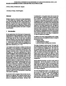
become poor10. Hence, in the present study, the root tips were collected between 11.30 to 12 noon from the roots of terminal node and results also showed that young root tips were containing more metaphase cells and stained better than matured root tips including the roots from the second nodes. The disadvantage of matured root tips probably was due to hard particles in the roots and limited cell divisions in the matured cells. The Carnoy’s fixative showed a better resolution for chromosome count in H. pinifolia. Though, 1N HCl played a subtle role for good cell separation at 60oC for 10min hydrolyzed samples (Fig. 1A & 1B). The sample hydrolyzed lesser than 10 min showed poor separation whereas above 10 min to 60 min result excessive cell break down (Fig. 2 & 3). Various authors recommended that the pectinase solution adjusted to an acidic pH makes it much easier to separate the cells from one another10. It is especially valuable for different materials such as the grasses11. This fixation is very good for chromosome morphology but makes the roots harder and the cells more difficult to separate. However, in the present study, 1N HCl for 10 min showed a good cell separation with higher rate of cell visibility.
Figure 1- Mitotic chromosome analysis in Halodule pinifolia 2n= 44 in 10min HCl hydrolyzation in iron alum heametoxylin staining method: A. 1000X magnification B. Enlarged C. Corrected image
Figure 2- Halodule pinifolia root tip squash stained with iron alum heametoxylin in different time intervals of HCl hydrolyzation: A. 5 min, B. 20 min, C. 30 min, D. 40 min and E. 60 min (1000X magnification)
Figure 3. Halodule pinifolia root tip squash digested 10 min in HCl for other stains: A. Aceto orcein, B. Acid alcoholic carmine and C. Shiff’s reagent (1000X magnification)
VANITHA et al: IMAGE INTERPRETATION TECHNIQUES FOR COUNTING CHROMOSOMES
Image analysis was performed in Adobe Photoshop ver. 9 CS2. It showed good 3D view of chromosome shape in the mitosis stage of H. pinifolia. 3D image of separated chromosome was prepared using filter menu sketch option. In the Photoshop tool palette, crop tool and eraser tool used for improve the clear
1979
separated chromosomes. The chromosome image analysis improved H. pinifolia chromosome shape there by made easy to enumerate the chromosome numbers clearly. The picture revealed clear separated chromosomes, when comparing to raw image (Fig 1C).
Table 1. Halodule pinifolia root tip squash stained with iron alum heametoxylin at different time intervals of HCl digestion and their properties
S. No. 1. 2. 3. 4. 5. 6.
Acid digestion (1N HCl) Time interval 5 10 20 30 40 60
Stain (2 hrs incubation)
Properties
Iron alum heametoxylin Iron alum heametoxylin Iron alum heametoxylin Iron alum heametoxylin Iron alum heametoxylin Iron alum heametoxylin
Lack of cell separation, unstained chromosomes Clear cell separation, Clearly stained chromosomes Excessive cell break down, Clearly stained chromosomes Excessive cell break down, over stained chromosomes Excessive cell break down, over stained chromosomes Excessive cell break down, over stained chromosomes
Table 2. Halodule pinifolia root tip squash stained with three different stains at different time intervals of HCl digestion and their properties
S. No
Acid digestion (1N HCl) time interval
Stains (2 hrs incubation) Properties
1.
5
1 Aceto orcein
2 Acid alcoholic carmine
3 Shiff’s reagent
2.
10
Aceto orcein
Acid alcoholic carmine
Shiff’s reagent
3.
20
Aceto orcein
Acid alcoholic carmine
Shiff’s reagent
4.
30
Aceto orcein
Acid alcoholic carmine
Shiff’s reagent
5.
40
Aceto orcein
Acid alcoholic carmine
Shiff’s reagent
6.
60
Aceto orcein
Acid alcoholic carmine
Shiff’s reagent
Regarding the different staining methods assayed, the best results were obtained when the root tips were stained in iron alum heamatoxylin for 2 hrs incubation, whereas, other staining methods not expressed visible chromosome (Table 1 & 2) even at the better cell separation. In the present study, iron alum heamatoxylin staining protocol resulted better resolution of chromosomes in the different stages of mitosis in root tip cells of Halodule pinifolia. Similarly, early morning or late afternoon collection showed poor results and confirmed 11.30 to 12 noon as the better times for sampling of seagrasses of cytogenetic studies. In the present study, the chromosome analysis
Lack of cell separation, unstained chromosome Clear cell separation, unstained chromosomes Excessive cell break down, unstained chromosome Excessive cell break down unstained chromosome Excessive cell break down unstained chromosome Excessive cell break down unstained chromosome
confirmed that H. pinifolia have diploid set (2n= 44) (Fig.1 A and B). These results are in agreement with previous findings reported for H. wrightii1 and H. pinifolia5. Acknowledgement Authors are grateful to the Director and Dean, Centre of Advance Study in Marine Biology and the authorities of Annamalai University for facilities and to the Ministry of Environment and Forests, Government of India for financial support. References 1
Hartog C.D., Loenhoud, P.J.V., Roelofs, J.G.M. & Van de Sande, J.C.P.M., Chromosome numbers of three seagrasses from the Netherlands Antilles, Aquat. Bot., 7(1979) 267-271.
1980 2
3
4
5
6
INDIAN J. MAR. SCI., VOL. 43 ,NO.10, OCTOBER 2014
Hartog, C.D., Hennen, J., Noten, M.P.A. & Wijk, R.J.V., Chromosome numbers of the European seagrasses, Plant Syst. Evol., 156(1987) 55–59. Keighery, G.J. & Coates, D.J., Chromosome Counts in Posidonia (Posidoniaceae), Plant. Syst. Evol., 137(1981) 221222. Kuo, J., James, S.H., Kirkman, H. & Hartog, C.D., Chromosome numbers and their systematic implications in australian marine angiosperms: the Posidoniaceae, Plant Syst. Evol., 171(1990) 199-204. Ito, Y. & Tanaka, N., Hybridisation in a tropical seagrass genus, Halodule (Cymodoceaceae), inferred from plastid and nuclear DNA phylogenies, Telopea, 13(2) (2011) 219–231. Snow, R., Alcoholic hydrochloric acid-carmine as a stain for chromosomes in squash preparations, Stain Technol.,
38(1963) 9-13. Marimuthu, K.M. & Subramaniam, M.K., A haematoxylin squash method for the root tips of Dolichos lablab L., Curr. Sci., 29(1960) 482-493. 8 Martinez-Gomez P., Improved technique for counting chromosome in almond, Sci. Hortic., 105(2005) 139-143. 9 Martens, M.H.R. & Reisch, B.I., An improved technique for counting chromosomes in grape, Hortscience, 23(1988) 896899. 10 Setterfield, G., Schreiber, R. & Woodard, J., Mitotic frequency in root tips, Stain Technol., 29(1954) 113-120. 11 Pienaar, R. & De, V., Combinations and variation of technique for improved chromosome studies in the Gramineae. J. S. Afr. Bot., 21(1955) 1-8. 7