image contributor and ins)tu)on, and commercial IVOCT vendor for each figure is provided in the. Online Supplementary Material. *Guide-âwire ar)fact. Abbrevia) ...
Peter Barlis MBBS MPH PhD FESC FCSANZ FACC FRACP
Northern & St Vincent’s Hospitals, Melbourne Associate Professor of Medicine The University of Melbourne
Outline • OCT: Brief Overview • OCT: beau3ful images but what do they all mean?? • Case illustra3ons and reference to the Consensus Document from the Interna3onal Working Group on intravascular OCT imaging • Quiz
• In March 2014 we celebrated 5 years of OCT in Australia
• • • • • • • • •
In 2009 Ques+ons that I was being asked What is OCT? Is OCT safe? How do you perform OCT? Which is be:er, IVUS or OCT? Comments I was hearing “We don’t need TCO, we use IVUS” “I don’t see a need for OCT as our lab doesn’t do CTO’s”
2014 – OCT Landscape • • • • •
30+ Systems across Australia and New Zealand 1500+ Systems globally Key questions being asked in 2014: How do you interpret the OCT images? What clinical role does OCT have over and above its research u3lity?
400
350
300
250
200
150
100
50
0
2013 2012 2011 2010 2009 2008 2007 2006 2005 2004 2003 2002 2001 2000 1999 1998 1997 1996
Key Areas for intracoronary imaging • Plaque/lesion assessment • Pre and Post-‐interven3on op3misa3on • Complica3on evalua3on • Restenosis / thrombosis evalua3on
Coronary flush – 3ps for op3mal clearance and imaging • Ensure guiding catheter is intubated into the LM or RCA • Close/lock the Y – connector • If manual injec3ng -‐ minimise the length of tubing • Adjust flush rates of contrast depending on the calibre of the vessel
Importance of getting image interpretation right § The detec3on of TCFA, par3cularly in stable pa3ents is desirable and may principally allow for early interven3on and preven3on of adverse events. § More and more studies are incorpora3ng 3 vessel OCT to examine non-‐culprit plaques § Mastering OCT images does take 3me and pa3ence and preferably the support of a mentor
Consensus Document JACC, 2012 • Interna3onal Working Group (IWG) on OCT standardisa3on – 60 % academic – 40 % industry
• (Academic )Wri3ng commigee produced this document aher nine mee3ngs of IWG.
Types of plaques • • • •
Fibrous plaque Fibrocalcific plaque Fibroatheroma Unstable lesions – Thin cap fibroatheroma – Ruptured plaques – Plaque erosion – Thrombus – High cap macrophage content
Normal artery
Normal artery – three layer appearance Hematoxylin-‐Eosin stain
OCT
ElasAca-‐van Gieson stain external media elasAc lamina adven33a
adven33a
Masson-‐trichrome stain
in3ma In3ma-‐media complex
IVUS
T. Akasaka, “New horizon of coronary intervenAon: histological plaque diagnosis and treatment.”
A spectrum of OCT findings can be iden3fied in a single pullback , therefore analysis is 3me consuming
Normal vessel appearance
Atherosclerotic plaque
Coronary stents
67 yr old female, single OCT pullback in the LCx artery
40 MHz - IVUS
Fibrous Plaques
OCT
Smooth, bright, homogeneous plaques, shown lower propensity to be unstable but as they develop, do cause luminal constric3on and typically stable coronary syndromes
a
A
B
a-‐2
a-‐1 40 MHz - IVUS
OCT Image
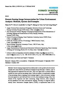
Fibrous and fibrocalcific plaques (A) Fibrous plaque with IEM (green arrow) and EEM (yellow arrow). (B) Plaque without IEM or EEM (white arrow). The EEM (yellow arrow) and IEM (green arrow) can be visualized opposite to the main lesion. (C) Fibrocalcific plaque showing circumferen3al signal-‐poor heterogeneous region with well-‐ delineated borders. (D) Mixed plaque with focal calcific deposit comprising regions with sharply delineated borders, consistent with calcium (red arrow), and adjacent signal-‐poor areas with poorly delineated borders, sugges3ve of lipid (yellow arrows). Scale bars represent 500 m. IVOCT technology, image contributor and ins3tu3on, and commercial IVOCT vendor for each figure is provided in the Online Supplementary Material. *Guide-‐wire ar3fact. Abbrevia3ons as in Figure 1.
Unstable/vulnerable plaque
• A fibrous cap is a 3ssue layer, which is ohen signal-‐rich, overlying a signal-‐poor region • Fibroatheroma is a lesion with an IVOCT-‐ delineated fibrous cap and a necro3c core
71 year old man Admiged with chest pain over 4 hours ECG T wave inversion leads II, aVF Troponin-‐I 0.50 PHx – Diabetes, hypertension, ex/smoker No further chest pain in CCU however 3 brief runs of ventricular tachycardia • coronary angiography • • • • •
Distal RCA OCT Ruptured Plaque
• Lipid-‐rich plaques • Low agenua3on, dark, diffuse borders as dis3nct to calcified plaques • Plaque rupture common at shoulder • Pathological studies have shown that macrophages collect more at plaque shoulder • Poten3al mechanisms include stress / shear force