Improving ECG Classification Accuracy Using an Ensemble of Neural Network Modules Mehrdad Javadi1*, Reza Ebrahimpour2,3, Atena Sajedin1,3, Soheil Faridi1,3, Shokoufeh Zakernejad2 1 Islamic Azad University, South Tehran Branch, Tehran, Iran, 2 Brain and Intelligent Systems Research Lab, Department of Electrical and Computer Engineering, Shahid Rajaee Teacher Training University, Tehran, Iran, 3 School of Cognitive Sciences (SCS), Institute for Research in Fundamental Sciences (IPM), Tehran, Iran
Abstract This paper illustrates the use of a combined neural network model based on Stacked Generalization method for classification of electrocardiogram (ECG) beats. In conventional Stacked Generalization method, the combiner learns to map the base classifiers’ outputs to the target data. We claim adding the input pattern to the base classifiers’ outputs helps the combiner to obtain knowledge about the input space and as the result, performs better on the same task. Experimental results support our claim that the additional knowledge according to the input space, improves the performance of the proposed method which is called Modified Stacked Generalization. In particular, for classification of 14966 ECG beats that were not previously seen during training phase, the Modified Stacked Generalization method reduced the error rate for 12.41% in comparison with the best of ten popular classifier fusion methods including Max, Min, Average, Product, Majority Voting, Borda Count, Decision Templates, Weighted Averaging based on Particle Swarm Optimization and Stacked Generalization. Citation: Javadi M, Ebrahimpour R, Sajedin A, Faridi S, Zakernejad S (2011) Improving ECG Classification Accuracy Using an Ensemble of Neural Network Modules. PLoS ONE 6(10): e24386. doi:10.1371/journal.pone.0024386 Editor: Ioannis P. Androulakis, Rutgers University, United States of America Received April 18, 2011; Accepted August 8, 2011; Published October 26, 2011 Copyright: ß 2011 Javadi et al. This is an open-access article distributed under the terms of the Creative Commons Attribution License, which permits unrestricted use, distribution, and reproduction in any medium, provided the original author and source are credited. Funding: This work was supported by a grant from Islamic Azad University, South Tehran Branch. The funders had no role in study design, data collection and analysis, decision to publish, or preparation of the manuscript. Competing Interests: The authors have declared that no competing interests exist. * E-mail:
[email protected]
different coarse signals. Wavelet coefficients obtained from the decomposition process are considered as the filtered signal in the sub bands. Features extracted from these coefficients can efficiently represent the characteristics of the original signal in different details [20–21]. Researchers have also demonstrated that the feature extraction methods such as Fourier Transform [22], Principal Component Analysis [23] and Independent Component Analysis [24] can be successfully employed to extract appropriate features for classification tasks. As for classifiers, Artificial Neural Networks have been used in a great number of medical diagnostic decision support system functions obtained after dilatation and translation of an analyzing wavelet [25–27]. Among them, the Multi Layer Perceptrons (MLPs) [16–19] and Radial Basis Function [3] [28–29] neural networks are probably the most popular. Combining classifiers to achieve higher accuracy is an active field of research with application in the area of ECG beat classification. Essentially, the idea behind combining classifiers is based on the so-called divideand-conquer principle, according to which a complex computational task is solved by dividing it into a number of computationally simple tasks and then combining the solutions of those tasks ¨ beyli [33] has demonstrated that the [30–32]. For example U combined eigenvector methods (RNN approach) can be useful in analyzing the ECG beats. Osowski et al. [34] have used an ensemble of neural networks for recognition and classification of arrhythmia. The implementation of Multiclass Support Vector Machine with the Error Correcting Output Codes is presented for classification of electrocardiogram (ECG) beats in ref [35]. There are two main strategies in combining classifiers: fusion and selection [36]. In classifier fusion, it is supposed that each ensemble
Introduction Accurate and computationally efficient means of classifying electrocardiography (ECG) arrhythmias has been the subject of considerable research effort in recent years. Electrocardiography deals with the electrical activity of the heart [1]. Monitored by placing sensors at the limb extremities of the subject, electrocardiogram (ECG) is a record of the origin and the propagation of the electrical potential through cardiac muscles. It provides valuable information about the functional aspects of the heart and cardiovascular system. Early detection of heart diseases/abnormalities can prolong life and enhance the quality of living through appropriate treatment. Therefore, numerous research works analyzing the ECG signals have been reported [2–4]. For effective diagnostics, the study of ECG pattern and heart rate variability signal may have to be carried out over several hours. Thus, the volume of the data becomes enormous which then results in a tedious and time consuming study. Naturally, the possibility for the analyst to miss (or misread) vital information is high. Therefore, computer-based analysis and classification of diseases can be very helpful in diagnostics [5–10]. Several algorithms have been developed for the detection and classification of the ECG signals [11–14]. ECG features can be extracted in time domain [15–18], in frequency domain [18–19], or represented as statistical measures [12]. The results of the studies have demonstrated that the Wavelet Transformation is the most promising method to extract features from the ECG signals [2] [5– 6] [10]. Wavelet Transformation opens a category of methods that represent the signal in different translations and scales. Moreover, the Discrete Wavelet Transformation decomposes a signal into PLoS ONE | www.plosone.org
1
October 2011 | Volume 6 | Issue 10 | e24386
ECG Classification by Neural Network Ensembles
member is trained on the whole problem space [37], whereas in classifier selection, each member is assigned to learn a part of the problem space [38–40]. This way, in the former strategy, the final decision is made considering the decisions of all members, while in the latter strategy, the final decision is made by aggregating the decisions of one or a few of experts [41–42]. Combining classifiers based on the fusion of outputs of a set of different classifiers has been developed as a method for improving the recognition rate of classification problems [43–45]. The general framework using an ensemble of neural classifiers in two levels is often referred to as Stacked Generalization [46]. In the first level, various neural classifiers are used to learn different models from the original dataset. The decisions of the first level classifiers and the corresponding target class of the original input data are then used as the input and target to learn the second level classifier, respectively. In this paper, we propose a new combination method for classifying normal heartbeats, Premature Ventricular Contraction (PVC) and other abnormalities. In the preprocessing module, an Undecimated Wavelet Transform is used to provide an informative representation that is both robust to noise and tuned to the morphological characteristics of the waveform features. For feature extraction, we have used a suitable set of features that consists of both morphological and temporal features. This way we can keep both spatial and temporal information of signals. For classification we have used a number of diverse MLPs neural networks as the base classifiers that are trained by Back Propagation algorithm. Then we employed and compared different combination methods. In our proposed method, unlike the Stacked Generalization, the second level classifier (combiner) receives the input pattern directly adding on the base classifiers outputs. In fact, in the learning phase, the combiner learns the expertise areas for each base classifier. In the test phase, based on spatial position of the input data, and by considering the expertise areas of all base classifiers, the combiner specifies the weights for optimal combination of the decisions from the base classifiers. Therefore, we expect that the Modified Stacked Generalization method to be able to use both fusion and selection mechanisms for various test samples, proportional to the position of the sample in the problem space. We used 10 different combination methods: Max, Min, Average, Product, Majority Voting, Borda Count, Decision Templates, Weighted Averaging based on Particle Swarm Optimization, Stacked Generalization and Modified Stacked Generalization. Experimental results indicate that our proposed combining method performs better than other combining methods.
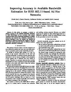
Figure 1. waveform of ECG signal: normal beat. doi:10.1371/journal.pone.0024386.g001
an important role in characterizing these types of arrhythmias. Hence, we exploit the instantaneous RR interval as another feature component, which is defined as the time elapse between the current and previous R peaks [15–17]. This paper investigates the detection and classification of PVC arrhythmias. In Figure 2, ECG signals of three classes are shown. The MIT–BIH arrhythmia database [47] was used as the data source in this study. The database contains 48 recordings each of which has a duration of 30 minutes and includes two leads; the modified limb lead II and one of the modified leads V1, V2, V4 or V5. The sampling frequency is 360 Hz; the data are band-pass filtered at 0.1–100 Hz and the resolution is 200 samples per mV. Twenty-three of the recordings are intended to serve as a representative sample of routine clinical recordings and 25 recordings contain complex ventricular, junctional and supra ventricular arrhythmias. There are over 109,000 labeled ventricular beats from 15 different heartbeat types. There is a large difference in the number of examples in each heart beat type. The largest class is ‘‘Normal beat’’ with about 75,000 examples and the smallest class is ‘‘Supra ventricular premature beat’’ with just two examples. The database is indexed both in timing information and beat classification. We used a total of seven records marked as: 100, 101, 102, 104, 105, 106, and 107 in the database. We extracted a total of 15,566 beats: 8390 normal beats, 627 abnormal PVC arrhythmia beats, and 6549 other arrhythmia beats. We used the database index files from database to locate beats in ECG signals. Of all these 15566 beats, we used 450 beats for training, 150 beats for validation and the other 14966 for testing our networks. This way we assigned equal number of samples to each class in our training and validation phases (150 for training and 50 for validation for each class). The objectives of preprocessing stage are the omission of highfrequency noise and the enhancement of signal quality to obtain appropriate features. ECG signal is measured on static conditions since various types of noise including muscle artifacts and electrode moving artifacts are coupled in dynamic environment. To remove such noises an advanced signal processing method, such as discrete wavelet transform denoising technique [20] should be used. This method has been emerged over recent years as a powerful time–frequency analysis and signal coding tool favored
Materials and Methods Data preparation An ECG consists of three basic waves: the P, QRS, and T. These waves correspond to the far field induced by specific electrical phenomena on the cardiac surface, namely, a trial depolarization (P wave), ventricular depolarization (QRS complex), and ventricular repolarization (T wave). One of the most important ECG components is the QRS complex [12]. Figure 1 shows a waveform of normal signal. Among the various abnormalities related to functioning of the human heart, premature ventricular contraction (PVC) is one the most important arrhythmias. PVC is the contraction of the lower chambers of the heart (the ventricles) that occur earlier than usual, because of abnormal electrical activity of the ventricles. PVC is related to premature heart beats that provide shorter RR intervals than other types of ECG signals. Changes in the RR intervals play PLoS ONE | www.plosone.org
2
October 2011 | Volume 6 | Issue 10 | e24386
ECG Classification by Neural Network Ensembles
Figure 2. ECG signals: (a) Normal Sinus rhythm beats; (b) Premature Venticular contraction beats; (c) other beats (non conducted Pwave and right bundle branch block beats respectively). doi:10.1371/journal.pone.0024386.g002
wavelet algorithms [49]. Even if a signal is not well represented by one member of the Daubechies family, it may still be efficiently represented by another. Selecting a wavelet function which closely matches the signal to be processed is of utmost importance in wavelet applications [50]. For example Rafiee et.al have shown that db44 is the most similar function for Electromyographic, Electroencephalographic and Vaginal Pulse Amplitude biomedical signals [48]. Daubechies wavelet family are similar in shape to QRS complex and their energy spectra are concentrated around low frequencies.
for the interrogation of complex signals. However, Discrete Wavelet Transformation is not a time-invariant transform. To solve this problem, we used the Stationary Wavelet Transform which is also known as the Undecimated Wavelet Transform or translation-invariant wavelet transform. Undecimated Wavelet Transform uses the average of several denoised signals that are obtained from the coefficients of e-decimated Discrete Wavelet Transformation [20]. Figure 3 overleaf shows a color-coded visualization of the Undecimated Wavelet Transform coefficients for an ECG beat. We can see that the Undecimated Wavelet Transform coefficients can capture the joint time-frequency characteristics of the ECG waveform, particularly the QRS complex. Suppose the signal S[L2 (