INFECTION AND IMMUNITY, Aug. 2006, p. 4655–4665 0019-9567/06/$08.00⫹0 doi:10.1128/IAI.00322-06 Copyright © 2006, American Society for Microbiology. All Rights Reserved.
Vol. 74, No. 8
Inactivation of a Two-Component Signal Transduction System, SaeRS, Eliminates Adherence and Attenuates Virulence of Staphylococcus aureus Xudong Liang,1 Chuanxin Yu,1 Junsong Sun,1 Hong Liu,1 Christina Landwehr,1 David Holmes,2 and Yinduo Ji1* Department of Veterinary and Biomedical Sciences, College of Veterinary Medicine, University of Minnesota, 1971 Commonwealth Ave., St. Paul, Minnesota 55108,1 and Anti-Infective Research, GlaxoSmithKline Research and Development, 1250 S. Collegeville Rd., Collegeville, Pennsylvania 194262 Received 27 February 2006/Returned for modification 28 April 2006/Accepted 9 May 2006
Staphylococcus aureus is a major human and animal pathogen. During infection, this organism not only is able to attach to and enter host cells by using its cell surface-associated factors but also exports toxins to induce apoptosis and kill invaded cells. In this study, we identified the regulon of a two-component signal transduction system, SaeRS, and demonstrated that the SaeRS system is required for S. aureus to cause infection both in vitro and in vivo. Using microarray and real-time reverse transcriptase PCR analyses, we found that SaeRS regulates the expression of genes involved in adhesion and invasion (such as those encoding fibronectin-binding proteins and fibrinogen-binding proteins) and genes encoding ␣-, -, and ␥-hemolysins. Surprisingly, we found that SaeRS represses the Agr regulatory system since the mutation of saeS up-regulates agrA expression, which was confirmed by using an agr promoter-reporter fusion system. More importantly, we demonstrated that inactivation of the SaeRS system significantly decreases the bacterium-induced apoptosis and/or death of lung epithelial cells (A549) and attenuates virulence in a murine infection model. Moreover, we found that inactivation of the SaeRS system eliminates staphylococcal adhesion and internalization of lung epithelial cells. We also found that both a novel hypothetical protein (the SA1000 protein) and a bifunctional protein (Efb), which binds to extracellular fibrinogen and complement factor C3, might partially contribute to bacterial adhesion to and invasion of epithelial cells. Our results indicate that activation of the SaeRS system may be required for S. aureus to adhere to and invade epithelial cells. Staphylococcus aureus is an important community- and hospital-acquired pathogen that can cause serious disease, including skin and soft tissue lesions, as well as life-threatening infections such as pneumonia, endocarditis, and toxic shock syndrome (38, 49). This organism’s ability to cause such a variety of diseases partially depends on the expression of its many virulence factors, such as surface-associated adhesins (17), a polysaccharide capsule, and a range of extracellular cytotoxins, proteases, DNases, and enterotoxins (27). Most of the virulence factors have been found to be controlled differentially by different two-component signal transduction regulatory systems (TCS), such as Agr (45), ArlRS (18, 37), and SaeRS (20), and global regulators, such as SarA (9), Rot (41, 53), and Mgr (39). Therefore, TCSs have been implicated together with other regulators to play an important role in bacterial pathogenesis (45). The well-studied regulatory system in S. aureus is the Agr quorum sensing system, which is composed of the AgrBDCA structural genes and RNAIII, the effector molecule of the agr locus (45). The agrB gene encodes a membrane-associated protease required for modifying the prepropeptide of AgrD and generating small peptide signaling molecules. The peptide
signal molecules can be recognized by the membrane-associated sensor kinase (AgrC), which subsequently activates the response regulator (AgrA), which up-regulates RNAIII production (28, 62). The expression of RNAIII is temporal, with maximal expression occurring in the transition from the postexponential to the stationary phase. RNAIII is a dual regulator of staphylococcal virulence factors, including exoproteins and surface proteins (e.g., protein A, coagulase, and some adhesins) (45). Recent studies suggested that Agr positively regulates cap5 expression both in vitro and in vivo (57). Other TCS loci, such as arlRS (18), also positively regulate the expression of virulence factors, such as Ser-Asp-rich bone sialoprotein-binding proteins, and repress some exported proteins, including cysteine protease, serine protease, the hla gene product, -hemolysin, and leukotoxins (37). The srrAB system is involved in the adaptation to anaerobic growth of S. aureus and in the regulation of virulence factors such as toxic shock syndrome toxin 1 and protein A (61). In this paper, we investigate the SaeRS system, another important two-component signal transduction system involved in the control of virulence gene expression (20, 23, 55). The response regulator SaeR is similar to other regulatory proteins, such as DrrA from Thermotoga maritima and ResD and PhoP from Bacillus subtilis (20). The N-terminal region of SaeR contains a highly conserved aspartate phosphorylation site commonly found in response regulators (6). The C terminus of the histidine protein kinase SaeS has an autophosphorylated
* Corresponding author. Mailing address: Department of Veterinary and Biomedical Sciences, College of Veterinary Medicine, University of Minnesota, 1971 Commonwealth Ave., St. Paul, MN 55108. Phone: (612) 624-2757. Fax: (612) 625-5203. E-mail:
[email protected]. 4655
4656
LIANG ET AL.
histidine residue that is similar to other sensors of histidine kinase, such as PhoR and YkoH from B. subtilis and VanS from Enterococcus faecium (20). The N-terminal region of the SaeS protein possesses two transmembrane domains that are also found in other sensor proteins (6). It has been reported that the expression of sae is repressed in the presence of glucose as a consequence of changes in the pH (46). The SaeRS system up-regulates the transcription of hla, hlb, and coa (20) but has no effect on the expression of agr or sarA in vitro (21). Moreover, the SaeRS system also controls hla expression in vivo, as the level of hla transcripts is significantly decreased in sae mutant strains during infection (22, 23). The SaeRS system might function independently as a regulator since SaeRS-dependent and Agr/SarA-independent activation of hla was found in exudates accumulated in a guinea pig model of infection (22, 23). It was reported that mutation of sae in S. aureus strain Newman eliminates the transcription and expression of fnbA and increases the expression of CP5; it also leads to a significant decrease of the internalization of S. aureus by endothelial cells (56). Moreover, the role of the SaeRS system as a virulence regulator has been demonstrated in several animal models of infection (3, 23, 51). Therefore, the SaeRS regulatory system appears to play an important role in the modulation of virulence gene expression during certain types of infection. In this study, we aimed to examine more specifically the roles of the SaeRS system in bacterial pathogenesis. We created a saeS gene replacement mutant in an S. aureus isolate from a human and identified the regulon of saeRS using Affymetrix S. aureus oligonucleotide arrays. We also examined the impact of the mutation of saeS on bacterial adherence and internalization in epithelial cells, S. aureus-induced apoptosis and death, and survival and/or virulence by using a murine infection model. Our microarray data indicate that SaeRS may affect the expression of a bifunctional protein (Efb) which binds to both extracellular fibrinogen (48) and complement factor C3 (36) and a gene encoding a hypothetical fibrinogenbinding protein (the SA1000 protein). In order to understand whether these proteins are involved in pathogenesis, we constructed allelic gene replacement mutants and examined the impact of mutation on bacterial adhesion and invasion. MATERIALS AND METHODS Bacterial strains, media, and growth conditions. S. aureus strain RN4220 was utilized as the primary recipient for allelic exchange constructs, together with strain WCUH29 (NCIMB40771), a virulent clinical isolate, as a secondary recipient for phage transduction. S. aureus strain 15981 and the 15981 ⌬saeRS strain were kindly provided by I. Lasa (56). Escherichia coli DH10B (Invitrogen) served as the host for all in vitro recombinant DNA. Bacteria were grown in tryptic soy broth (TSB) (Difco) and on tryptic soy agar (TSA) at 37°C. Bacterial cell cultures were incubated at 37°C with shaking at 200 rpm. The Sa371ko mutant was maintained on TSA plates with tetracycline (Tc) at a concentration of 5 g/ml. Construction of saeS, SA1000, and efb gene replacement null mutants, saeS, SA1000, and efb gene complementary strains, and agrA promoter-gfp reporter fusion system. The S. aureus vector pSA7755 was used to generate gene replacement mutants as described previously (15). A cassette containing the tet gene (flanked by chromosomal fragments from upstream and downstream of the saeS, SA1000, or efb gene to be replaced) was constructed in pBluescript and inserted into pSA7755. An S. aureus strain, RN4220, carrying this hybrid plasmid was grown at a restrictive temperature (40°C) for pSA7755 replication in the presence of 5 g/ml tetracycline. These conditions allow the growth of only cells with tet inserted into the chromosome by homologous recombination. To obtain gene
INFECT. IMMUN. replacement, the mutated locus was transduced into the wild-type strain WCUH29 by 11 transduction. We determined that allelic replacement had occurred and resulted in the saeS null mutant strain Sa371ko, the SA1000 mutant strain Sa1000ko, and the efb null mutant strain Efbko by selecting for tetracycline resistance and screening for the loss of the erythromycin resistance marker carried by the vector. The mutation in saeS, SA1000, or efb was verified by PCR using primers specific to the saeS, SA1000, efb, or tet gene. The results showed that no PCR product from the saeS, SA1000, or efb gene was obtained from the mutant strains, whereas there was a PCR product of the expected size obtained from the wild-type strain using primers specific to saeS, SA1000, or efb (data not shown). In contrast, a PCR product was obtain from the mutant strains (but not the wild-type strain) by using a primer specific to tet. To further confirm the saeS, SA1000, or efb mutation, we performed Southern blot analysis using digoxigenin (DIG)-labeled probes and found that no DNA hybridized with the saeS, SA1000, or efb probe in the mutant chromosomal DNA (Fig. 1). In order to examine whether the expression of saeS, SA1000, or efb in trans can complement the effect of mutation of the corresponding endogenous gene, we constructed the recombinant plasmids pYH4/saeS, pYH4/SA1000, and pYH4/efb by cloning the saeS, SA1000, and efb coding regions (obtained by PCR), respectively, into the AscI and PmeI sites of pYH4 (31) and electroporated them into the Sa371ko, Sa1000ko, and Efbko strains, resulting in the Sa371com, Sa1000com, and Efbcom strains, respectively. The recombinant plasmid DNAs of pYH4/saeS, pYH4/SA1000, and pYH4/efb were isolated from the complementary strains and confirmed by PCR and DNA sequencing (data not shown). In order to confirm whether the mutation of saeS has an impact on agrA expression, we utilized an agrA promoter-gfp reporter fusion system, which was a kind gift from Philip Hill. Plasmid DNA was purified and electroporated into S. aureus WCUH29 and Sa371ko, resulting in the WCUH29/pCY1006 and Sa371ko/pCY1006 strains, respectively. gfp expression was determined by Western blotting as described previously (31), using a green fluorescent protein (GFP) antibody (Abcam Inc., MA). RNA isolation and purification. Overnight cultures of S. aureus were inoculated into 5% TSB medium and grown to the mid-exponential (3 h) phase of growth. Cells were harvested by centrifugation, and RNAs were isolated by use of an RNAPrep kit (Promega, MI). Contaminating DNA was removed with a DNA-free kit (Ambion), and the RNA yield was determined spectrophotometrically at 260 nm. cDNA synthesis, cDNA fragmentation, and labeling. The integrity of the RNA preparations was analyzed by electrophoresis in 1.2% agarose-0.66 M formaldehyde gels. The 23S and 16S rRNA bands were clear, without any obvious smearing patterns. Briefly, a total of 10 g of RNA was reverse transcribed to generate cDNA, using Superscript II reverse transcriptase (RT) and random primers (Invitrogen). The RNA was then removed by treatment at 65°C for 30 min with NaOH. The cDNA was purified by using a QIAquick PCR purification kit (QIAGEN). The purified cDNA was digested with DNase I and labeled with biotin-ddUTP (Roach). Hybridization and scanning. After determining that the fragmented cDNAs were labeled with biotin, the fragmented biotinylated cDNAs were hybridized to S. aureus chips (Affymetrix) containing probe sets for S. aureus genomic open reading frames (ORFs), and hybridization intensities for each of the genes/ transcripts were collected from the scanned images. Microarray analysis. The S. aureus array (Affymetrix) contained probe sets for over 3,300 S. aureus open reading frames based on the updated S. aureus genomic sequences of strains N315, Mu50, NCTC 8325, and COL. Additionally, the array also contained probes to study both the forward and reverse orientations of over 4,800 intergenic regions throughout the S. aureus genome. A subsequent analysis suggested that these probes represent approximately 2,738, 2,668, 2,773, and 2,810 individual genes of the S. aureus COL, N315, NTC8325, and Mu50 genomes, respectively. To identify genes with significantly altered expression, microarray analyses and a series of statistical analyses (filtering) were performed as described previously (37). We selected the genes with significant differential expression (P ⬍ 0.05). Those genes negatively regulated by the SaeRS system were identified as ORFs with transcript titers at least 1.8-fold higher in Sa371ko (saeS negative) than in WCUH29. Genes whose transcript levels were at least 1.8-fold higher in WCUH29 than in Sa371ko (saeS negative) were categorized as being positively regulated by SaeRS. RT-PCR and quantitative real-time RT-PCR analysis. In order to examine whether the complementary strains expressed the knockout genes in trans, we performed RT-PCR, using the saeS-, SA1000-, or efb-specific primers listed in Table 1, as described previously (29). In order to confirm the results obtained from the microarray analyses, we employed quantitative real-time RT-PCR to compare the RNA levels that showed significant changes of expression in the
EFFECTS OF SaeRS INACTIVATION ON S. AUREUS
VOL. 74, 2006
4657
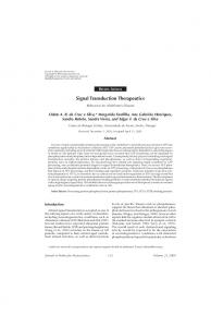
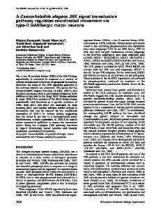
FIG. 1. Schematic diagrams and Southern blot analysis of allelic gene replacement mutants. (A) Chromosomal DNAs isolated from WCUH29 (lane 2) and Sa371ko (lane 3) were digested with XbaI (X) and probed with DIG-labeled saeS. As expected, a 2.8-kb DNA fragment from WCUH29 was detected in the Southern blot. (B) Chromosomal DNAs isolated from WCUH29 (lane 2) and Sa1000ko (lane 3) were digested with HindIII (H) and probed with DIG-labeled SA1000. As expected, a 1.9-kb DNA fragment from WCUH29 was detected in the Southern blot. (C) Chromosomal DNAs isolated from WCUH29 (lane 2) and Efbko (lane 3) were digested with PstI (P) and probed with DIG-labeled efb. As expected, a 2.5-kb DNA fragment from WCUH29 was detected in the Southern blot. Lanes M, 1-kb DNA markers labeled with DIG.
microarray assay (37). The first-strand cDNA was synthesized using reverse transcriptase with a SuperScript III Platinum two-step qRT-PCR kit (Invitrogen). For each RNA sample, duplicate reverse transcription reactions were performed, as well as a control without reverse transcriptase, in order to determine the level of DNA contamination. PCRs were set up in triplicate by using SYBR green PCR master mix (Bio-Rad). Real-time sequence-specific detection
TABLE 1. Primers used in real-time RT-PCR Primer
Sequence (5⬘–3⬘)
SA1000for ................GTATCAACGTTTGCCGGTGAATCTC SA1000rev ................CAGCTCTTTGTGCTTTACGGTGTGTT SA1003for (Efb)......GTACAATGATGGTACTTTTAAATATCAAT CTAGAC SA1003rev (Efb) .....GTTCTTTTTTAATAGTTGCATCAGTTTT CGCT SA1004for ................AAGGGAATAAAGCAGATGCAAGTAGTCT SA1004rev ................GTGCCGCTTTAGCTCTATATTCATTCAT SA1007for ................CAACTGATAAAAAAGTAGGCTGGAAAG TGAT SA1007rev ................CTGGTGAAAACCCTGAAGATAATAGAG SA1844agrAfor........GTGAAATTCGTAAGCATGACCCAGTTG SA1844agrArev .......TGTAAGCGTGTATGTGCAGTTTCTAAAC SA2290 fnbBfor.......GCAGTGAGCGACCATACAACAGTT SA2290 fnbBrev ......CAATCACGCCATAATTACCGTGACCA 16S rRNAfor ...........CTGTGCACATCTTGACGGTA 16S rRNArev ...........TCAGCGTCAGTTACAGACCA
and relative quantitation were performed with the Stratagene Mx3000P real-time PCR system. Gene-specific primers were designed to yield ⬃100-bp specific products (Table 1). Relative quantification of the product was calculated using the comparative cycle threshold method as described for the Stratagene Mx3000P system. The housekeeping 16S rRNA gene was used as an endogenous control (37). All samples were analyzed in triplicate and normalized against 16S rRNA gene expression. The experiments were repeated at least twice and analyzed for correlation to the microarray results. Cell culture and epithelial cell adhesion and invasion assay. A549 human lung epithelial cells (ATCC CCL 185) were cultured in RPMI 1640 medium supplemented with 10% fetal bovine serum (FBS; Invitrogen). Cultures of A549 cells were maintained in a medium containing penicillin (5 g/ml) and streptomycin (100 g/ml) (Sigma). Assays of bacterial invasion and adherence were performed as previously described (1, 10, 57). Assays were performed in RPMI 1640 medium supplemented with of 10% FBS (RPMI-FBS). We used 0.025% Triton X-100 for better cell lysis. Briefly, 1 day prior to infection, approximately 2 ⫻ 105 cells were seeded in each well of 24-well plates and incubated overnight at 37°C in a CO2 incubator. Monolayers of A549 cells (2 ⫻ 105 cells/well) were infected by adding 0.5 ml RPMI containing approximately 5 ⫻ 105 CFU of bacteria, followed by centrifugation at 100 ⫻ g for 5 min, and were incubated for 1 h at 37°C in 5% CO2. To measure bacterial adherence, the culture medium was removed from monolayers 1 h after infection and discarded. The monolayer cells were then washed three times with phosphate-buffered saline (PBS; pH 7.4) to remove nonadherent bacteria. Epithelial cells were dispersed by the addition of 150 l of 0.25% trypsin-1 mM EDTA (Invitrogen) and then lysed by the addition of 400 l of 0.025% Triton X-100. The numbers of bacterial CFU released from the lysed epithelial cells were determined by plating of diluted lysates on TSA plates. For invasion assays, the culture medium was collected from wells used for total
4658
LIANG ET AL.
INFECT. IMMUN.
counts and discarded from wells used for invasion assays after 2 h of incubation. The monolayer cells were washed three times with PBS (pH 7.4), followed by the addition of 1 ml of RPMI-10% FCS containing 100 g/ml gentamicin and 5 g/ml lysostaphin to invasion wells and 1 ml of RPMI-10% FBS to total wells, and were then incubated. After 2 h, the supernatants in the wells were removed and discarded. All wells were washed three times with 1 ml PBS. A total of 1 ml RPMI-10% FBS containing100 g/ml gentamicin and 5 g/ml lysostaphin was added to the invasion wells to kill outside bacteria, and 1 ml of RPMI-10% FBS was added to the wells used for total counts. The supernatant was removed from each well after 1 h of incubation. All wells were washed three times with 1 ml warm PBS, and then 150 l of 0.25% trypsin-EDTA was added. After 5 min, the cells in each well were carefully collected, and then 400 l of 0.025% Triton X-100 was added to the tubes on ice. The numbers of bacterial CFU released from the lysed epithelial cells were determined by plating of diluted lysates on TSA plates. The bacterial adhesion in each well was determined as the CFU that adhered to and invaded into the cells and is expressed as a percentage of the CFU in the inoculum (58). The controls were wells pretreated with medium alone (RPMI) or were the wild-type control strains, considered to have 100% adhesion. Adhesion and invasion were then normalized against controls (1, 58). Each experiment was repeated three times, and all of the relative adhesion and invasion values were calculated and statistically analyzed by Student’s t test, using Microsoft Excel 2003 software. P values of ⬍0.05 were considered significant. DNA fragmentation assay. Infected and control cells were collected, lysed with lysis buffer, and treated with RNase (10 mg/ml) and proteinase K (20 mg/ml). DNAs were purified from the epithelial cells 24 h after infection with the saeS mutant or its parent strain by using phenol, precipitated with ethanol, and washed with 75% ethanol. DNAs were dissolved in 100 l Tris-EDTA buffer, followed by electrophoresis using 1.2% agarose gels (43). Cytotoxicity assays. All cells were grown in 96-well plates to 70% confluence. Inhibitors were diluted serially in complete medium and applied to the cells. The supernatants from the overnight cultures of WCUH29 and Sa371ko were immediately applied to the cells, and the treated cells were incubated at 37°C with 5% CO2 overnight (16 h). At the end of the experiment, cell viability was determined by using the CellTiter 96 aqueous nonradioactive cell proliferation assay (Promega) according to the manufacturer’s instructions. Hematogenous pyelonephritis infection. Overnight cultures of bacteria were started from single colonies in 10 ml of TSB and grown at 37°C with shaking. Cells were centrifuged and washed twice in PBS. The A600 was adjusted to approximately 0.2. Female CD-1 mice (18 to 20 g) were inoculated with 0.2 ml of this suspension (containing approximately 7 log10 CFU bacteria) by tail vein injection (29, 30). Mice were monitored twice daily for signs of illness, and any which appeared moribund were euthanized prior to the end of the experiment. All remaining animals were euthanized by carbon dioxide overdose at 3 days postinoculation. Both kidneys were removed, using aseptic technique, and homogenized in 1 ml PBS before enumeration of viable bacteria following plating on TSA plates.
RESULTS Construction and characterization of saeS gene replacement mutant of a human clinical S. aureus isolate. In order to examine the effect of the SaeRS system on bacterial pathogenesis, we created a saeS gene replacement mutant of the human clinical isolate WCUH29, designated Sa371ko, by the homologous gene recombination strategy described in Materials and Methods. To confirm the allelic replacement, chromosomal DNAs were isolated from the mutant Sa371ko and wild-type WCUH29 strains, and diagnostic PCR analysis (see Materials and Methods) and Southern blot analysis were used to confirm the mutation (Fig. 1A). The results showed that the saeS gene was knocked out by allelic gene replacement (Fig. 1A). To determine whether the allelic mutation of saeS had any impact on bacterial growth and morphology, we measured the growth curve of the saeS mutant strain in nutrient-rich TSB medium with Tc and the CFU on TSA-Tc. The results showed that there was no difference in growth pattern and CFU between the saeS knockout strain and its parent strain. We did
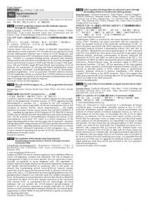
not observe obvious differences in the morphological phenotype, such as colony size and shape, after the mutation of saeS. The SaeRS system regulates the expression of genes involved in adhesion, invasion, and toxicity in vitro. Previous studies have demonstrated that the SaeRS system is involved in regulating the expression of several virulence genes, including hla, hlb, and coa (21, 22). In order to comprehensively understand the saeRS regulon, we performed a microarray analysis to examine the effect of the mutation of saeS on gene transcription, using Affymetrix S. aureus oligonucleotide chips as described in Materials and Methods. Unlike other two-component signal transduction systems, such as agr (12) and arl (37), where the mutation of agr or arl has a global effect on the expression of numerous genes, our microarray data showed that the mutation of saeS significantly affected the expression of fewer than 20 genes (Table 2). The microarray data indicated that the SaeRS system is involved in positive regulation of genes encoding fibronectin-binding proteins (FnBPs) and fibrinogen-binding proteins, such as fnbB, fnb, and coa (the mRNA levels in the wild type were ⬎10-, ⬎3-, and ⬎7-fold higher, respectively, than those in the null mutant), as well as genes encoding a putative hypothetical protein, the SA1000 protein, and a bifunctional protein (Efb) (the mRNA levels in the wild type were ⬎50- and ⬎35-fold higher, respectively, than those in the null mutant) (Table 2). To validate these results, a real-time RT-PCR was performed, and the results demonstrated that the levels of fnbB, SA1000, efb, and SA1004 mRNAs in the wild-type strain were significantly higher than those in the saeS mutant strain (Table 3). We also found that SaeRS is involved in the regulation of genes encoding toxins, including hla, hlb, hlgC, and set-15 (the levels of mRNA in the wild type were ⬎23-, ⬎4-, ⬎1.7-, and ⬎75-fold higher, respectively, than those in the null mutant) (Table 2). The observation that SaeRS positively affected hla and set-15 expression was confirmed by a real-time RT-PCR analysis in which RNA samples from the saeS mutant and the wild-type strain were taken at the mid-exponential growth phase. The results demonstrated that hla and set-15 mRNAs were significantly higher in the wild-type strain than in the saeS mutant strain (Table 3). In addition, we found that SaeRS positively affects several genes with unknown functions (Table 2). Importantly, our microarray and RT-PCR results indicate that even the magnitudes of the changes of the identified gene mRNA levels in the saeS mutant strain were highly consistent between the microarray and real-time RT-PCR data. Very surprisingly, the microarray results indicated that the SaeRS system negatively affects agrA (Table 2). In addition, the mutation of saeS increased agrB (1.5- fold) and agrC (1fold) expression (data not shown). Real-time RT-PCR analysis confirmed that the agrA mRNA level was significantly lower in the wild-type strain than in the null mutant (Table 3). To further confirm the effect on agr expression, we introduced an agrA promoter-gfp reporter fusion and determined the impact of the mutation in saeS on agr expression by monitoring the reporter gene. The results showed that the production of GFP was significantly up-regulated in the saeS null mutant (Sa371ko/pCY1006) compared to that in the wild-type control (WCUH29/pCY1006) (Fig. 2). Taken together, these data indicate that SaeRS may repress the agrA regulatory system in strain WCUH29.
EFFECTS OF SaeRS INACTIVATION ON S. AUREUS
VOL. 74, 2006
4659
TABLE 2. S. aureus genes regulated by SaeRS N315 ORF
Description
Change (fold)a
agr, sar, rot, and/or sigB effectb
coa fnbB fnb efb
Coagulase precursor Fibronectin-binding protein B Homologue of FnbA Extracellular fibrinogen-binding protein (36)
⫹23.4 ⫹10.3 ⫹3.2 ⫹35.1
⫹ (agr, sarA, rot, and sigB) ⫹ (sarA) ⫹ (sarA and sigB)
set-15
⫹75 ⫹3 ⫹23.4 ⫹4.6 ⫹1.7
Gene
Cell-associated protein genes SA0222 SA2290 SA2291 SA1003 Exported protein genes SA0393 SA0746 SA1007 SA1752 SA2208
hla hlb hlgC
Exotoxin 15 Staphylococcal nuclease Alpha-hemolysin Beta-hemolysin Gamma-hemolysin component C
Regulatory system protein genes SA0660 SA1844
saeS agrA
Histidine kinase sensor Response regulator
Hypothetical protein genes SA0394 SA0743 SA0841 SA1000 SA1004 SA1813 SA1804
⫹ (agr, sarA, rot, and sigB)
⫹176 ⫺2.2
Hypothetical protein Similar to staphylocoagulase precursor Similar to cell surface protein MapW Hypothetical fibrinogen-binding protein Hypothetical protein Similar to leukocidin LukM precursor Hypothetical transcriptional regulator
⫹27 ⫹4.5 ⫹2.4 ⫹58.7 ⫹16.9 ⫹5.7 ⫺34
a
Normalized values for the wild-type strain over those for the saeS null mutant. agr and sar effects were described by Dunman et al. (12), rot effects were described by Saı¨d-Salim et al. (53), and sigB effects were described by Bischoff et al. (4). ⫹, up-regulation; ⫺, down-regulation. b
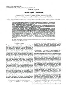
Inactivating the SaeRS system eliminates staphylococcal adhesion and internalization of epithelial cells. It has been reported that the SaeRS system plays an important role in staphylococcal adhesion and internalization into endothelial cells (55). Our genomic mRNA analysis also indicated that the SaeRS system regulates gene products that are associated with bacterial adhesion and/or invasion. In order to more specifically characterize the role of the SaeRS system in the bacterium’s interaction with epithelial cells, we examined the impact of the saeS null mutation on adhesion to and/or internalization of lung epithelial cells. We found that the adherence of the saeS mutant strain was significantly less than that of the parent strain (Fig. 3A). Moreover, compared to the parent cells, less than 10% of the saeS mutant cells were able to internalize into
TABLE 3. Real-time RT-PCR analysis of gene expression regulated by SaeRS Change (fold)a N315 ORF
SA2290
Gene
fnbB
SA1000 SA1003
efb (36)
SA1004 SA0390 SA1007 SA1844
set-15 hla agrA
Description
Fibronectin-binding protein B Hypothetical fibrinogenbinding protein Extracellular fibrinogenbinding protein Hypothetical protein Exotoxin Alpha-hemolysin Two-component response regulator
RT-PCR
Microarray
⫹5.2
⫹10.3
⫹69.6
⫹58.7
⫹33.6
⫹35.1
⫹22.6 ⫹89.3 ⫹4.8 ⫺11.2
⫹16.9 ⫹75 ⫹23.4 ⫺2.2
a Normalized values for the wild-type strain over those for the saeS null mutant. Positive numbers denote up-regulation in the wild-type strain.
epithelial cells after 2 h of infection (Fig. 3B). To further confirm the role of SaeRS in bacterial adherence and invasion, we examined the effect of the saeRS null mutation on adhesion to and invasion of epithelial cells. The results were similar to those for the saeS null mutant in that both adhesion and internalization of the null mutant (strain 15981 ⌬saeRS) were significantly less than those of the parent strain (15981; data not shown). To investigate whether the expression of saeS in trans can complement the effect of the mutation of endogenous saeS, we constructed a recombinant plasmid, pYH4/saeS, containing the saeS coding region and transformed it into the Sa371ko strain. First, we examined saeS expression by RT-PCR using saeSspecific primers. The results showed that a specific saeS PCR product was yielded with RT; in contrast, no PCR product was detected in the reaction without RT (Fig. 3C). This indicates that there is saeS expression in the pYH4/saeS complementary construct. We then examined the influence of the plasmidborne saeS gene on adherence and internalization. The results
FIG. 2. Western blot analysis of gfp expression in an agrA promotergfp fusion. S. aureus strains were incubated in TSB. The same amounts of bacterial cells were harvested from cultures at an optical density at 600 nm of 0.5 (log phase) by centrifugation. Whole-cell lysates were prepared, and the same amounts of protein were loaded into 12% sodium dodecyl sulfate-polyacrylamide gels and probed with rabbit anti-Gfp antiserum in a Western blot assay. Lane 1, Sa371ko/ pCY1006; lane 2, WCUH29/pCY1006; lane 3, WCUH29.
4660
LIANG ET AL.
INFECT. IMMUN.
FIG. 3. (A and B) Effects of SaeRS on adherence to and internalization of S. aureus by epithelial cells. The bacterial cells were collected from overnight cultures (wild-type strain WCUH29, saeS null mutant strain Sa371ko, and the complementary strain Sa371com with plasmid-borne saeS), washed with PBS, and diluted and resuspended in RPMI 1640 medium with 10% FBS just prior to infection of monolayer cells. (A) Staphylococcal adherence to A549 cells. (B) Intracellular invasion. Relative adherence and relative invasion were calculated as described in Materials and Methods. Data are the means ⫾ standard errors of the means for nine infected monolayers of cells (three experiments, each performed in triplicate). Asterisks indicate significant differences between the wild type and the mutant (P ⬍ 0.01). (C) RT-PCR detection of saeS expression in trans. S. aureus strains were incubated overnight in TSB with appropriate antibiotics, and total RNAs were purified from the above cultures and treated with DNA-free kits. RT-PCR was performed using saeS- and 16S rRNA-specific primers. RT-PCR products from 16S rRNA were used as positive controls. The negative controls were samples prepared without RT or template DNA. M, 100-bp DNA marker.
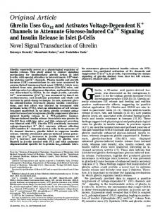
showed that the bacterium’s capacity to adhere to and invade epithelial cells was restored fully (Fig. 3A and B). A putative fibrinogen-binding protein and the exported fibrinogen-binding protein are involved in adherence and internalization. It is well known that FnBPs are required for the internalization of S. aureus by endothelial cells (47). We selected two genes (SA1000 and efb), encoding a hypothetical protein and a bifunctional protein, Efb (36) (which binds extracellular fibrinogen and complement factor C3), respectively, due to their apparent regulation by SaeRS from our microarray data and investigated whether they are involved in bacterial adhesion and invasion. We created SA1000 and efb allelic gene replacement null mutants as described in Materials and Methods and confirmed the replacement mutations by PCR (see Materials and Methods for details) and Southern blot analysis (Fig. 1B and C). We then examined the adhesion to and invasion of epithelial cells by the mutant strains Sa1000ko and Efbko. The results showed that both adhesion and internalization of the null mutant strains (Sa1000ko and Efbko) were significantly less than those of the parent strain (Fig. 4A to D). These results indicate that the putative hypothetical protein and Efb may be involved in adhesion to and invasion of epithelial cells. To investigate whether the expression of SA1000 or efb in trans can complement the effect of the mutation of endogenous SA1000 or efb, we constructed the recombinant plasmids pYH4/SA1000 and pYH4/efb and transformed them into the Sa1000ko and Efbko strains, respectively. The RT-PCR results showed that specific SA1000 and efb PCR products were
yielded with RT; in contrast, no PCR products were detected in the reaction mixtures without RT (data not shown). Moreover, the adhesion and invasion results showed that the bacteria’s capacity to adhere to and invade epithelial cells was restored fully (Fig. 4A to D). These results indicate that the putative SA1000 protein and the extracellular fibrinogen-binding protein Efb also affect pathogen-host cell interactions, albeit with an effect that is not as pronounced as that of the saeS knockout. Inactivation of the SaeRS system inhibits S. aureus-induced apoptosis of human lung epithelial cells. It has been reported that alpha-toxin is involved in S. aureus-induced apoptosis and cell death through certain host cell signaling pathways (2, 14, 24, 33). Previous studies as well as our microarray results demonstrated that the SaeRS system regulates hla gene expression (21, 22). Thus, it is likely that the SaeRS regulatory system may be involved in the S. aureus-induced apoptosis of host cells. To test this hypothesis, we examined the effect of the null mutation in saeS on S. aureus-induced apoptosis, using a standard DNA fragmentation assay. We observed no ladder fragmentation pattern of chromosomal DNA for cells infected with the saeS null mutant strain (Fig. 5). In contrast, distinctive ladder patterns of chromosomal DNA were observed for cells infected with the parent strain (Fig. 5). We then quantitatively measured the cell death induced by S. aureus, using a standard cytotoxic assay. The results showed that ⬃55% of the epithelial cells infected by the wild-type strain survived 26 h of infection, whereas ⬎95% of the epithe-
VOL. 74, 2006
EFFECTS OF SaeRS INACTIVATION ON S. AUREUS
4661
FIG. 4. Effects of the putative fibrinogen-binding SA1000 protein (A and B) and the extracellular fibrinogen-binding protein Efb (C and D) on adherence to and internalization of a human clinical S. aureus isolate by epithelial cells. The bacterial cells were collected from overnight cultures (wild-type strain WCUH29, the SA1000 null mutant strain Sa1000ko, the SA1000 complementary strain Sa1000com, the efb null mutant Efbko, and the efb complementary strain Efbcom), washed with PBS, and diluted and resuspended in RPMI 1640 medium with 10% FBS just prior to infection of monolayer cells. (A and C) Staphylococcal adherence to A549 cells. (B and D) Intracellular invasion. Relative adherence and relative invasion were calculated as described in Materials and Methods. Data are the means ⫾ standard errors of the means for nine infected monolayers of cells (three experiments, each performed in triplicate). Asterisks indicate significant differences between the wild-type and mutant strains (P ⬍ 0.05).
lial cells infected by the saeS mutant strain survived the same period of infection (Fig. 6A). To determine whether the higher survival rate of the epithelial cells infected by the mutant may be due at least in part to the reduced levels of exported toxins, we examined the effect of SaeRS on cytotoxicity, using the supernatants of S. aureus cultures. In correlation with the whole-cell infection results, the supernatants of the saeS mutant strain showed no detect-
FIG. 5. Effect of SaeRS on DNA fragmentation characteristic of apoptosis of epithelial cells (A549) induced by S. aureus. Lanes: M, 1-kb DNA laddering marker; 1, control without treatment; 2, cells incubated overnight with wild-type S. aureus WCUH29; 3, cells incubated overnight with saeS null mutant strain Sa371ko.
able toxicity to epithelial cells, whereas the supernatants of the parent strain caused ⬎50% cell death 24 h after being exposed to the cells (Fig. 6B). To investigate whether the expression of saeS in trans can complement the effect of the mutation of endogenous saeS, we examined the influence of the plasmid-borne saeS gene on S. aureus-induced cell death. The results showed that the bacterium’s capacity to cause the death of epithelial cells was restored fully (Fig. 6A and B, Sa371com). Taken together, the above results demonstrate that the SaeRS system is important for S. aureus-induced death of epithelial cells (A549). The SaeRS system significantly affects bacterial survival during infection. To investigate the role of SaeRS in pathogenesis, we chose a murine model of hematogenous pyelonephritis. This model represents a localized kidney infection from which bacteria can be recovered and quantitated and has been used successfully to examine the essentiality of genes in vivo (29, 30). We infected mice via the tail vein, removed their kidneys at 3 days postinfection, and examined the bacterial loads. We compared the null mutant strain with the wild type as a control, using identical bacterial CFU. The results showed that approximately 4 log10 CFU of the saeS null mutant were recovered from infected kidneys, whereas, ⬃6 log10 CFU of the wild-type strain were recovered from kidneys (Fig. 7). These results indicate that the mutation in saeS significantly
4662
LIANG ET AL.
INFECT. IMMUN.
FIG. 6. Effects of SaeRS on S. aureus-induced death of epithelial cells (A549) by bacterial cells (A) and supernatants (5 l/ml) of overnight cultures (B). Cell viability was measured after overnight treatment and is expressed as the average for at least three experiments ⫾ standard deviation. Asterisks indicate significant differences between the wild-type and mutant strains (P ⬍ 0.05).
attenuated the virulence of S. aureus in the murine model of hematogenous pyelonephritis. DISCUSSION Microarray analysis has been widely used to identify genes regulated by different regulators in S. aureus, including AgrA and SarA (12), Rot (53), Sigma B (4), and ArlRS (37). In this study, we demonstrated that a two-component signal transduction system, SaeRS, is a predominant regulator of virulence factors in S. aureus. Using an Affymetrix oligonucleotide array, we identified the genes that are directly and/or indirectly reg-
FIG. 7. S. aureus recovered from infected kidneys. Five mice per group were infected with about 107 CFU of bacteria via an intravenous injection of 0.2 ml of bacterial suspension into the tail vein, using a tuberculin syringe. The mice were sacrificed by carbon dioxide overdose 3 days after infection. Kidneys were removed aseptically and homogenized in 1 ml of PBS for enumeration of viable bacteria. This experiment was repeated three times with similar results. **, P ⬍ 0.01.
ulated by the SaeRS system. Our microarray analysis showed that inactivating the SaeRS system dramatically down-regulates the expression of virulence genes that encode both cell wall-associated proteins (including fibronectin- and fibrinogenbinding proteins) and exported proteins, such as toxins. These data are highly consistent with our data from the semiquantitative RT-PCR analysis presented in this study, in which we saw a decrease in saeS expression and a decrease in mRNA expression of coa, fnbA, fnbB, efb, hla, and other genes. Our results are also consistent with previous findings that SaeRS controls the expression of coa, fnb, and hla (20, 21), since no fnbA, fnbB, or coa mRNA was detectable in the saeS mutant strain (55). We also found that SaeRS may control the transcription of a gene encoding a hypothetical fibrinogen-binding protein (SA1000). Therefore, our results indicate that the SaeRS system is likely an important regulator of virulence factors in S. aureus. Surprisingly, our microarray and RT-PCR results indicated that the SaeRS system negatively affects the expression of agrA, which was confirmed by using an agrA promoter-gfp reporter fusion. To investigate whether this negative impact is a direct effect is beyond the scope of this study. Our result is inconsistent with a previous report that Agr acts upstream of sae (46), indicating that the two regulatory systems may interact with each other. In addition, our studies did not reveal significant differences in expression of the extracellular adherence protein (Eap) and the extracellular matrix protein-binding protein (Emp) (26) after inactivation of the SaeRS system. This is inconsistent with some previous reports (21, 24), which may be due either to the different sensitivities of different approaches or to the use of different S. aureus isolates (5). However, genes that can bind to a dephosphorylated response regulator, SaeR, may have been missed, since we examined only the effects of knocking out the sensor of histidine kinase on gene expression.
VOL. 74, 2006
EFFECTS OF SaeRS INACTIVATION ON S. AUREUS
Also, microarray expression analyses are limited by the fact that short-lived and unstable transcripts are often not measured, as microarrays are essentially a “snapshot” of transcriptional activity occurring at a fixed point in time. Therefore, some genes that are differentially regulated by the SaeRS regulator during different phases of growth may go undetected. Because the microarray data represent the steady-state average levels of mRNA, whether these increased levels result from a direct or indirect effect of SaeRS cannot be discerned in this study. Our results indicate that the SaeRS system regulates the expression of genes required for S. aureus to initiate infection, as we found that interrupting the signaling pathway of SaeRS significantly diminished bacterial adherence to and internalization into epithelial cells. These results are consistent with the significantly lower transcription levels of fnbA and coa in the saeS null mutant in this study and with previous findings that the SaeRS system plays an important role in bacterial adhesion to and invasion of human endothelial cells (55). S. aureus expresses a series of adhesins which can facilitate the organism’s adherence to and/or invasion of nonphagocytic cells by interacting with extracellular matrix components of the host, such as collagen, fibrinogen, and fibronectin (17). Although there are some contradictory results from different models of infection (7, 8, 16, 35, 40, 44, 48), fibronectin-binding proteins are the main surface-associated proteins that function as adhesins and invasins by assembling the extracellular matrix protein Fn, which bridges to host cell receptors, such as ␣51integrin (13, 19, 54). It has been demonstrated that fibronectinbinding proteins play a critical role in S. aureus infective endocarditis (35, 50, 60) and osteomyelitis (32, 42). The prevalence of FnBPs in clinical isolates also indicates the importance of FnBPs for S. aureus infection (47, 48, 52). In this study, we not only demonstrated that the SaeRS system regulates the production of FnBPs but also found that it controls a putative fibrinogen-binding protein (the SA1000 protein) and Efb (36), which also contributes in part to the bacterial adhesion to and invasion of epithelial cells. It should be pointed out that we named the ORF SA1000 based on the S. aureus N315 annotated genome. However, we are not sure if the SA1000 protein is able to bind fibrinogen. Further studies to investigate potential mechanisms of the effects of the SA1000 protein and Efb on adhesion and invasion are in progress. Our results also demonstrate that the SaeRS system modulates the expression of genes required for S. aureus to cause severe infections, since inactivation of the SaeRS system reduced the staphylococcal capability of inducing apoptosis and cell death. These results are consistent with the eliminated transcription of hla expression in vitro in this study and previous reports (11, 22). Our unpublished data and studies by other investigators have demonstrated that alpha-toxin is necessary for S. aureus to cause different types of cell apoptosis and death via different signaling transduction pathways (2, 14, 25, 33). Furthermore, in this study we demonstrated that the SaeRS system plays an important role in S. aureus pathogenesis in a murine model of infection. The contribution of sae to virulence has also been demonstrated using a mouse intraperitoneal infection model (3). These observations are consistent with other findings showing that alpha-toxin plays an important role in pathogenesis in different models of infection (29, 34). Thus,
activation of the SaeRS system is crucial for S. aureus to induce the death of infected cells, which in turn may promote the spread of infection. Previous studies have demonstrated that surface-associated proteins, including FnBPs, are produced in great numbers in the early log phase of growth in vitro and during the early stage of infection; in contrast, the exported alpha-toxin is generated dramatically in the stationary phase of growth in vitro and during later stages of infection (11, 22). The expression of coa and hla is regulated by a series of global regulators, including Agr, SarA, Sigma B, and Rot, while the transcription of fnb is regulated by SarA and Sigma B (4, 12, 45). Moreover, the global regulators Agr and SarA have been demonstrated to play a role in the induction of apoptosis in epithelial cells by S. aureus, as agr and sar mutants are internalized but do not induce apoptosis (59). Therefore, the SaeRS system might coordinate with these and/or other global regulators to differentially control the expression of virulence genes both in vitro and in vivo during infection. In conclusion, in this study we identified the genes that are directly and/or indirectly regulated by SaeRS by using microarray analysis. Our results demonstrated that inactivation of the SaeRS system dramatically eliminates the capability of S. aureus to adhere to and/or invade epithelial cells and to trigger apoptosis and death of epithelial cells. Moreover, we demonstrated that a novel hypothetical fibrinogen-binding protein (the SA1000 protein) and a well-studied extracellular fibrinogen-binding protein, Efb (which are regulated by SaeRS and revealed in our microarray assay), are also involved in adhesion and invasion during pathogen-host interactions. These data indicate that activation of the SaeRS system is required for S. aureus to adhere to and invade epithelial cells.
4663
ACKNOWLEDGMENTS We thank Li Zheng for his technical assistance, Aaron Becker for his assistance with microarray analysis, and M. Rosenberg, K. Matchett, and the anonymous reviewers for critical readings of the manuscript. Y. Ji’s laboratory is supported by grant AI057451 from the National Institute of Allergy and Infectious Disease and by AHC Faculty Research Development grant 03-02 at the University of Minnesota. REFERENCES 1. Agerer, F., A. Michel, K. Ohlsen, and C. R. Hauck. 2003. Integrin-mediated invasion of Staphylococcus aureus into human cells requires Src family protein-tyrosine kinases. J. Biol. Chem. 278:42524–42531. 2. Bantel, H., B. Sinha, W. Domschke, G. Peters, K. Schulze-Osthoff, and R. U. Ja ¨nicke. 2001. ␣-Toxin is a mediator of Staphylococcus aureus-induced cell death and activates caspases via the intrinsic death pathway independently of death receptor signaling. J. Cell Biol. 155:637–647. 3. Benton, B. M., J. Zhang, S. Bond, C. Pope, T. Christian, L. Lee, K. Winterberg, M. B. Schmid, and J. M. Buysse. 2004. Large-scale identification of genes required for full virulence of Staphylococcus aureus. J. Bacteriol. 186: 8478–8489. 4. Bischoff, M., P. Dunman, J. Kormanec, D. Macapagal, E. Murphy, W. Mounts, B. Berger-Ba ¨chi, and S. Projan. 2004. Microarray-based analysis of the Staphylococcus aureus B regulon. J. Bacteriol. 186:4085–4099. 5. Blevins, J. S., K. E. Beenken, M. O. Elasri, B. K. Hurlburt, and M. S. Smeltzer. 2002. Strain-dependent differences in the regulatory roles of sarA and agr in Staphylococcus aureus. Infect. Immun. 70:470–480. 6. Bronner, S., H. Monteil, and G. Prevost. 2004. Regulation of virulence determinants in Staphylococcus aureus: complexity and applications. FEMS Microbiol. Rev. 28:183–200. 7. Brouillette, E., G. Grondin, L. Shkreta, P. Lacasse, and B. G. Talbot. 2003. In vivo and in vitro demonstration that Staphylococcus aureus is an intracellular pathogen in the presence or absence of fibronectin-binding proteins. Microb. Pathog. 35:159–168. 8. Brouillette, E., B. G. Talbot, and F. Malouin. 2003. The fibronectin-binding
4664
9. 10. 11.
12.
13.
14.
15.
16. 17. 18. 19.
20. 21. 22.
23.
24. 25.
26. 27. 28. 29. 30.
31. 32.
LIANG ET AL.
proteins of Staphylococcus aureus may promote mammary gland colonization in a lactating mouse model of mastitis. Infect. Immun. 71:2292–2295. Cheung, A. L., J. M. Koomey, C. A. Butler, S. J. Projan, and V. A. Fischetti. 1992. Regulation of exoprotein expression in Staphylococcus aureus by a locus (sar) distinct from agr. Proc. Natl. Acad. Sci. USA 89:6462–6466. Cue, D., P. Dombek, H. Lam, and P. P. Cleary. 1998. Streptococcus pyogenes serotype M1 encodes multiple pathways for entry into human epithelial cells. Infect. Immun. 66:4593–4601. Da Silva, M., J. Zahm, D. Gras, O. Bjolet, M. Abely, J. Hinnrasky, M. Milliot, M. de Assis, C. Hologne, N. Bonnet, M. Merten, M. Plotkowski, and E. Puchelle. 2004. Dynamic interaction between airway epithelial cells and Staphylococcus aureus. Am. J. Physiol. Lung Cell Mol. Physiol. 287:L453– L551. Dunman, P. M., E. Murphy, S. Haney, D. Palacios, G. Tucker-Kellogg, S. Wu, E. L. Brown, R. J. Zagursky, D. Shlaes, and S. J. Projan. 2001. Transcription profiling-based identification of Staphylococcus aureus genes regulated by the agr and/or sarA loci. J. Bacteriol. 183:7341–7353. Dziewanowska, K., J. M. Patti, C. F. Deobald, K. W. Bayles, W. R. Trumble, and G. A. Bohach. 1999. Fibronectin binding protein and host cell tyrosine kinase are required for internalization of Staphylococcus aureus by epithelial cells. Infect. Immun. 67:4673–4678. Essmann, F., H. Bantel, G. Totzke, I. Engels, B. Sinha, K. Schulze-Osthoff, and R. U. Janicke. 2003. Staphylococcus aureus alpha-toxin-induced cell death: predominant necrosis despite apoptotic caspase activation. Cell Death Differ. 10:1260–1272. Fan, F., R. D. Lunsford, D. Sylvester, J. Fan, H. Celesnik, S. Iordanescu, M. Rosenberg, and D. McDevitt. 2001. Regulated ectopic expression and allelicreplacement mutagenesis as a method for gene essentiality testing in Staphylococcus aureus. Plasmid 46:71–75. Flock, J. I., S. A. Hienz, A. Heimdahl, and T. Schennings. 1996. Reconsideration of the role of fibronectin binding in endocarditis caused by Staphylococcus aureus. Infect. Immun. 64:1876–1878. Foster, T. J., and M. Ho ¨o ¨k. 1998. Surface protein adhesins of Staphylococcus aureus. Trends Microbiol. 6:484–488. Fournier, B., A. Klier, and G. Rapoport. 2001. The two-component system ArlS-ArlR is a regulator of virulence gene expression in Staphylococcus aureus. Mol. Microbiol. 41:247–261. Fowler, T., E. R. Wann, D. Joh, S. Johansson, T. J. Foster, and M. Ho ¨o ¨k. 2000. Cellular invasion by Staphylococcus aureus involves a fibronectin bridge between the bacterial fibronectin-binding MSCRAMMs and host cell 1 integrins. Eur. J. Cell Biol. 79:672–679. Giraudo, A., A. Calzolari, A. Cataldi, C. Bogni, and R. Nagel. 1999. The sae locus of Staphylococcus aureus encodes a two-component regulatory system. FEMS Microbiol. Lett. 177:15–22. Giraudo, A. T., A. L. Cheung, and R. Nagel. 1997. The sae locus of Staphylococcus aureus controls exoprotein synthesis at the transcriptional level. Arch. Microbiol. 168:53–58. Goerke, C., U. Fluckiger, A. Steinhuber, W. Zimmerli, and C. Wolz. 2001. Impact of the regulatory loci agr, sarA and sae of Staphylococcus aureus on the induction of ␣-toxin during device-related infection resolved by direct quantitative transcript analysis. Mol. Microbiol. 40:1439–1447. Goerke, C., U. Fluckiger, A. Steinhuber, V. Bisanzio, M. Ulrich, M. Bischoff, J. M. Patti, and C. Wolz. 2005. The role of Staphylococcus aureus global regulators sae and B in virulence gene expression during device-related infection. Infect. Immun. 73:3415–3421. Harraghy, N., J. Kormanec, C. Wolz, D. Homerova, C. Goerke, K. Ohlsen, S. Qazi, P. Hill, and M. Herrmann. 2005. sae is essential for expression of the staphylococcal adhesins Eap and Emp. Microbiology 151:1789–1800. Haslinger, B., K. Strangfeld, G. Peters, K. Schulze-Osthoff, and B. Sinha. 2003. Staphylococcus aureus ␣-toxin induces apoptosis in peripheral blood mononuclear cells: role of endogenous tumor necrosis factor-␣ and the mitochondrial death pathway. Cell Microbiol. 5:729–741. Hussain, M., K. Becker, C. von Eiff, G. Peters, and M. Herrmann. 2001. Analogs of Eap protein are conserved and prevalent in clinical Staphylococcus aureus isolates. Clin. Diagn. Lab. Immunol. 8:1271–1276. Iandolo, J. J. 1990. The genetics of staphylococcal toxins and virulence factors, p. 399–426. In B. H. Iglewski and V. L. Clark (ed.), Molecular basis of bacterial pathogenesis. New York Academic Press, New York, N.Y. Ji, G., R. Beavis, and R. Novick. 1995. Cell density control of staphylococcal virulence mediated by an octapeptide pheromone. Proc. Natl. Acad. Sci. USA 92:12055–12059. Ji, Y., A. Marra, M. Rosenberg, and G. Woodnutt. 1999. Regulated antisense RNA eliminates alpha-toxin virulence in Staphylococcus aureus infection. J. Bacteriol. 181:6585–6590. Ji, Y., B. Zhang, S. Van Horn, P. Warren, M. Burnham, G. Woodnutt, and M. Rosenberg. 2001. Identification of critical staphylococcal genes using conditional growth phenotypes generated by antisense RNA. Science 293: 2266–2269. Ji, Y., D. Yin, B. Fox, D. Holmes, D. Payne, and M. Rosenberg. 2004. Validation of antibiotic mechanism of action by regulated antisense RNA expression in Staphylococcus aureus. FEMS Microbiol. Lett. 231:177–184. Johansson, A., J. I. Flock, and O. Svensson. 2001. Collagen and fibronectin
INFECT. IMMUN.
33.
34.
35.
36.
37.
38. 39. 40.
41.
42.
43. 44.
45. 46.
47.
48.
49.
50.
51.
52.
53.
54.
55.
56.
binding in experimental staphylococcal osteomyelitis. Clin. Orthop. 382:241– 246. Jonas, D., I. Walev, T. Berger, M. Liebetrau, M. Palmer, and S. Bhakdi. 1994. Novel path to apoptosis: small transmembrane pores created by staphylococcal ␣-toxin in T lymphocytes evoke internucleosomal DNA degradation. Infect. Immun. 62:1304–1312. Kernodle, D. S., R. Voladri, B. E. Menzies, C. C. Hager, and K. M. Edwards. 1997. Expression of an antisense hla fragment in Staphylococcus aureus reduces alpha-toxin production in vitro and attenuates lethal activity in a murine model. Infect. Immun. 65:179–184. Kuypers, J. M., and R. A. Proctor. 1989. Reduced adherence to traumatized rat heart valves by a low-fibronectin-binding mutant of Staphylococcus aureus. Infect. Immun. 57:2306–2312. Lee, L., X. Liang, M. Hook, and E. Brown. 2004. Identification and characterization of the C3 binding domain of the Staphylococcus aureus extracellular fibrinogen-binding protein (Efb). J. Biol. Chem. 279:50710–50716. Liang, X., L. Zheng, C. Landwehr, D. Lunsford, D. Holmes, and Y. Ji. 2005. Global regulation of gene expression by ArlSR, a two-component signal transduction system of Staphylococcus aureus. J. Bacteriol. 187:5486–5492. Lowy, F. D. 1998. Staphylococcus aureus infections. N. Engl. J. Med. 339: 520–532. Luong, T. T., S. Newell, and C. Y. Lee. 2003. Mgr, a novel global regulator in Staphylococcus aureus. J. Bacteriol. 185:3703–3710. McElroy, M. C., D. J. Cain, C. Tyrrell, T. J. Foster, and C. Haslett. 2002. Increased virulence of a fibronectin-binding protein mutant of Staphylococcus aureus in a rat model of pneumonia. Infect. Immun. 70:3865–3873. McNamara, P., K. Milligan-Monroe, S. Khalili, and R. A. Proctor. 2000. Identification, cloning, and initial characterization of rot, a locus encoding a regulator of virulence factor expression in Staphylococcus aureus. J. Bacteriol. 182:3197–3203. Menzies, B. E. 2003. The role of fibronectin binding proteins in the pathogenesis of Staphylococcus aureus infections. Curr. Opin. Infect. Dis. 16:225– 229. Menzies, B. E., and I. Kourteva. 2000. Staphylococcus aureus ␣-toxin induces apoptosis in endothelial cells. FEMS Immunol. Med. Microbiol. 29:39–45. Mongodin, E., O. Bajolet, J. Cutrona, N. Bonnet, F. Dupuit, E. Puchelle, and S. de Bentzmann. 2002. Fibronectin-binding proteins of Staphylococcus aureus are involved in adherence to human airway epithelium. Infect. Immun. 70:620–630. Novick, R. P. 2003. Autoinduction and signal transduction in the regulation of staphylococcal virulence. Mol. Microbiol. 48:1429–1449. Novick, R. P., and D. Jiang. 2003. The staphylococcal saeRS system coordinates environmental signals with agr quorum sensing. Microbiology 149: 2709–2717. Peacock, S. J., N. P. Day, M. G. Thomas, A. R. Berendt, and T. J. Foster. 2000. Clinical isolates of Staphylococcus aureus exhibit diversity in fnb genes and adhesion to human fibronectin. J. Infect. 41:23–31. Peacock, S. J., C. E. Moore, A. Justice, M. Kantzanou, L. Story, K. Mackie, G. O’Neill, and N. P. Day. 2002. Virulent combinations of adhesin and toxin genes in natural populations of Staphylococcus aureus. Infect. Immun. 70: 4987–4996. Projan, S., and R. Novick. 1997. The molecular basis of pathogenicity, p. 55–81. In G. Archer and K. Crossley (ed.), Staphylococci in human diseases. Churchill Livingstone, New York, N.Y. Que, Y. A., P. Franc¸ois, J. A. Haefliger, J. M. Entenza, P. Vaudaux, and P. Moreillon. 2001. Reassessing the role of Staphylococcus aureus clumping factor and fibronectin-binding protein by expression in Lactococcus lactis. Infect. Immun. 69:6296–6302. Rampone, H., G. L. Martinez, A. T. Giraudo, A. Calzolari, and R. Nagel. 1996. In vivo expression of exoprotein synthesis with a sae mutant of Staphylococcus aureus. Can. J. Vet. Res. 60:237–240. Rice, K., M. Huesca, D. Vaz, and M. J. McGavin. 2001. Variance in fibronectin binding and fnb locus polymorphisms in Staphylococcus aureus: identification of antigenic variation in a fibronectin binding protein adhesin of the epidemic CMRSA-1 strain of methicillin-resistant S. aureus. Infect. Immun. 69:3791–3799. Saı¨d-Salim, B., P. M. Dunman, F. M. McAleese, D. Macapagal, E. Murphy, P. J. McNamara, S. Arvidson, T. J. Foster, S. J. Projan, and B. N. Kreiswirth. 2003. Global regulation of Staphylococcus aureus genes by rot. J. Bacteriol. 185:610–619. Sinha, B., P. P. Franc¸ois, O. Nu ¨sse, M. Foti, O. M. Hartford, P. Vaudaux, T. J. Foster, D. P. Lew, M. Herrmann, and K. H. Krause. 1999. Fibronectinbinding protein acts as Staphylococcus aureus invasin via fibronectin bridging to integrin-␣51. Cell. Microbiol. 1:101–117. Steinhuber, A., C. Goerke, M. G. Bayer, G. Do ¨ring, and C. Wolz. 2003. Molecular architecture of the regulatory locus sae of Staphylococcus aureus and its impact on expression of virulence factors. J. Bacteriol. 185:6278–6286. Toledo-Arana, A., N. Merino, M. Vergara-Irigaray, M. De´barbouille´, J. R. Penade´s, and I. Lasa. 2005. Staphylococcus aureus develops an alternative, ica-independent biofilm in the absence of the arlRS two-component system. J. Bacteriol. 187:5318–5329.
VOL. 74, 2006
EFFECTS OF SaeRS INACTIVATION ON S. AUREUS
57. van Wamel, W., Y. Q. Xiong, A. S. Bayer, M. R. Yeaman, C. C. Nast, and A. L. Cheung. 2002. Regulation of Staphylococcus aureus type 5 capsular polysaccharides by agr and sarA in vitro and in an experimental endocarditis model. Microb. Pathog. 33:73–79. 58. Wang, B., R. Yurecko, S. Dedhar, and P. P. Cleary. 2006. Integrin-linked kinase is an essential link between integrins and uptake of bacterial pathogens by epithelial cells. Cell. Microbiol. 8:257–266. 59. Wesson, C. A., L. E. Liou, K. M. Todd, G. A. Bohach, W. R. Trumble, and K. W. Bayles. 1998. Staphylococcus aureus Agr and Sar global regulators influence internalization and induction of apoptosis. Infect. Immun. 66: 5238–5243.
60. Xiong, Y. Q., A. S. Bayer, M. R. Yeaman, W. van Wamel, A. C. Manna, and A. L. Cheung. 2004. Impacts of sarA and agr in Staphylococcus aureus strain Newman on fibronectin-binding protein A gene expression and fibronectin adherence capacity in vitro and in experimental infective endocarditis. Infect. Immun. 72:1832–1836. 61. Yarwood, J. M., J. K. McCormick, and P. M. Schlievert. 2001. Identification of a novel two-component regulatory system that acts in global regulation of virulence factors of Staphylococcus aureus. J. Bacteriol. 183:1113–1123. 62. Zhang, L., L. Gray, R. P. Novick, and G. Ji. 2002. Transmembrane topology of AgrB, the protein involved in the post-translational modification of AgrD in Staphylococcus aureus. J. Biol. Chem. 277:34736–34742.
Editor: V. J. DiRita
4665