Journal of Immunological Methods 256 Ž2001. 121–140 www.elsevier.comrlocaterjim
Recombinant Technology
Recombinant fusion proteins for haemagglutination-based rapid detection of antibodies to HIV in whole blood Amita Gupta, Sanjay Gupta, Vijay K. Chaudhary ) Department of Biochemistry, UniÕersity of Delhi South Campus, Benito Juarez Road, New Delhi-110 021, India Received 6 April 2001; received in revised form 30 May 2001; accepted 30 May 2001
Abstract Recombinant fusion proteins, consisting of a monovalent anti-human RBC monoclonal antibody B6, and conserved immunodominant peptide of HIV-1 envelope glycoprotein gp41 or HIV-2 envelope glycoprotein gp36, have been designed and purified after over-expression in E. coli. These fusion proteins are Fab-based and were obtained by assembling the light chain with Fd Žvariable domain and the first constant domain of the heavy chain. or Fd fusions containing HIV-derived peptide, and following a protocol of in vitro denaturation of inclusion bodies and subsequent renaturation to assemble functional Fab. Using a multistep column chromatographic procedure, monomeric Fab and Fab fusion proteins containing HIV-derived peptide were purified to high degree, free of aggregates. The yield of various proteins on the laboratory scale Ž1–2 l of shake flask culture. was in the range of tens of milligram. Purified anti-human RBC Fab fusion proteins containing sequences derived from HIV-1 gp41 and HIV-2 gp36 were highly specific for detection of antibodies to HIV-1 and HIV-2, respectively. The described design, expression and purification protocols will make it possible to produce specific recombinant reagents in large quantities for agglutination-based rapid detection of antibodies to HIV in whole blood. q 2001 Elsevier Science B.V. All rights reserved. Keywords: Agglutination; Diagnosis; HIV
AbbreÕiations: B6, an anti-human RBC monoclonal antibody; cmyc, decapeptide recognised by monoclonal antibody 9E10; env13, 31 amino acids Ž590–620. of HIV-1 envelope glycoprotein Žgp41.; env24, 31 amino acids Ž581–611. of HIV-2 envelope glycoprotein Žgp36.; E. coli, Escherichia coli; Fd, fragment consisting of variable domain ŽVH . and the first constant domain ŽC H 1. of the heavy chain of antibody; Fab, antigen binding fragment; Fdenv, Fd fragment carrying 31 amino acid peptide from envelope glycoprotein of HIV-1 or HIV-2; Fabenv, Fab fusion protein carrying 31 amino acids from envelope glycoprotein of HIV-1 or HIV-2; IgG, immunoglobulin type G; IPTG, isopropyl-b-D-thiogalactoside; LC, light chain of an antibody; MAb, monoclonal antibody; PBS, phosphate buffered saline Ž20 mM phosphate buffer, pH 7.2 containing 145 mM NaCl.; RT, room temperature; RBC, human red blood cell. ) Corresponding author. Tel.: q91-11-467-4157r58; fax: q91-11-688-5270. E-mail address:
[email protected] ŽV.K. Chaudhary.. 0022-1759r01r$ - see front matter q 2001 Elsevier Science B.V. All rights reserved. PII: S 0 0 2 2 - 1 7 5 9 Ž 0 1 . 0 0 4 3 5 - 5
122
A. Gupta et al.r Journal of Immunological Methods 256 (2001) 121–140
1. Introduction
The high specificity of antibodies and antibody fragments has been exploited for a variety of diagnostic and therapeutic applications ŽKemp et al., 1988; Pastan et al., 1992; Brinkmann and Pastan, 1994.. For this, antibodies are used either directly or modified to impart new functions ŽNeuberger et al., 1984; Williams and Neuberger, 1986.. Initially, chemical conjugation methods were used for modifications but these methods are now being replaced by recombinant methods ŽChaudhary et al., 1989; Wilson et al., 1991; Coia et al., 1996.. This recombinant approach has been possible due to the availability of simple methods to obtain DNA encoding variable domains of antibodies ŽOrlandi et al., 1989; Chaudhary et al., 1990a., and expression systems to produce antibody fragments such as single chain Fv ŽscFv., disulfide-linked Fv ŽdsFv. and Fab in E. coli and other procaryotic and eucaryotic hosts ŽPluckthun, 1992.. These fragments are modified by gene fusion to encode fusion proteins having new effector functions. They include immunotoxins for targeted therapy of cancer and AIDS ŽBrinkmann and Pastan, 1994., antibody conjugates ŽCho et al., 1997. in therapeutics, and antibody enzyme fusions in diagnostics ŽMuller et al., 1999.. In an interesting application, Kemp et al. Ž1988. designed a novel bifunctional molecule for the detection of anti-HIV antibodies in the blood of an infected individual. This molecule was produced by chemical conjugation of a non-agglutinating anti-human RBC MAb with a peptide derived from HIV-1 envelope gp41. When added to whole blood from HIV-infected individual, this molecule resulted in agglutination of RBCs within 2 min. In this assay, RBCs in the blood of an HIV-infected person get coated with the bifunctional molecule and cross-linking of coated RBCs is effected by the naturally occurring bivalent antiHIV antibodies in the blood resulting in agglutination visible to naked eyes. Later, this technology was developed for the detection of a variety of antigens and antibodies ŽRylatt et al., 1990.. Chemical conjugation techniques were refined but remained cumbersome and resulted in low yields ŽWilson et al.,
1991.. Recombinant methods were then employed for expression of monovalent scFv fragment of antiRBC antibody fused to HIV antigens ŽLilley et al., 1994; Coia et al., 1996.. The fusion protein was localised in the culture medium and was purified by affinity chromatography. The yield of purified protein was not reported although the culture supernatant was estimated to contain 1–5 mg of fusion protein per liter. Later, the same group produced a Fab fusion protein carrying immunodominant epitopes of HIV-1 and HIV-2 fused to the C-terminus of Fd and LC in Fab ŽDolezal et al., 1995.. However, the yield of this protein was very low Ž50 mgrl culture.. It was obvious that use of recombinant molecules for such a novel approach of immunoassay was hampered by the low yields of fusion proteins obtained. An alternate approach to high level production of functional monovalent antibody fragments relies upon cytosolic expression as inclusion bodies followed by denaturation and in vitro renaturation procedure ŽBuchner and Rudolph, 1991.. This procedure has also been effectively used for producing recombinant immunotoxins ŽBuchner et al., 1992a,b. in large quantities. This method has been successful in the production of both Fv- and Fab-based proteins ŽBuchner et al., 1992a; Choe et al., 1994.. The objective of the present study was to develop recombinant, monovalent, Fab-based bifunctional antibody molecules for detection of anti-HIV antibodies in whole blood. For this, HIV antigens derived from immunodominant region of HIV-1 gp41 and HIV-2 gp36 have been separately fused at the Cterminus of Fd chain of an anti-RBC MAb, B6. MAb B6 was isolated by conventional hybridoma technology using spleen cells from mice immunised with O RhD negative human RBCs. The universal reactivity of this antibody was established by screening more than 1000 random blood samples. The DNA encoding LC and Fd of B6 was cloned using PCR-based methods Žour unpublished data.. In the present paper, we describe designing, high level expression in E. coli, purification and characterisation of B6Fab-based fusion proteins containing HIV antigens, for their use in the rapid detection of anti-HIV antibodies in whole blood.
A. Gupta et al.r Journal of Immunological Methods 256 (2001) 121–140
2. Materials and methods 2.1. Materials MAb B6 was isolated by conventional hybridoma technology in our laboratory Žour unpublished results.. The DNA encoding LC and Fd of B6 were cloned using PCR-based methods Žour unpublished data.. Q-Sepharose fast flow, SP-Sepharose fast flow, Sephacryl S-200 Žhigh resolution., Sephadex G-25 Žmedium. and chromatography columns, XK50r20, XK16r20, XK26r70, XK26r100, HR10r10 and HR16r10 were purchased from Amersham Pharmacia Biotech, Buckinghamshire, England. Toyobutyl gel ŽM. was obtained from Tosohaas, Montgomeryville, PA, USA. Human serum samples were obtained from Post Graduate Institute of Medical Education and Research ŽPGI., Chandigarh, India and Centre for Disease Control ŽCDC., Atlanta, USA. These were previously collected serum samples that had been characterised for HIV infection using immunoassays. For this study, they were provided as coded samples and identity of the donor was not disclosed. 2.2. Expression Õector A high copy number T7 promoter-based vector, pVCCD41140, was used for the cloning and expression of various fusion proteins. This vector was constructed by cloning a PCR product encoding the first 176 amino acids of human CD4 as Nde I–EcoR I insert into pVC45fq t ŽChaudhary et al., 1990a,b.. In this vector, the initiation codon is part of the Nde I site ŽCATATG.. The CD4 encoding DNA is flanked by Nhe I and Mlu I site and is followed by codons for decapeptide tag, cmyc. The vector utilizes BL21Ž lDE3. host that carries l-lysogen encoding T7 RNA polymerase gene under the control of lacUV5 promoter inducible by IPTG ŽStudier et al., 1990.. DNA encoding LC or Fd was cloned as Nhe I–Mlu I fragment in this vector. pVCB6Fd1140 contains DNA sequence encoding Fd region of anti-RBC monoclonal antibody, B6, as Nhe I–Mlu I fragment followed by DNA encoding a 10-amino acid tag, cmyc. This plasmid expresses Fd
123
fragment of B6 with decapeptide cmyc at the Cterminus ŽB6Fd.. pVCB6Fdenv131240 is similar to pVCB6Fd1140 but contains in its Mlu I site, a sequence encoding 31 amino acids Ž590–620. of HIV-1 gp41 envelope ŽRatner et al., 1985.. This plasmid was constructed by cloning a synthetic oligonucleotide duplex having Mlu I compatible ends in a manner that upon cloning in the correct orientation Mlu I site between Fd and the env sequence is destroyed, but the site between env and cmyc sequence is regenerated. This plasmid expresses a fusion protein of B6Fd carrying 31 amino acids of HIV-1 gp41 with cmyc sequence at the C-terminus ŽB6Fdenv13.. pVCB6Fdenv241240 was constructed by cloning into Mlu I site of pVCB6Fd1140, an oligonucleotide duplex encoding 31 amino acids Ž581–611. of HIV-2 envelope gp36 ŽClavel et al., 1986.. In this construct, the Mlu I site between Fd and env2 sequence is regenerated, but the site between env2 and cmyc is destroyed. This plasmid expresses a fusion protein of B6Fd carrying 31 amino acids of HIV-2 gp36 and C-terminal cmyc tag ŽB6Fdenv24.. pVCB6LC1140 is similar to pVCB6Fd1140 but contains DNA sequence encoding light chain of B6 ŽB6LC. as Nhe I–Mlu I fragment with a stop codon preceding Mlu I site.
2.3. Expression and isolation of inclusion bodies BL21 Ž lDE3. cells were transformed with various plasmids and grown on LB plates containing ampicillin for 16 h at 32 8C. Transformed cells from six plates Žapproximately 2000 colonies each. were scrapped and inoculated in 1-l superbroth containing ampicillin, and grown at 30 8C with vigorous shaking. At OD600 nm of 2.5–3.0, the culture was induced with IPTG Žfinal concentration, 0.25 mM. and grown at 30 8C. After 2 h of induction, the culture was chilled over ice. Cells were harvested by centrifugation at 4000 = g ŽGSA rotor, Sorvall RC5C. for 10 min at 4 8C. Inclusion bodies were extracted following a protocol as described earlier ŽBuchner and Rudolph, 1991.. Briefly, the cell pellet from 1-l culture Žcontained in one 250-ml GSA bottle. was suspended in 135 ml of
124
A. Gupta et al.r Journal of Immunological Methods 256 (2001) 121–140
T50 E 20 solution Ž50 mM Tris pH 8.0, 20 mM EDTA. using tissuemiser with large probe ŽModel T25, IKA, Germany. at 20,500 rpm for 2 min. Forty-milligram lysozyme in 5 ml of T50 E 20 was added and the suspension was incubated on an orbital shaker for 60 min at room temperature. To this suspension, 15 ml of 5 M NaCl and 15 ml of 25% Triton X-100 were added. The bottles were shaken vigorously and kept on a reciprocating shaker for 1 h at 37 8C. The samples were centrifuged at 23,500 = g ŽGSA rotor, Sorvall RC5C. for 1 h at 4 8C. The supernatant was discarded and the pellet was suspended in 100 ml 1% Triton X-100 solution in T50 E 20 , using tissuemiser, as described above. The samples were centrifuged at 23,500 = g for 50 min at 4 8C. The pellet was washed by suspending it in 100-ml T50 E 20 using tissuemiser and centrifugation at 23,500 = g for 50 min at 4 8C. This step was repeated four times. Finally, the pellet was suspended in 30-ml T50 E 20 using tissuemiser, transferred to 50-ml Oak ridge tube and centrifuged at 27,000 = g for 50 min at 4 8C ŽSS34 rotor, Sorvall RC5C.. An aliquot Ž30 ml. of the suspension was removed before centrifugation, mixed with 30 ml of 2 = Laemmli buffer Žreducing., boiled at 110 8C for 3–5 min and analysed on 0.1% SDS–12.5% PAGE. The pellet containing inclusion bodies in Oak ridge tubes was stored at y70 8C until further use. 2.4. Assembly of Fab molecules Inclusion bodies isolated from 1-l culture each of B6Fd or B6Fdenv fusion protein and B6LC were thawed and 10-ml solubilisation buffer Ž0.1 M Tris– HCl, pH 8.0, 2 mM EDTA, 6 M guanidine HCl. was added to each tube. Inclusion bodies were solubilised properly using tissuemiser with small probe at 9500 rpm for 4–5 min. Total protein of solubilised FdrFd fusion protein and LC was estimated by Bradford method using BSA as standard. The final protein concentration was adjusted to 10 mgrml by adding solubilisation buffer. Equimolar amounts of LC and Fd or Fdenv fusion proteins were taken and solid dithioerythritol ŽDTE. was added to a final concentration of 10 mgrml and the tubes were kept at room temperature for 2 h. After this, LC and Fd solutions were mixed and centrifuged at 93,000 = g, at 4 8C,
for 30 min Ž70.1 Ti rotor, L7-65 Ultracentrifuge, Beckman.. The supernatant was collected and was diluted 100 fold in cold renaturation buffer Ž0.1 M Tris–HCl, pH 8.0, 2 mM EDTA, 0.5 M arginine HCl, 0.9 mM oxidised glutathione. by adding it drop wise to the renaturation buffer while stirring. This material was kept undisturbed at 10 8C for 70 h for renaturation. The renatured material was dialysed against 3–4 volumes of 20 mM Tris, pH 8.0, containing 100 mM urea. Dialysis was done for 72 h with four buffer changes. Conductivity of buffer was measured at each change to monitor dialysis. The final dialysis was done in buffer containing 50 mM urea. The dialysed material was filtered through 2.3- and 0.4mm filters and then used to purify renatured monomeric FabrFabenv fusion proteins. 2.5. Purification of monomeric FabenÕ fusion proteins After renaturation, the assembled monomeric Fab fusion protein was separated from oligomerised Fab fusion protein, unassembled LC and Fd molecules and other contaminants using a series of column chromatographic steps. These steps were common for various fusion proteins. The procedure to purify B6Fabenv24 Žmade by assembling B6LC and B6Fdenv24. is described below in detail. The pH of the dialysed and filtered solution containing Fab fusion protein was adjusted to ; 5.0 with 5% acetic acid and the samples were kept undisturbed at 4 8C for 12 h. After centrifugation at 23,500 = g ŽGSA, Sorvall., 4 8C for 10 min, the supernatant was filtered through 0.4-mm filter. Using FPLC ŽAmersham Pharmacia Biotech, USA., the filtrate Ž3.5 l. was applied at a flow rate of 12 mlrmin on a 50-ml SP-Sepharose column ŽXK 50r20. pre-equilibrated with 20 mM acetate buffer, pH 5.0 ŽBuffer A.. All the chromatography steps were carried out at 2–8 8C. The column was washed with 100 ml of Buffer A containing 100 mM NaCl at a flow rate of 8 mlrmin and the protein was eluted with a 250-ml linear gradient of NaCl Ž100–600 mM. in Buffer A. The elution was monitored at 280 nm and fractions were collected. The fractions Ž12 ml each. were neutralized immediately with 120 ml
A. Gupta et al.r Journal of Immunological Methods 256 (2001) 121–140
1 M Tris solution. The fractions containing the desired protein Žanalysed by SDS–PAGE under non-reducing conditions and by RBC agglutination assay described in Section 2.7. were pooled. The pool of fractions obtained from SP-Sepharose column was subjected to hydrophobic column chromatography on Toyobutyl resin. Average salt concentration of this pool was estimated from the chromatogram and solid NaCl was added to the pool to a final concentration of 2 M. The pH was made to ; 7.2 by adding 1 M Tris solution and the sample was loaded on a 25-ml Toyobutyl column ŽXK 16r20. pre-equilibrated with 20 mM phosphate buffer, pH 7.2 ŽBuffer B. containing 2 M NaCl. The column was washed with 50 ml of Buffer B containing 2 M NaCl at a flow rate of 3 mlrmin, followed by 50 ml of Buffer B containing 1.0 M NaCl. The protein was eluted with a 100-ml step gradient of Buffer B containing 0.6 M NaCl. The elution was monitored at 280 nm and 8-ml fractions were collected and analysed by agglutination assay. Fractions containing monomeric Fab fusion protein were pooled and iodoacetamide was added to a final concentration of 50 mM. This protein sample was incubated at 25 8C for 15 min and then loaded on a 325-ml Sephadex G-25 desalting column ŽXK 26r70. pre-equilibrated with 20 mM Tris buffer, pH 8.5 ŽBuffer C.. After loading, the column was developed with Buffer C at a flow rate of 5 mlrmin with monitoring of absorbance at 280 nm and collection of 12-ml fractions. Based on absorbance values, fractions containing protein were pooled. This pool was loaded on a 20-ml Q-Sepharose column ŽHR 16r20. pre-equilibrated with Buffer C. The column was washed with 40 ml of Buffer C at a flow rate of 4 mlrmin, and the protein was eluted with a 120-ml linear gradient of NaCl Ž0–350 mM. in Buffer C. The elution was monitored at 280 nm and 4-ml fractions were collected. The fractions were analysed by SDS–PAGE under non-reducing conditions and by agglutination assay. The fractions containing desired protein were pooled ŽQ-Sepharose pool.. The Q-Sepharose pool was loaded on 500-ml Sephacryl S-200 column ŽXK 26r100.. The column was developed with PBS at a flow rate of 1 mlrmin. The elution was monitored at 280 nm and 4-ml fractions were collected. The fractions were analysed by SDS–PAGE under non-reducing conditions and
125
by agglutination assay. Fractions containing active, monomeric Fab fusion protein were pooled. 2.6. Purification of B6 Fab protein B6Fab protein Žwithout HIV-antigen peptide. was purified using protocol similar to that described in Section 2.5. The dialysed material was loaded on SP-Sepharose column, eluted and after analysis, relevant fractions were pooled and loaded on Toyobutyl column. However, Fab protein did not bind to Toyobutyl column and was present in flow through collected during loading. The flow through was treated with iodoacetamide, desalted on Sephadex G-25 column and then purified on Q-Sepharose column. Fractions eluted from Q-Sepharose were pooled after analysis and this preparation was used for further work. 2.7. Agglutination assay of Õarious samples to check for the presence of actiÕe, monomeric Fab Sixty microliters of column fraction or an appropriate dilution Žincluding sample before column and flow through obtained during loading. was taken in a 96-well round-bottom microtitre plate ŽWells 1–12.. Two-fold serial dilution were made in PBS-BSA ŽPBS containing 0.2% BSA. ŽWells A–H.. To each well, 30 ml of 2% human RBC suspension Žwashed RBC pellet reconstituted vrv to 2% in PBS–BSA. was added. After mixing, the plate was incubated at 37 8C for 1 h. Agglutination was read visually and also using microplate reader ŽShowscan software, Cerus 900, Biotek Instruments.. The maximum dilution of sample that gave agglutination Žend point. was recorded. Agglutination in any well at this stage indicates the presence of aggregates in that sample. The plate was centrifuged at 800 = g at RT for 5 min. The supernatant was discarded by inversion and the pellet was suspended in remaining buffer by vortexing. Sixty microliters of anti-cmyc MAb Ž1:1000 dilution of 9E10 ascitic fluid in PBS–BSA. was added per well. After mixing, the plate was incubated at 37 8C for 1 h. Agglutination was read as above. Samples, which gave agglutination only after addition of 9E10 antibody, contain active monomeric FabrFab fusion protein.
126
A. Gupta et al.r Journal of Immunological Methods 256 (2001) 121–140
A. Gupta et al.r Journal of Immunological Methods 256 (2001) 121–140
127
Fig. 2. Diagrammatic representation of proteins encoded by various plasmids along with their molecular weight. Fd, antibody fragment consisting of variable domain ŽVH . and first constant domain ŽC H 1. of the heavy chain; LC, light chain of antibody. Details of B6Fd, B6LC, cmyc, env13 and env24 are as given in Fig. 1.
2.8. QuantitatiÕe microtitre plate agglutination assay to detect anti-HIV antibodies in serumr plasma Human RBCs of O RhD negative blood were washed twice with PBS–BSA. To 2% RBC suspension ŽSection 2.7., purified B6FabrB6Fabenv fusion protein was added to a final concentration of 1 mgrml. The contents were mixed by tapping and inversion and incubated at 37 8C for 1 h with intermittent mixing, to produce coated RBC suspension. Separately, two-fold serial dilution of human sera Žstarting from initial dilution of 100 fold. were made in PBS–BSA in a microtitre plate ŽWells A–H.. To each well containing serum dilution, 30 ml of coated RBC suspension was added. After mixing, the plate
was incubated at 37 8C for 1 h. Agglutination was read using microplate reader and end point Žmaximum dilution of serum sample that produced visible agglutination. was recorded. 2.9. QualitatiÕe slide agglutination assay to detect anti-HIV antibodies Human serumrplasma samples were mixed with washed RBCs ŽSection 2.7. from O RhD negative blood collected from a normal individual in 1:1 ratio to make reconstituted blood for testing. Alternatively, blood collected in an anti-coagulant can be used. B6Fab, B6Fabenv13, B6Fabenv24 were diluted to 10 mgrml in PBS–BSA.
Fig. 1. Diagrammatic representation of various plasmids encoding LC, Fd, and Fd fusion proteins. All the plasmids carry insert between Nhe I ŽGCTAGC. and Mlu I ŽACGCGT. restriction sites and are under the control of T7 promoter. ATG of Nde I site ŽCATATG. is the initiation codon. Only relevant genes and restriction sites are shown. The map is not to the scale. T7, T7 promoter; RBS, Shine–Dalgarno sequence of gene 10 of phage T7; env13, 31 amino acids Ž590–620. of HIV-1 envelope gp41; env24, 31 amino acids Ž581–611. of HIV-2 envelope gp36; cmyc, decapeptide recognized by monoclonal antibody, 9E10; T, T7 transcription terminator; F q , phage M13 origin of replication; Amp r, b-lactamase gene; Ori, ColE1 origin of replication. B6Fd, antibody fragment consisting of variable domain ŽVH . and the first constant domain of the heavy chain ŽC H 1. of MAb B6; B6LC, light chain of MAb B6 consisting of variable domain ŽVL . and the constant domain ŽC L .. Amino acids are shown in single letter code below the nucleotide sequence. Restriction enzyme sites are shown above the nucleotide sequence. ) —translation stop. ŽA. High copy number T7 promoter-based expression vector for B6Fd. Details of region marked as ‘a’ and ‘b’ by double-headed arrows are shown in Aa and Ab, respectively. ŽAa. Sequence showing ribosome binding site and initiation codon Žin bold letters.. ŽAb. Sequence showing end of C H 1 domain of B6 fused to cmyc in pVCB6Fd1140. The cysteine residue in C H 1, which makes disulfide bond with LC, is shown in bold. ŽB. Segment of expression vector pVCB6Fdenv131240 showing the cassette between T7 promoter and cmyc, which contains B6Fd sequence fused to env13 sequence. Details of region marked as ‘a’ by double-headed arrows is shown in Ba. ŽBa. Sequence showing end of C H 1 domain fused to env13 and cmyc in pVC B6Fdenv131240. The cysteine residues in env13, which make intra-molecular disulfide bond, are shown in bold. ŽC. Segment of expression vector pVCB6Fdenv241240 showing the cassette between T7 promoter and cmyc, which contains B6Fd sequence fused to env24 sequence. Details of region marked as ‘a’ by double-headed arrows is shown in Ca. ŽCa. Sequence showing end of C H 1 domain fused to env24 and cmyc in pVC B6Fdenv241240. The cysteine residues in env24, which make intra-molecular disulfide bond, are shown in bold. ŽD. Segment of expression vector pVCB6LC1140 showing T7 promoter and B6LC. Details of region marked as ‘a’ by double-headed arrows is shown in Da. ŽDa. Sequence at the C-terminus of LC. The cysteine residue in LC, which makes disulfide bond with cysteine in C H 1 domain of B6Fd, is shown in bold.
128
A. Gupta et al.r Journal of Immunological Methods 256 (2001) 121–140
On a glass slide Žhaving three marked spots., a drop of reconstitutedrwhole blood sample Žapproximately 20 ml. was dispensed on each spot. Twenty microliters of B6Fab was added on spot control, 20 ml of B6Fabenv13 on spot ‘HIV-1’ and 20 ml of B6Fabenv24 on spot ‘HIV-2’. The contents on each spot were mixed using plastic stick. The slide was kept undisturbed for 3 min at RT Ž25–30 8C., and agglutination Žclumping of RBCs. was recorded visually while rotating the slide.
formed with various plasmids were grown and expression of the desired protein was induced with IPTG. Cells from induced cultures were analysed for expression by SDS–PAGE under reducing con-
3. Results 3.1. Vector for expression of Õarious proteins A T7 promoter-based expression vector, pVCCD41140, was employed for cloning DNA fragments as Nhe I–Mlu I inserts. pVCB6Fd1140 ŽFig. 1A. carries sequence encoding amino acids 1–219 of the heavy chain ŽFd. of an anti-RBC monoclonal antibody, B6, as Nhe I–Mlu I insert. Nhe I site in the vector is preceded by Nde I site ŽCATATG. whose ATG constitutes the initiation codon ŽFig. 1Aa.. Mlu I site is followed by sequence for a 10-amino acid tag, cmyc that is recognised by MAb, 9E10 ŽFig. 1Ab.. In pVCB6Fdenv131240, following Fd region, a sequence encoding 31 amino acids Ž590–620. of HIV-1 gp41 Ženv13. has been inserted in a way that Mlu I site is regenerated between env13 and cmyc sequence ŽFig. 1B,Ba.. In pVCB6Fdenv241240, following Fd, a sequence encoding 31 amino acids Ž581–611. of HIV-2 gp36 Ženv24. has been inserted so that Mlu I site is regenerated between Fd and env24 ŽFig. 1C,Ca.. pVCB6LC1140 carries sequence encoding the light chain Žamino acids 1–219. of B6 as Nhe I–Mlu I insert ŽFig. 1D.. This plasmid contains a stop codon after the coding sequence of B6LC and preceding Mlu I site ŽFig. 1Da.. All the constructs encode a protein with three additional amino acids ŽMAS. at their N-terminus ŽFig. 1Aa.. 3.2. Expression of Õarious fusion proteins Proteins encoded by different plasmids are shown diagrammatically in Fig. 2. BL21Ž lDE3. cells trans-
Fig. 3. Coomassie blue R-250-stained gel of total cell ŽTC. pellet and inclusion bodies ŽIB. of various B6 proteins. Aliquots equivalent to 10-ml culture ŽTC. and 50-ml culture ŽIB. were subjected to 0.1% SDS–12.5% PAGE under reducing conditions. Lane M, molecular weight marker with molecular weight shown in kDa; Panel A: Lanes 1 and 2, B6LC; Lanes 3 and 4, B6Fdenv13; Panel B: Lanes 1 and 2, B6Fd; Lanes 3 and 4, B6Fdenv24. In Panels A and B, Lanes 1 and 3 are total cell ŽTC. samples and Lanes 2 and 4 are inclusion bodies ŽIB. samples.
A. Gupta et al.r Journal of Immunological Methods 256 (2001) 121–140
ditions. pVCB6Fd1140 expresses a protein of approximately 25 kDa, which constitutes ; 20% of total cellular protein ŽFig. 3B; Lane 1.. pVCB6Fdenv131240 and pVCB6Fdenv241240 express fusion proteins B6Fdenv13 ŽFig. 3A; Lane 3. and B6Fdenv24 ŽFig. 3B; Lane 3., respectively, of approximately 29 kDa each. These two fusion proteins also constitute ; 20% of total cellular protein. On Western Blot, B6Fd, B6Fdenv13 and B6Fdenv24 reacted with MAb 9E10 showing the presence of cmyc tag Ždata not shown.. pVCB6LC1140 expressed a protein of approximately 24 kDa, but the expression was very poor as compared to that of B6Fd proteins Ždata not shown.. Analysis of B6LC sequence revealed that arginine residues at four positions Ž24, 58, 59 and 82 residues of LC. were coded by minor codons ŽAGA and AGG. which might be resulting in poor expression ŽKane, 1995.. Therefore, codons at positions 24, 58 and 59 were changed to major codons ŽCGT and CGG. using two separate oligonucleotides and following the method for sitedirected mutagenesis as described by Kunkel Ž1985.. The expression of mutant LC increased several fold and was similar to that of B6 Fd fusion proteins ŽFig. 3A; Lane 1.. Mutant B6LC was used for further work. 3.3. Preparation and purification of inclusion bodies and assembly of Fab molecules Localization studies showed that various fusion proteins produced insoluble mass of protein in the
129
cytosol, which could be isolated as inclusion bodies Ždata not shown.. The recombinant fusion protein constituted approximately 70–80% of total protein in the purified inclusion body preparations ŽFig. 3A,B; Lanes 2 and 4.. For the assembly of various B6Fab derivatives, a protocol comprising of in vitro denaturation of FdrFdenv and LC, and their subsequent renaturation and association was optimised. B6Fd derivatives and B6LC were completely denatured and reduced and then mixed in equimolar ratio. The assembly of Fab was initiated by diluting the mixed Fd and LC in a buffer containing oxidised glutathione. This created proper redox conditions for the refolding of Fd and LC, formation of intramolecular disulfide bonds, association of Fd with LC, and formation of intermolecular disulfide bond to form Fab. The renatured material was dialysed to remove denaturants. This was followed by a series of column chromatography steps to purify monomeric Fab derivatives. 3.4. Purification of Õarious B6FabenÕ fusion proteins A four-step protocol consisting of cation exchange, hydrophobic, anion exchange and gel filtration chromatography was developed to purify B6Fabenv13 and B6Fabenv24. Fractions obtained at various chromatography steps were analysed for level of purity Žby SDS–PAGE. and for presence of functional and monomeric Fab Žby agglutination assay.. Fab molecules are monovalent in nature and, there-
Fig. 4. Diagrammatic representation of various Fab molecules. Recombinant B6Fd or B6Fd fusion protein makes inter-chain disulfide bond with B6 light chain ŽLC.. Intra-chain disulfide bonds of LC and Fd are not shown. C, cysteine; a —yield corresponds to calculated amount of Fab Žin mg. obtained from 4 l of renatured material Žcontaining 400 mg of total protein.; b —yield in percentage from starting material containing 400 mg total protein.
130
A. Gupta et al.r Journal of Immunological Methods 256 (2001) 121–140
Fig. 5. SP-Sepharose chromatography of B6Fabenv24. ŽA. Chromatogram showing elution profile of protein. Sample Ž3.4 l. was loaded on 50 ml SP-Sepharose Žpacked in XK50r20. column at 12 mlrmin. Elution was carried out at 6 mlrmin with continuous monitoring of absorbance at 280 nm, and 12-ml fractions were collected. ŽB. Coomassie blue-stained gel of the fractions eluted from SP-Sepharose column subjected to 0.1% SDS–10% PAGE. Samples were boiled in equal volume of 2 = Laemmli sample buffer Žnon-reducing.. Each lane contains sample equivalent to 10 ml of fraction. Molecular weight of Fab fusion protein is shown in kDa; Lane D, dialysed renatured material; Lanes 19–33, fractions 19–33 from SP-Sepharose column; Lane C, a recombinant Fab protein purified by other protocol Žcontrol.; arrows indicate the position of B6Fabenv24 protein and B6LC. Aggn., end point of agglutination recorded as maximum dilution of each sample, which gave visible agglutination. ‘0’ indicates no visible agglutination with undiluted sample Žneat.. 9E10, a MAb which recognizes a decapeptide tag, cmyc at the C-terminus of Fd in FabrFab fusion proteins.
A. Gupta et al.r Journal of Immunological Methods 256 (2001) 121–140
fore, can bind to RBCs but cannot cross-link RBCs to give visible agglutination. However, these Fabcoated RBCs can be cross-linked by MAb 9E10, which binds to cmyc tag present at the C-terminus of all Fab molecules. Therefore, all samples containing functional Fab molecules will show agglutination of RBCs on addition of 9E10, and the extent of agglutination will be directly proportional to the amount of active Fab in that sample. However, if a sample shows agglutination before addition of 9E10, it indicates that the Fab molecules in that sample are not monovalent but have aggregated to become multivalent. Such samples are not processed further. The yields obtained for B6Fabenv proteins are shown in Fig. 4. The following section describes purification of B6Fabenv24 in detail. After renaturation of B6LC and B6Fdenv24 for assembly of B6Fabenv24, the renatured material was dialysed and pH of the protein solution was adjusted to 5.0, which led to some precipitation. On analysis by SDS–PAGE, the precipitate was found to contain mainly high molecular weight aggregates Ždata not shown.. The precipitate was removed by centrifugation and filtration. The clear protein solution was loaded on SP-Sepharose column and bound proteins were eluted with a linear gradient of NaCl. Each fraction was analysed by SDS–PAGE and for RBC agglutination. Absorbance profile shows that a large peak eluted between 300 and 700 mM NaCl concentration with shoulder at 400 mM and also at 550 mM NaCl concentration ŽFig. 5A.. SDS–PAGE showed that fractions 21–27 contained a band of ; 53 kDa corresponding to calculated molecular weight of B6Fabenv24 ŽFigs. 4 and 5B.. These fractions also contained band corresponding to B6LC and high molecular weight contaminants. Fractions 18–20 Žcorresponding to the shoulder at 400 mM NaCl. contained mainly free B6LC with very small quantity of Fab ŽFig. 5B, data shown only for fraction 19.. In agglutination assay, fractions 27–33 Žcorresponding to the shoulder at 550 mM NaCl. showed agglutination before the addition of MAb 9E10 indicating the presence of aggregated B6Fabenv24 ŽFig. 5B.. Fractions 21–25 showed strong agglutination only after addition of MAb 9E10 indicating the presence of monomeric Fab; therefore, fractions 21– 26 were pooled for further purification. The NaCl concentration of pool was estimated to be 0.4 M
131
Žcalculated from the chromatogram based on salt concentration of fractions pooled. and was made to 2 M by adding solid NaCl. The pH was adjusted to 7.2 with 1 M Tris solution, and the pool was subjected to chromatography on Toyobutyl column. After loading the sample, the column was washed with 1.0 M NaCl, and the elution was effected with 0.6 M NaCl. The protein eluted as a broad peak ŽFig. 6.. Finally, passage of buffer containing no NaCl led to elution of a small amount of protein. The fractions were analysed for agglutination activity. All the fractions of the large peak Želuted at 0.6 M NaCl. showed agglutination only after addition of MAb 9E10 indicating the presence of monomers Ždata not shown.. The aggregates bound more tightly to the column and eluted only in buffer containing no salt Žfractions 19–20. and showed agglutination of RBCs before the addition of MAb 9E10 Ždata not shown..
Fig. 6. Toyobutyl chromatography of B6Fabenv24. Chromatogram showing elution profile of protein. Salt concentration of pool after SP-Sepharose column was made 2M by adding NaCl and this was loaded on 25 ml Toyobutyl gel Žpacked in XK16r20 column.. The column was developed with a step gradient with continuous monitoring of absorbance at 280 nm, and fractions of 12 ml were collected. Fab fusion protein, B6Fabenv24 eluted at 0.6M NaCl concentration. Elution of contaminants and aggregates is shown. Fractions collected were analysed by agglutination Ždata not shown. and pooled.
132
A. Gupta et al.r Journal of Immunological Methods 256 (2001) 121–140
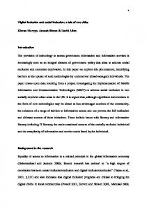
Fig. 7. Q-Sepharose chromatography of B6Fabenv24. ŽA. Chromatogram showing elution profile of protein. Sample after desalting on Sephadex G-25 Ž; 130 ml. was loaded on a 20-ml Q-Sepharose column Žpacked in HR 16r10. at 4 mlrmin. Elution was done at 3 mlrmin with continuous monitoring of absorbance at 280 nm, and 4-ml fractions were collected. ŽB. Coomassie blue-stained gel of the fractions eluted from Q-Sepharose column. Samples were processed and electrophoresed as in Fig. 5. Molecular weight of Fab fusion protein is shown in kDa; Lane P2, pool after Sephadex G-25 column; Lanes 26–37, fractions 26–37 from Q-Sepharose column. Arrows indicate the position of B6Fabenv24 protein and aggregates. Aggn., as in Fig. 5.
A. Gupta et al.r Journal of Immunological Methods 256 (2001) 121–140
133
Fig. 8. Sephacryl S-200 chromatography of B6Fabenv24. ŽA. Chromatogram showing elution profile of protein. Sample Ž40 ml. was loaded on 500 ml Sephacryl S-200 Žpacked in XK26r100. column at 0.5 ml per minute. Elution was carried out at 1 mlrmin with continuous monitoring of absorbance at 280 nm, and 4-ml fractions were collected. ŽB. Coomassie blue-stained gel of the fractions eluted from Sephacryl S-200 column. Samples were processed and electrophoresed as in Fig. 5. Molecular weight of Fab fusion protein is shown in kDa; Lane P1, pool after SP-Sepharose column; Lane P2, pool after Sephadex G-25 column; Lane P3, pool after Q-Sepharose column; Lanes 66–79, fractions 66–79 from Sephacryl S-200 column Žpeak fractions were not analysed.. Arrow indicates the position of B6Fabenv24 protein. Aggn., as in Fig. 5.
134
A. Gupta et al.r Journal of Immunological Methods 256 (2001) 121–140
The pool of fractions from hydrophobic column, free of most aggregated proteins, was treated with iodoacetamide to block free sulfhydryl groups present in any incorrectly folded Fab molecules and contaminating free LC and Fd chains. The excess iodoacetamide and salt were then removed from the sample by desalting on Sephadex G-25 column before loading on a 20-ml Q-Sepharose anion exchange column. The Q-Sepharose column was developed with a linear gradient of NaCl. B6Fabenv24 eluted between 100- and 250-mM gradient of NaCl as a symmetrical peak ŽFig. 7A.. SDS–PAGE of frac-
tions showed that fractions 26–32 contained a band of 53 kDa corresponding to calculated molecular weight of B6Fabenv24 protein, while fractions 34–37 contained some amount of Fab but also contained bands corresponding to high molecular weight aggregates ŽFig. 7B.. In agglutination assay, fractions 34–37 showed agglutination before addition of 9E10 antibody indicating the presence of aggregates. After addition of 9E10, strong agglutination was observed in fractions 26–32. Fractions 26–33 were pooled and loaded on Sephacryl S-200 gel filtration column to remove any remaining aggregates ŽFig. 8.. Fractions
Fig. 9. Purification of B6Fab. Coomassie blue-stained gel of the eluted fractions of B6Fabfrom ŽA. SP-Sepharose and ŽB. Q-Sepharose column. Samples were processed and electrophoresed as in Fig. 5. Molecular weight of Fab fusion protein is shown in kDa; Panel A: Lane D, dialysed renatured material; Lanes 14–28, fractions 14–28 from SP-Sepharose column; Lane C, B6Fab protein purified by another protocol Žcontrol.; Panel B: Lane P1, pool after SP-Sepharose column; Lane P2, pool after Toyobutyl and Sephadex G-25 column; Lanes 26–33, fractions 26–33 from Q-Sepharose column; Lane E, BSA 10 mg Žcontrol.. Arrows indicate the position of B6Fab protein, free B6Fd and free B6LC. Aggn., as in Fig. 5.
A. Gupta et al.r Journal of Immunological Methods 256 (2001) 121–140
66–79 of Sephacryl S-200 column showed band corresponding to B6Fabenv24 protein on SDS– PAGE with purity of ; 95% ŽFig. 8B.. In agglutination assay, none of these fractions showed agglutination before addition of 9E10 antibody indicating the absence of aggregates. After addition of 9E10, agglutination was observed in fractions 66–80. Fractions 68–80 were pooled. B6Fabenv24 protein Ž15.1 mg. was obtained from 3.4 l of renatured material containing 340 mg total protein. B6Fabenv13 was purified following an identical scheme and set of columns. The elution from hydrophobic column was carried out using 0.7 M NaCl concentration. Elution profiles were also similar to that of B6Fabenv24. Highly purified B6Fabenv13 Ž18.2 mg, ; 95% purity. was obtained from 4.5 l of renatured material containing 450 mg total protein. 3.5. Purification B6Fab A three-step protocol consisting of cation exchange, hydrophobic and anion-exchange column
135
chromatography was developed for purification of Fab protein containing no envelope fusion. For the assembly of B6Fab, denatured inclusion bodies of B6LC and B6Fd were mixed in equimolar ratio and renatured. The renatured material was dialysed to remove denaturants and B6Fab was purified from the dialysed material following column chromatography procedures described below. The pH of the dialysed material Ž2.7 l. was adjusted to ; 5.0 and after filtration, it was loaded on 50-ml SP-Sepharose column ŽXK 50r20.. The SP-Sepharose column was developed with a linear gradient of NaCl and protein eluted between 250and 600-mM gradient of NaCl with elution profile similar to that for B6Fabenv24 ŽSection 3.4.. SDS– PAGE analysis of peak fractions showed that fractions 16–24 contained a 50-kDa band corresponding to B6Fab protein ŽFig. 9A.. In agglutination assay, none of the fractions showed agglutination before addition of 9E10 antibody indicating the absence of aggregates. After addition of 9E10, strong agglutination was observed in fractions 18–26 ŽFig. 9A.; these fractions were pooled. The concentration of
Table 1 Activity of various B6Fab fusion proteins Identity of samples is based on data provided by the source of sample as described in Section 2. Serum
1 2 3 4 5 6 7 8 9 10 11 12 13 14 15 16
71r97 81r97 108r97 119r97 123r97 172r97 654r97 575r97 JA6-0905-0002 266r97 288r97 GM7-1109-0004 IM6-0304-0020 871 872 9E10
Source of sample sample
Identity
PGI PGI PGI PGI PGI PGI PGI PGI CDC PGI PGI CDC CDC PGI PGI MAb
HIV-1 HIV-1 HIV-1 HIV-1 HIV-1 HIV-1 HIV-1 HIV-2 HIV-2 HIV-1 q 2 HIV-1 q 2 HIV-1q 2 HIV-1 Negative Negative Control
Plate agglutinationa B6Fab B6Fab env13 0 0 0 0 0 0 0 0 nd 0 0 nd nd 0 0 25,600
) 51,200 6400 25,600 ) 51,200 ) 51,200 ) 51,200 51,200 0 nd ) 51,200 ) 51,200 nd nd 0 0 25,600
B6 Fab env24
Slide agglutinationb B6Fab B6Fab Žcontrol. env13 ŽHIV-1.
B6 Fab env24 ŽHIV-2.
0 0 0 0 0 0 0 ) 12,800 nd 3200 1600 nd nd 0 0 25,600
0 0 0 0 0 0 0 0 0 0 0 0 0 0 0 nd
0 0 0 0 0 0 0 3q 3q 2q 2q 2q 0 0 0 nd
4q 3q 3q 4q 3q 4q 4q 0 0 4q 3q 3q 4q 0 0 nd
9E10, a monoclonal antibody which binds to decapeptide tag, cmyc; nd, not done. ‘0’ indicates no agglutination with 1:100 dilution of sample. a Numbers indicate the end point of agglutination recorded as maximum dilution of each sample that gave visible agglutination. b The agglutination was visibly recorded with ‘0’ as no agglutination and ‘4 q ’ as the highest agglutination, as seen in Fig. 10.
136
A. Gupta et al.r Journal of Immunological Methods 256 (2001) 121–140
NaCl in the pool was estimated to be approximately 0.3 M Žcalculated from the chromatogram. and was made to 2 M by adding NaCl. The pH was adjusted to 7.2 with 1 M Tris solution, and the sample was loaded on 10-ml Toyobutyl column. B6Fab did not bind to the column and eluted in flow through during loading Ždata not shown.. The flow through was treated with iodoacetamide to block any free sulfhydryl groups. The excess iodoacetamide and salt were then removed from the sample by desalting on Sephadex G-25 column. The protein pool obtained from G-25 column was loaded on a 20-ml Q-Sepharose column ŽHR16r10., and the column was developed with a linear gradient of NaCl. B6Fab eluted between 100- and 230-mM gradient of NaCl. SDS–PAGE analysis showed that fractions 26–31 contained band corresponding to B6Fab with purity of ; 95% ŽFig. 9B.. Faint bands of high molecular weight were also present. However, these bands did not react with MAb 9E10 on Western blot, indicating that they were not multimers of B6Fab Ždata not shown.. In agglutination assay, none of the fractions showed agglutination before addition of 9E10 antibody. After addition of 9E10, agglutination was observed in fractions 25–33. Fractions 25–32 were pooled. Forty-one milligrams of B6Fab protein was obtained from 2.5 l of renatured material containing 250 mg total protein, which corresponds to 16.5% recovery of total protein used for renaturation ŽFig. 4..
with B6Fabenv13-coated RBCs. Samples positive for both HIV-1 and HIV-2 showed agglutination with both the proteins. The serum samples could be diluted to very high degree indicating the sensitivity of detection. B6Fab-coated RBCs did not show agglutination with any of the serum samples except for control MAb 9E10 indicating the high specificity of the reagents. Since these proteins are being developed for the detection of HIV antibodies in a simple format that should not require any sophisticated instruments or pipetting devices, agglutination assay was performed in a slide format. In this assay, O RhD negative RBCs were mixed with serum from HIV-infected persons to mimic whole blood, and a drop of this reconstituted blood was added to a drop of one of the proteins Ž10 mgrml. on a glass slide. The solution was mixed with a disposable stick and kept undisturbed. After 3 min, the slide was rotated and agglutination was read against white light. The agglutination was visibly recorded on a scale from 0 to 4 q ,
3.6. Characterization of purified B6 Fab and B6FabenÕ fusion proteins Immunoreactivity of various purified proteins was studied using two normal, eight HIV-1 positive, two HIV-2 positive and three HIV-1q 2 positive serum samples following a quantitative microtitre plate agglutination assay and a qualitative but simple slide agglutination assay as described in Section 2. The identity of the serum samples was based on several immunoassays, which are able to discriminate between antibodies to HIV-1 and HIV-2. In microtitre plate assay, HIV-1 positive samples showed strong agglutination with B6Fabenv13-coated RBCs and no reactivity with B6Fabenv24-coated RBCs ŽTable 1.. Similarly, HIV-2 positive samples reacted strongly with B6Fabenv24-coated RBCs and gave no reaction
Fig. 10. Slide agglutination assay. The assay was performed as described in Section 2.9. Appearance of clumps of RBCs on spot HIV-1 andror HIV-2 indicates the presence of anti-HIV antibodies in the sample. No agglutination is expected in control. ŽA. Sample IM6-0304-0020 ŽHIV-1.. ŽB. Sample JA6-0905-0002 ŽHIV-2.. ŽC. Sample GM7-1109-0004 ŽHIV-1q2.. Identity of HIV infection in the sample is indicated in the parenthesis.
A. Gupta et al.r Journal of Immunological Methods 256 (2001) 121–140
where ‘0’ indicates no agglutination and ‘4 q ’ indicates the highest agglutination. B6Fab Žcontrol. did not show any agglutination of RBCs ŽTable 1; Fig. 10. with any serum sample. B6Fabenv13 ŽHIV-1. showed agglutination with HIV-1 and HIV-1q 2 positive samples ŽFig. 10A,C; Table 1. while B6Fabenv24 ŽHIV-2. showed agglutination with HIV-2 and HIV-1q 2 samples ŽFig. 10B,C; Table 1.. These results highlight the specificity of the proteins on their use in a simple slide assay format.
4. Discussion The data presented here clearly demonstrates that recombinant fusion proteins can be designed and obtained in purified form in large quantities for use in haemagglutination-based detection of antibodies to HIV in whole blood. There were many important considerations while developing these recombinant proteins. Since the proteins comprise of chimeric molecules consisting of anti-RBC monovalent fragment fused to immunodominant epitopes of HIV-1 and HIV-2, attention was given to designing of both the components. Selection of anti-human RBC antibody was based on the premise that they should possess high affinity, bind to all types of human RBCs irrespective of variation in major and minor blood group antigens, and the molecule recognised by them should be present in high density on RBC surface. Similarly, the immunodominant antigens of HIV should be conserved and not be subtype-specific. A chimeric protein made from these two components should give high yield using simple purification procedures should not aggregate, should be stable for long duration at 4 8C and ambient temperatures, and should be able to detect antibodies against HIV in serum from individuals infected with any subtype of HIV, with high sensitivity and specificity. Finally, the format of testing should be easy and result should be readable without requirement of any sophisticated instrument. The following discussion highlights the development of these molecules in line with the above considerations. The present study employs Fab fragment of an anti-RBC MAb, B6, which was isolated in our laboratory by immunizing mice with a pool of O RhD
137
negative human RBCs, and following conventional hybridoma technology. The evaluation of its binding characteristics to human RBC was based on initial screening of clones on 20 different blood samples followed by final screening of a selected clone, B6, for reactivity to approximately 1000 randomly collected blood samples Žour unpublished results.. The DNA sequence encoding Fd and LC was obtained by PCR-based techniques Žsequence of B6Fd and B6LC will be published elsewhere.. Selection of HIV epitopes was based on the alignment of available HIV genome sequence of different subtypes to identify conserved regions and information available regarding their immunodominant nature ŽGnann et al., 1987a,b; Broliden et al., 1991.. A 31-amino acid sequence in envelope glycoprotein gp41 of HIV-1 Žamino acids 590–620. is highly conserved in most subtypes of HIV-1 and is known to be immunodominant. Antibodies to this region have been reported to be present in most cases of HIV-1 infection ŽGnann et al., 1987a,b; Broliden et al., 1991.. Similarly, a 31-amino acid sequence in HIV-2 envelope glycoprotein, gp36 Žamino acids 581–611 of gp36., analogous to gp41 sequence is highly conserved and has been shown to be immunodominant. These regions of gp41 and gp36 were therefore selected for making B6Fdenv fusion proteins. The method of preparing Fab and Fabenv fusion proteins was based on denaturation of FdrFdenv and LC, their mixing in equimolar ratio followed by in vitro renaturation by dilution in a buffer creating appropriate redox conditions. Since assembly of Fab requires formation of many intra-molecular and inter-molecular disulfide bonds, after refolding, it was necessary to evolve a strategy to isolate monomeric, correctly folded Fab free from aggregates and unassembled Fd and LC chains. The B6Fabenv fusion proteins are likely to make more intra- and inter-molecular interactions due to hydrophobic nature of the envelope sequences and the two cysteine residues present in env sequence, which make a disulfide bond. Keeping the above factors in consideration, a multistep protocol involving cation exchange, hydrophobic, anion-exchange and gel filtration chromatography was developed to isolate highly pure, monomeric Fab and Fabenv fusion proteins. All the three proteins, B6Fab, B6Fabenv13 and B6Fabenv24, bound tightly to SP-Sepharose and
138
A. Gupta et al.r Journal of Immunological Methods 256 (2001) 121–140
eluted with NaCl Ž400–500 mM NaCl.. The presence of monomeric protein was tested in an RBC agglutination assay where monomeric proteins show agglutination only after the addition of MAb 9E10 which binds to a 10-amino acid tag, cmyc present at the C-terminus of each protein. The aggregated proteins show agglutination of RBCs before the addition of MAb 9E10. For further processing, fractions containing monomeric protein were pooled. This step of cation exchange chromatography primarily concentrates the large volume of renatured protein and also separates monomers from majority of free LC and aggregates. However, the eluate contains high molecular aggregates and other contaminants visible as a smear on the gel ŽFig. 8B, Lane P1.. When this eluate was loaded on hydrophobic Toyobutyl column, both B6Fabenv13 and B6Fabenv24 bound at 2 M NaCl concentration and the desired protein was eluted with lower salt concentration, 0.7 M NaCl for B6Fabenv13 and 0.6 M NaCl for B6Fabenv24. This step removed remaining aggregates, which eluted from the column at zero salt concentration. Since B6Fab does not carry the HIV-envelope sequences, it is less hydrophobic and, therefore, did not bind to the hydrophobic matrix and came in the flow through. Aggregates and most of the contaminants eluted at zero salt concentration Ždata not shown., indicating that this step was essential to remove hydrophobic impurities and other aggregates. Fd and LC molecules contain cysteine residues, which make intrachain and interchain disulfide bonds to assemble functional Fab molecule. Besides, these disulfide-forming cysteine residues, B6Fabenv13 and B6Fabenv24, also contain cysteine residues in envelope sequence that make a disulfide bond. There is always a possibility that any uncoupled cysteine can make disulfide bonds with cysteine of another molecule or within the same improperly folded molecule that may lead to incorrect inter- or intramolecular disulfide bond formation. Also, unassociated LC and Fd contain free sulfhydryl groups that could interact with Fab thereby disrupting its structure. To eliminate such interactions, free sulfhydryl groups were blocked irreversibly with iodoacetamide during purification. During optimisation of this protocol, it was observed that B6Fabenv13 and B6Fabenv24 purified without iodoacetamide treatment aggregated upon storage, even though the final
purified protein prior to storage was monomeric. This indicated that this preparation had incorrectly folded molecules with reactive groups, which resulted in aggregation of stored protein Ždata not shown.. This aggregation was not observed after pre-treatment with iodoacetamide. After iodoacetamide treatment, anion exchange chromatography was performed. On Q-Sepharose column, high molecular weight proteins were found to elute at higher salt concentration in the case of Fabenv proteins ŽFig. 7B; Lanes 34–37.. No such aggregates were observed in Fab proteins, which contained no envelope sequence. Therefore, Fab protein was used without any further purification, but the Fabenv fusion proteins were further purified on a gel-filtration column for complete removal of all high molecular weight species. The envelope sequences influence the folding of Fd and its association with LC to form Fab. Also, these sequences, probably due to their hydrophobic nature and presence of cysteine residues, are more prone to incorrect intermolecular interactions, resulting in an increased proportion of molecules undergoing aggregation. This is also reflected in the yields obtained for various proteins. While 66 mg Ž; 16.5% of the starting protein. of B6Fab can be obtained from a 4-l renaturation mix containing 400 mg of denatured LC and Fd isolated from 1-l culture each, approximately 15–18 mg Ž; 5%. of B6Fabenv fusion proteins were obtained from same amount of starting material ŽFig. 4.. The purified proteins were stable for 7–10 days at 37 8C. However, for use in agglutination assay, a mixture of sugars and protease inhibitors was added to these proteins to enhance their stability. Under these conditions, the proteins were stable for more than 30 days at 37 8C and for 6 months at 4 8C Žour unpublished results.. The original description of rapid whole blood agglutination assay for HIV antibodies employed chemical conjugation procedures to produce a monomeric bifunctional molecule, which was marketed as Simpli-Red whole blood immunoassay system ŽAGEN Biomedical Limited, Australia.. Later, efforts were made to produce these reagents in E. coli either as scFv or Fab fusion proteins. However, the yields of these proteins were only in hundreds of microgram and were not enough for practical use.
A. Gupta et al.r Journal of Immunological Methods 256 (2001) 121–140
The present work breaks that barrier and yields tens of milligrams of fusion proteins on the laboratory scale using simple column chromatography steps. The good yield is probably due to the intracellular over-expression followed by in vitro renaturation strategy for Fab assembly while earlier workers depended upon a secretion-based strategy where hydrophobic envelope sequences might have affected the secretion of the molecules. The in vitro renaturation protocol to assemble Fab molecules is very efficient with reported recovery of up to 40% ŽBuchner and Rudolph, 1991.. The recovery of refolded and purified B6Fab was approximately 17%. The lower recovery of envelope fusion protein Žapproximately 5%. could be due to the presence of hydrophobic envelope sequences, as mentioned above. The recovery would also depend upon the folding characteristics of the Fab molecule, which in turn depend on the primary sequence of the antibody. In case of B6, we have identified a mutation in the framework region of B6VH , which results in approximately two-fold improvement in the yield of B6Fab protein Žour unpublished results.. Further optimisation by varying the concentration of L-arginine and oxidised glutathione might also improve refolding ŽBuchner et al., 1992a.. Moreover, it may be possible to select a variant B6 with improved binding and folding characteristics using phage display technology ŽHoogenboom, 1997.. Quantitative Žmicrotitre plate assay. and qualitative Žslide agglutination. format of evaluation of these proteins shows that envelope fusion proteins produce strong agglutination reaction with high specificity, and can detect very low levels of antibody in human serum as revealed by high dilution of serum sample, which could produce visible agglutination in microtitre plate assay. The qualitative slide agglutination format is for use on whole blood with no need of dilution. Detection of individuals infected with HIV is a vital key in attempts to curtail the spread of the virus, particularly by way of contaminated blood products. Simple, rapid, inexpensive tests, which can be performed without the need of any expensive and sophisticated instruments, are desired not only for the developing and under-developed countries but also for developed countries in clinical settings such as emergency rooms where obtaining immediate re-
139
sults could be beneficial. The above mentioned data demonstrates the potential of the described fusion proteins for the development of sensitive and specific whole blood-based agglutination assay for detection of antibodies to HIV-1 and HIV-2. A HIV diagnostic kit for antibody detection may not possess desired sensitivity if the reagents contain only one immunodominant region of HIV antigens, as the antibodies to this immunodominant region may not be present in all samples from different stages of disease. Moreover, with the emergence of new subtypes having differences in sequence, identification and inclusion of other immunodominant epitopes becomes necessary. For this, fusion proteins with other immunodominant epitopes of HIV have been prepared in our laboratory and are under evaluation as cocktail with the fusion proteins described above. The technology described in this paper can also be used for development of similar molecules for detecting various infections, such as hepatitis B, hepatitis C and syphilis, which are relevant in blood transfusion.
Acknowledgements AG and SG were recipients of Research Fellowship from the Council of Scientific and Industrial Research, Government of India. We are thankful to Dr. Shobha Sehgal ŽPGI, Chandigarh, India. and Dr. R. Lal ŽCDC, Atlanta, USA. for serum samples. Mr. Rajiv Chawla is acknowledged for help in preparation of manuscript. This work was funded by the Department of Biotechnology, Government of India.
References Brinkmann, U., Pastan, I., 1994. Immunotoxins against cancer. Biochim. Biophys. Acta 1198, 27–45. Broliden, P.A., Ruden, U., Ouattara, A.S., de The, G., Solver, E., Trojnar, J., Wahren, B., 1991. Specific synthetic peptides for detection of and discrimination between HIV-1 and HIV-2 infection. J. Acquired Immune Defic. Syndr. 4, 952–958. Buchner, J., Rudolph, R., 1991. Renaturation, purification and characterization of recombinant Fab-fragments produced in Escherichia coli. BiorTechnology 9, 158–162. Buchner, J., Pastan, I., Brinkmann, U., 1992a. A method for increasing the yield of properly folded recombinant fusion
140
A. Gupta et al.r Journal of Immunological Methods 256 (2001) 121–140
proteins: single-chain immunotoxins from renaturation of bacterial inclusion bodies. Anal. Biochem. 205, 263–270. Buchner, J., Brinkmann, U., Pastan, I., 1992b. Renaturation of a single-chain immunotoxin facilitated by chaperones and protein disulfide isomerase. BiorTechnology 10, 682–685. Chaudhary, V.K., Queen, C., Junghans, R.P., Waldmann, T.A., FitzGerald, D.J., Pastan, I., 1989. A recombinant immunotoxin consisting of two antibody variable domains fused to Pseudomonas exotoxin. Nature 339, 394–397. Chaudhary, V.K., Batra, J.K., Gallo, M.G., Willingham, M.C., FitzGerald, D.J., Pastan, I., 1990a. A rapid method of cloning functional variable-region antibody genes in Escherichia coli as single-chain immunotoxins. Proc. Natl. Acad. Sci. U. S. A. 87, 1066–1070. Chaudhary, V.K., Jinno, Y., Gallo, M.G., FitzGerald, D., Pastan, I., 1990b. Mutagenesis of Pseudomonas exotoxin in identification of sequences responsible for the animal toxicity. J. Biol. Chem. 265, 16306–16310. Cho, B.K., Roy, E.J., Patrick, T.A., Kranz, D.M., 1997. Singlechain Fvrfolate conjugates mediate efficient lysis of folate-receptor-positive tumor cells. Bioconjugate Chem. 8, 338–346. Choe, M., Webber, K.O., Pastan, I., 1994. B3ŽFab.-PE38M: a recombinant immunotoxin in which a mutant form of Pseudomonas exotoxin is fused to the Fab fragment of monoclonal antibody B3. Cancer Res. 54, 3460–3467. Clavel, F., Guetard, D., Brun-Vezinet, F., Chamaret, S., Rey, M.A., Santos-Ferreira, M.O., Laurent, A.G., Dauguet, C., Katlama, C., Rouzioux, C. et al., 1986. Isolation of a new human retrovirus from West African patients with AIDS. Science 233, 343–346. Coia, G., Hudson, P.J., Lilley, G.G., 1996. Construction of recombinant extended single-chain antibody peptide conjugates for use in the diagnosis of HIV-1 and HIV-2. J. Immunol. Methods 192, 13–23. Dolezal, O., Coia, G., Guthrie, R.E., Lilley, G.G., Hudson, P.J., 1995. Escherichia coli expression of a bifunctional Fab-peptide epitope reagent for the rapid diagnosis of HIV-1 and HIV-2. Immunotechnology 1, 197–209. Gnann Jr., J.W., Nelson, J.A., Oldstone, M.B., 1987a. Fine mapping of an immunodominant domain in the transmembrane glycoprotein of human immunodeficiency virus. J. Virol. 61, 2639–2641. Gnann Jr., J.W., Schwimmbeck, P.L., Nelson, J.A., Truax, A.B., Oldstone, M.B., 1987b. Diagnosis of AIDS by using a 12amino acid peptide representing an immunodominant epitope of the human immunodeficiency virus. J. Infect. Dis. 156, 261–267. Hoogenboom, H.R., 1997. Designing and optimising library selection strategies for generating high-affinity antibodies. Tibtech 15, 62–70.
Kane, J.F., 1995. Effects of rare codon clusters on high-level expression of heterologous proteins in Escherichia coli. Curr. Opin. Biotechnol. 6, 494–500. Kemp, B.E., Rylatt, D.B., Bundesen, P.G., Doherty, R.R., McPhee, D.A., Stapleton, D., Cottis, L.E., Wilson, K., John, M.A., Khan, J.M. et al., 1988. Autologous red cell agglutination assay for HIV-1 antibodies: simplified test with whole blood. Science 241, 1352–1354. Kunkel, T.A., 1985. Rapid and efficient site-specific mutagenesis without phenotypic selection. Proc. Natl. Acad. Sci. U. S. A. 82, 488–492. Lilley, G.G., Dolezal, O., Hillyard, C.J., Bernard, C., Hudson, P.J., 1994. Recombinant single-chain antibody peptide conjugates expressed in Escherichia coli for the rapid diagnosis of HIV. J. Immunol. Methods 171, 211–226. Muller, B.H., Chevrier, D., Boulain, J.C., Guesdon, J.L., 1999. Recombinant single-chain Fv antibody fragment-alkaline phosphatase conjugate for one-step immunodetection in molecular hybridization. J. Immunol. Methods 227, 177–185. Neuberger, M.S., Williams, G.T., Fox, R.O., 1984. Recombinant antibodies possessing novel effector functions. Nature 312, 604–608. Orlandi, R., Gussow, D.H., Jones, P.T., Winter, G., 1989. Cloning immunoglobulin variable domains for expression by the polymerase chain reaction. Proc. Natl. Acad. Sci. U. S. A. 86, 3833–3837. Pastan, I., Chaudhary, V., FitzGerald, D.J., 1992. Recombinant toxins as novel therapeutic agents. Annu. Rev. Biochem. 61, 331–354. Pluckthun, A., 1992. Mono and bivalent antibody fragments produced in Escherichia coli: engineering, folding and antigen binding. Immunol. Rev. 130, 151–188. Ratner, L., Haseltine, W., Patarca, R., Livak, K.J., Starcich, B., Josephs, S.F., Doran, E.R., Rafalski, J.A., Whitehorn, E.A., Baumeister, K. et al., 1985. Complete nucleotide sequence of the AIDS virus, HTLV-III. Nature 313, 277–284. Rylatt, D.B., Kemp, B.E., Bundesen, P.G., John, M.A., O’Reilly, E.J., Cottis, L.E., Miles, S.J., Khan, J.M., Dinh, D.P., Stapleton, D., Hillyard, C.J., 1990. A rapid whole-blood immunoassay system. Med. J. Aust. 152, 75–77. Studier, F.W., Rosenberg, A.H., Dunn, J.J., Dubendorff, J.W., 1990. Use of T7 RNA polymerase to direct expression of cloned genes. Methods Enzymol. 185, 60–89. Williams, G.T., Neuberger, M.S., 1986. Production of antibodytagged enzymes by myeloma cells: application to DNA polymerase I Klenow fragment. Gene 43, 319–324. Wilson, K.M., Gerometta, M., Rylatt, D.B., Bundesen, P.G., McPhee, D.A., Hillyard, C.J., Kemp, B.E., 1991. Rapid whole blood assay for HIV-1 seropositivity using an Fab-peptide conjugate. J. Immunol. Methods 138, 111–119.