Accepted Manuscript Indirect reduction technique using a distraction support in minimally invasive percutaneous plate osteosynthesis of tibial shaft fractures Wen-Wei Dong, Zeng-Yuan Shi, Zheng-Xin Liu, Hai-Jiao Mao PII:
S1008-1275(16)30280-2
DOI:
10.1016/j.cjtee.2016.09.001
Reference:
CJTEE 174
To appear in:
Chinese Journal of Traumatology
Received Date: 6 August 2015 Revised Date:
20 January 2016
Accepted Date: 26 February 2016
Please cite this article as: Dong WW, Shi ZY, Liu ZX, Mao HJ, Indirect reduction technique using a distraction support in minimally invasive percutaneous plate osteosynthesis of tibial shaft fractures, Chinese Journal of Traumatology (2016), doi: 10.1016/j.cjtee.2016.09.001. This is a PDF file of an unedited manuscript that has been accepted for publication. As a service to our customers we are providing this early version of the manuscript. The manuscript will undergo copyediting, typesetting, and review of the resulting proof before it is published in its final form. Please note that during the production process errors may be discovered which could affect the content, and all legal disclaimers that apply to the journal pertain.
ACCEPTED MANUSCRIPT
Original article
percutaneous plate osteosynthesis of tibial shaft fractures
RI PT
Indirect reduction technique using a distraction support in minimally invasive
Wen-Wei Dong, Zeng-Yuan Shi*, Zheng-Xin Liu, Hai-Jiao Mao
SC
Department of Trauma Orthopaedics, Affiliated Hospital Medical School of Ningbo University Zhejiang, Ningbo 315020, China
Article history
TE D
Received August 6, 2015 Revised January 20, 2016 Accepted February 26, 2016
M AN U
*Corresponding author: Tel.: +8615967804880, Email: szy
[email protected]
Fund
This project was supported by Ningbo Social Development Projects Grant
AC C
ABSTRACT
EP
(2013C50020) and Ningbo Municipal Natural Science Foundation (2016A610129).
Purpose: To describe an indirect reduction technique during minimally invasive percutaneous plate osteosynthesis (MIPPO) of tibial shaft fractures with the use of a distraction support.
Methods: Between March 2011 and October 2014, 52 patients with a mean age of 48 years (16-72 years) sustaining tibial shaft fractures were included. All the patients
1
ACCEPTED MANUSCRIPT
underwent MIPPO for the fractures using a distraction support prior to insertion of the plate. Fracture angular deformity was assessed by goniometer measurement on
RI PT
preoperative and postoperative images. Results: Preoperative radiographs revealed a mean of 7.6°(1.2°-28°) angulation in coronal plane and a mean of 6.8°(0.5°-19°) angulation in sagittal plane.
SC
Postoperative anteroposterior and lateral radiographs showed a mean of 0.8°(0°
M AN U
-4.0°) and 0.6°(0°-3.6°) of varus/valgus and apex anterior/posterior angulation, respectively. No intraoperative or postoperative complications were noted. Conclusions: This study suggests that the distraction support during MIPPO of tibial shaft fractures is an effective and safe method with no associated complications.
Percutaneous plating
TE D
Key words: Tibial shaft fractures; Indirect reduction; Distraction support;
EP
1. Introduction
AC C
The management of tibial shaft fractures can be challenging because of the scarcity of soft tissue, their subcutaneous nature and poor vascularity.1,2 Locked intramedullary nailing is the gold standard for treating tibial shaft fractures.3 However, precise control of reduction at the proximal and distal quarters is difficult to achieve due to metaphyseal widening of the tibia associated with a long lever arm. Therefore, some authors4-6 advocate the use of plates to treat the fractures involving proximal or
2
ACCEPTED MANUSCRIPT
distal quarters of the tibia. Minimally invasive plating techniques use indirect reduction methods and allow
RI PT
stabilization of tibial fractures while reducing iatrogenic soft tissue injury and damage to bone vascularity, in addition to preserving the osteogenic fracture haematoma. However, closed reduction may be more difficult than open methods. There are a
SC
number of indirect reduction techniques have been described to facilitate fixation,
M AN U
including reduction clamps,7,8 temporary distracters,8-10 external fixators,11 manual manipulation techniques7,8,12 and sustained traction via fracture table.13 Although these techniques are effective, most of them are cumbersome, difficult to maneuver and their intraoperative application adds length to the time of the surgical procedure.
TE D
We describe our experience of an alternative method for minimally invasive percutaneous plate osteosynthesis (MIPPO) of tibial shaft fracture using a distraction support (patent number: 201210153980.x) which was designed for tibial nailing.14
EP
This technique has proven to be a reliable method for closed reduction of tibial
AC C
diaphyseal fractures.
2. Material and methods 2.1. Patients' data
Between March 2011 and October 2014, 312 patients who sustained 316 tibial
fractures were admitted to our hospital. A total of 52 patients were included in the study. There were 16 females and 36 males with a mean age of 48 years (16-72 years).
3
ACCEPTED MANUSCRIPT
The various mechanisms of injury included a road traffic crash (32 patients), a sports-related injury (6 patients), and a work-related incident (14 patients). Only one
RI PT
had a contralateral intertrochanteric fracture and none had polytrauma. There were 18 nonsmokers and 34 smokers in this patient group. Inclusion criteria were skeletally mature patients who had proximal or distal diaphyseal fractures with or without
SC
intraarticular extension into the ankle joint and skeletal immature middle shaft
M AN U
fracture. Exclusion criteria were tibial plateau fractures with an intraarticular extension, pathological fractures, mature middle shaft fractures, multi-fragmentary intraarticular pilon fractures requiring fine wire external fixation, and open fractures. All fractures were closed. There were three segmental fractures. According to
TE D
OTA/AO classification,15 diaphyseal fractures involved the proximal third in 15 cases, the middle third in 2 cases and the distal third in 35 cases. Among the fractures located in the distal third, 15 had an extension down to the tibial plafond, with
EP
articular involvement, which were classified as 43B1 because there was no
AC C
displacement at articular level, although the main component of the fracture was located in the diaphysis. The remaining fractures were classified as 42A in 23 cases, 42B in 9 and 42C in 5 (Table 1). Immediate temporary skeletal stabilization was achieved using an above-knee back slab. All patients had radiographs. Table1. Data of patients with closed tibial fractures
Items
n
4
ACCEPTED MANUSCRIPT
Gender Male Female
36 16
Injury mechanism
34 18
Location p/3# m/3& d/3*
15 2 35
OTA/AO Classification 42A 42B 42C 43B1
23 9 5 15
p/3: proximal third; &m/3: middle third; *d/3: distal third.
TE D
#
SC
Smoker Yes No
RI PT
32 6 14
M AN U
Traffic Sports Work
Depending on the skin condition, the mean delay between trauma and surgery
EP
was 5.5 ± 3.3 days (range, 3-9 days). All 52 patients underwent MIPPO with anatomic reduction achieved using a distraction support prior to insertion of the plate, which
AC C
was performed by a single surgeon with a special interest in these techniques. Fracture alignment and angular deformity were assessed by goniometer measurement obtained from preoperative and postoperative anteroposterior and lateral images for all subjects. Malalignment was defined as more than 5°of angulation in any plane.16 The study was approved by the institutional review board of the hospital. All patients signed an informed consent statement before the operation.
5
ACCEPTED MANUSCRIPT
2.2. Description of the device The distraction support consists of femoral tray, tibial tray and base (Fig. 1A).
RI PT
The tibial tray contains anterior nut on each side which has a left-hand thread in one end and a right-hand in the other. Thus both screws are moved out by turning the nut, causing greater traction intraoperatively. The knee angle can be easily adjusted as the
SC
tibial tray lies on a simple ratchet device of the base. The support can help different
M AN U
patients by adjusting the posterior nut on the femoral tray. The femoral tray connects the tibial tray and the base via a pivot respectively and allows the femoral tray, tibial tray and base to be folded in the same plane which facilitates antisepsis and storage when the support is not applied (Fig. 1B).
TE D
2.3. Surgical treatment
Under adequate anaesthesia, the patient was positioned in the supine position on a radiolucent operating table. The ipsilateral iliac crest and the entire lower limb were
EP
prepared and draped in the usual sterile fashion. A sterile tourniquet was usually
AC C
positioned proximally on the tight. The distraction support was placed on the operating table beneath the limb on the operative side. The femoral tray was placed under the patient’s thigh and positioned as close to the buttock as possible. A sterile drape was placed under the distal thigh thus creating a barrier between the support and the limb. Adjustment was then made to the length of the femoral tray, tibial tray as well as to the knee flexion angle. A separate sterile self-stick ankle strap can be used
6
ACCEPTED MANUSCRIPT
to secure the foot to the tibial tray (Fig. 2). Careful attention was paid to the rotational alignment of the limb. The surgeon examined the limb from above, generally planning
RI PT
to align the tibial tuberosity with the first web space. By turning the anterior nut on each side of the tibial tray, traction can then be applied to the fracture (the amount being sufficient to reduce the fracture). Intraoperative fluoroscopy was used to
SC
identify the fracture site and confirm the reduction (Fig. 3A-H). Selection of the
M AN U
appropriate plate length after reduction was achieved. The plate was then applied via the standard MIPPO technique according to the fracture pattern. Accurate plate positioning was confirmed by fluoroscopy. Another plate of similar length is aligned externally and acts as a guide through which stab incision is given. Subsequent screws
TE D
are inserted close to either side of the fracture (Fig. 3I-K). With fractures extending into the ankle joint, 4.5 mm cannulated screws were inserted through stab incisions which were sutured in the standard fashion (Fig. 4). We started ankle and knee range
EP
of motion exercises postoperatively. Non-weight-bearing walking was kept for 6-8
AC C
weeks. They were followed up at 4 weeks firstly and at 3, 6, 9 and 12 months after operation with clinical and radiological examinations (lateral and anteroposterior X-rays). A typical case was shown in Fig. 5. Once union was assessed in radiograph, weight bearing was permitted. 3. Results Totally, 52 patients with 52 tibial shaft fractures were treated with the described
7
ACCEPTED MANUSCRIPT
technique; 23 fractures were classified as 42A, 9 were 42B, 5 were 42C and 15 were 43B1. Preoperative radiographs revealed a mean of 7.6°(1.2°-28°) angulation in
RI PT
coronal plane and a mean of 6.8 ° (0.5 ° -19 ° ) angulation in sagittal plane. Postoperative radiographs were then evaluated to determine the final alignment. All the 52 patients were found to have acceptable alignment. Postoperative
SC
anteroposterior and lateral radiographs showed the distal segment returned to its
M AN U
anatomical alignment with a mean of 0.8°(0°-4.0°) angulation of varus/valgus and 0.6°(0°-3.6°) angulation of apex anterior/posterior (Fig.6). The minimum follow-up was 12 months (average, 20 months; range, 12-48 months) after surgery. No intraoperative or postoperative complications were noted in the study group. All
TE D
the patients had clinical and radiographic follow-up. Radiographic union was achieved with an average of 4 months (range, 3-6 months). No nonunion or delayed
4. Discussion
EP
union was found in this group.
AC C
MIPPO is a popular technique for the treatment of tibial fractures.17 Performing percutaneous plate osteosynthesis on a routine operation table presents some difficulties in closed reduction and obtaining quality pictures. Traction in the long axis of the leg facilitates the procedure. A variety of techniques have been described for closed tibial reduction18 which can be broadly divided into fracture table traction, manual traction and distractor techniques.
8
ACCEPTED MANUSCRIPT
The fracture table traction method provides excellent consistency of traction. Complications caused by calcaneal pin are rare but include subtalar encroachment,
RI PT
haemorrhage and pain at entry point due to neuroma. More commonly, oblique insertion of calcaneal pin will lead to varus or valgus deformation at the fracture site during traction. Furthermore, the use of traction table increases the set up time and
SC
reduces access to the contralateral lower extremity which makes assessment of
M AN U
rotational deformity difficult.19 Manual traction circumvents the problems of increased set up time and inadequate access. The major concern about manual traction is the accuracy of reduction, maintenance of reduction by manual traction alone can also be difficult.20 In order to aid fracture reduction and eliminate the use of a fracture
TE D
table, a number of distraction devices have been described. Though it enables patients to be “free-draped” on a radiolucent table and incurs many benefits over manual traction, application of distractor is an additional invasive procedure and increased set
EP
up time, typically quoted at around 20 min.21
AC C
We believe that distraction support is a simple and safe technique to aid and maintain anatomic reduction during MIPPO of tibial fractures. There are a number of significant advantages over fracture table traction, manual traction and distractor techniques. The support is simple and easy to maneuver which can be used for all fractures without increasing the operative or screening times.14 It is particularly useful in multifragmented fractures, where the reduction is otherwise difficult to hold.
9
ACCEPTED MANUSCRIPT
Maneuvers to achieve reduction and repetitive manipulation of the fracture are avoided. The support avoids the use of excessive traction by turning the anterior nuts
correct length, the frame construct will resist shortening.
RI PT
and length can be restored precisely under radiographic control. Once held at the
Careful control of the distal segment is critical in achieving acceptable reduction,
SC
which must be attained to prevent angular deformity and malunion.22 The foot is
M AN U
secured to the tibial tray using a self-stick strap in a neutral position. The support can provide axial force to align fragments by turning the nuts. The theoretical advantage of the bilateral uniplanar rectangular frame distraction over the unilateral uniplanar universal distractor is a better ability to control coronal plane angulation while still
TE D
maintaining ideal traction without over or under distraction. We believe that distraction support is a very useful tool to assist in obtaining and maintaining an intraoperative reduction without the extra assistant for tibia fractures.
EP
There are certain advantages in leaving the fracture site closed during MIPPO of
AC C
tibia. However, malreduction with minimally invasive approaches is a significant concern. Borg et al23 treated 21 patients with percutaneous plating of distal tibial fractures with manual traction or distractor; 4 had 6° to 10° of angular deformity postoperatively, 2 delayed union and 2 nonunion during the follow-up. Others5,24,25 also documented the risk of sagittal plane malreduction with percutaneous plating of these fractures. In the present study there were no patients with angular deformities
10
ACCEPTED MANUSCRIPT
greater than 5°. It has been shown that the quality of fracture reduction affects the rate of healing and incidence of delayed union or malunion.25,26 The good reduction was
delayed union or nonunion occurred in our group.
RI PT
achieved by the distraction support. The average union time was four months. No
Additionally, distraction support is noninvasive during reduction, avoiding
SC
iatrogenic injury to the patient. Another advantage is the absence of hardware in the
M AN U
radiographic region of the tibia. The construct as described optimizes the radiographic visualization of the tibia by keeping frame out of the radiographic field. It provides a simple and noninvasive method for indirect reduction. Distraction support for acute tibial fractures has now become the preferred method of limb positioning for the
TE D
majority of the surgeons working in our trauma units. The technique may not be applicable in patients who have fractures more than 10-14 days if shortening is a major component of the fracture deformity. In these situations, it is advisable to
EP
consider the temporary intraoperative external fixation if an attempt is being made to
AC C
correct shortening.
In conclusion, fracture reduction using the distraction support proved to be
successful. The combined use of this simple distraction support along with minimally invasive insertion of the AO tibial locking plates is a simple and reliable surgical strategy, which can be readily applied in these difficult injuries. References
11
ACCEPTED MANUSCRIPT
1.
Borrelli J Jr, Prickett W, Song E, et al. Extraosseous blood supply of the tibia and the effects of different plating techniques: a human cadaveric study. J Orthop
2.
RI PT
Trauma. 2002;16:691-695. Collinge CA, Sanders RW. Percutaneous plating in the lower extremity. J Am Acad Orthop Surg. 2000;8:211-216.
Attal R, Hansen M, Kirjavainen M, et al. A multicentre case series of tibia
SC
3.
M AN U
fractures treated with the Expert Tibia Nail (ETN). Arch Orthop Trauma Surg. 2012;132:975-984. doi: 10.1007/s00402-012-1502-y. 4.
Feng W, Fu L, Liu J, et al. Biomechanical evaluation of various fixation methods for proximal extra-articular tibial fractures. J Surg Res. 2012;178:722-727. doi:
5.
TE D
10.1016/j.jss.2012.04.014.
Tong DK, Ji F, Cai XB. Locking internal fixator with minimal invasive plate osteosynthesis for the proximal and distal tibial fractures. Chin J Traumatol.
Kwok CS, Crossman PT, Loizou CL. Plate versus nail for distal tibial fractures: a
AC C
6.
EP
2011;14:233-236.
systematic review and meta-analysis. J Orthop Trauma. 2014;28:542-548. doi: 10.1097/BOT.0000000000000068.
7.
Polat A, Kose O, Canbora K, et al. Intramedullary nailing versus minimally
invasive plate osteosynthesis for distal extra ‑ articular tibial fractures: a prospective randomized clinical trial. J Orthop Sci. 2015;20:695-701. doi:
12
ACCEPTED MANUSCRIPT
10.1007/s00776-015-0713-9. 8. Meena RC, Meena UK, Gupta GL, et al. Intramedullary nailing versus proximal
RI PT
plating in the management of closed extra-articular proximal tibial fracture: a randomized controlled trial. J Orthop Traumatol. 2015;16:203-208. 9.
Redfern DJ, Syed SU, Davies SJ. Fractures of the distal tibia: minimally invasive
SC
plate osteosynthesis. Injury. 2004;35:615-620. doi: 10.1016/j.injury.2003.09.005.
M AN U
10. Bhat R, Wani MM, Rashid S, et al. Minimally invasive percutaneous plate osteosynthesis for closed distal tibial fractures: a consecutive study based on 25 patients.
Eur
J
Orthop
10.1007/s00590-014-1539-4.
Surg
Traumatol.
2015;25:563-568.
doi:
TE D
11. Toms AD, McMurtie A, Maffulli N, et al. Percutaneous plating of the distal tibia. J Foot Ankle Surg. 2004;43:119-203. doi: 10.1053/j.jfas.2004.03.005. 12. Ronga M, Longo UG, Maffulli N. Minimally invasive locked plating of distal tibia
EP
fractures is safe and effective. Clin Orthop Relat Res. 2010;468:975-982. doi:
AC C
10.1007/s11999-009-0991-7.
13. Ehlinger M, Adam P, Bonnomet F. Minimally invasive locking screw plate fixation of non-articular proximal and distal tibia fractures. Orthop Traumatol Surg Res. 2010;96:800-809. doi: 10.1016/j.otsr.2010.03.025.
14. Dong W, Shi Z, Liu Z, et al. Design and clinical application of surgical device for closed reduction of tibial fracture. Zhongguo Xiu Fu Chong Jian Wai Ke Za
13
ACCEPTED MANUSCRIPT
Zhi. 2013;27:1281-1285. 15. Marsh JL, Slongo TF, Agel J, et al. Fracture and dislocation classification
RI PT
compendium - 2007: Orthopaedic Trauma Association classification, database and outcomes committee. J Orthop Trauma. 2007;21:S1-S133.
16. Freedman EL, Johnson EE. Radiographic analysis of tibial fracture malalignment
SC
following intramedullary nailing. Clin Orthop Relat Res. 1995;(315):25-33.
Injury. 2011;42:975-984.
M AN U
17. Newman SD, Mauffrey CP, Krikler S. Distal metadiaphyseal tibial fractures.
18. Beazley JC, Hull P. Temporary intra-operative reduction techniques for tibial fracture fixation: a review of the literature. Injury. 2010;41:1228-1233. doi:
TE D
10.1016/j.injury.2010.07.250.
19. Flierl MA, Stahel PF, Hak DJ, et al. Traction table-related complications in orthopaedic surgery. J Am Acad Orthop Surg. 2010;18:668-675.
EP
20. McKee MD, Schemitsch EH, Waddell JP, et al. A prospective, randomized clinical
AC C
trial comparing tibial nailing using fracture table traction versus manual traction. J Orthop Trauma. 1999;13:463-469.
21. Moorcroft CI, Thomas PB, Ogrodnik PJ, et al. A device for improved reduction of tibial fractures treated with external fixation. Proc Inst Mech Eng H. 2000;214:449-457. 22. Lowe JA, Tejwani N, Yoo B, et al. Surgical techniques for complex proximal tibial
14
ACCEPTED MANUSCRIPT
fractures. J Bone Joint Surg Am. 2011;93:1548-1559. 23. Borg T, Larsson S, Lindsjo U. Percutaneous plating of distal tibial fractures. results
in
21
patients.
Injury.
2004;35:608-614.
doi:
RI PT
Preliminary
10.1016/j.injury.2003.08.015
24. Maffulli N, Toms AD, McMurtie A, et al. Percutaneous plating of distal tibial
SC
fractures. Int Orthop. 2004;28:159-162. doi: 10.1007/s00264-004-0541-6.
M AN U
25. Khoury A, Liebergall M, London E, et al. Percutaneous plating of distal tibial fractures. Foot Ankle Int. 2002;23:818-824.
26. Shibuya N, Humphers JM, Fluhman BL, et al. Factors associated with nonunion, delayed union, and malunion in foot and ankle surgery in diabetic patient. J Foot
TE D
Ankle Surg. 2013;52:207-211. doi: 10.1053/j.jfas.2012.11.012.
Fig 1. An illustration of the distraction support: unfolded (A), folded (B).
EP
Fig 2. The affected extremity was fixed on the distraction support with a self-stick ankle strap.
AC C
Fig. 3. Contrast of radiographs before and after distraction: administration anteroposterior and lateral X-rays showing a displaced OTA 42C fractures (A, B); intraoperative anteroposterior fluoroscopy showing part reduction of the proximal and middle fractures compared to the administration X-rays when the affected extremity was fixed on the distraction support with the self-stick ankle strap (C, D); intraoperative anteroposterior fluoroscopy revealing a good reduction of the proximal and middle fractures by turning the anterior nut on each side of the tibial tray (E, F); lateral fluoroscopy showing a good reduction of the proximal and middle fractures after distraction(G, H). Intraoperative fluoroscopy showing an accurate plate position and internal fixation: a good position of the plate in lateral view (I); a good position of the internal fixation and reduction of the proximal and middle fractures in coronal planes (J, K).
15
ACCEPTED MANUSCRIPT
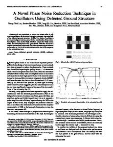
Fig. 4. One week postoperative picture showed a good recovery of minimally invasive skin incision.
AC C
EP
TE D
M AN U
SC
RI PT
Fig. 5. A 52-year-old male sustained a closed tibial fracture (OTA/AO classification, 42C) from a road traffic crash. He underwent minimal invasively percutaneous plate fixation using the distraction support seven days after injury. Four weeks postoperative radiographs showed a good reduction and internal fixation of the fracture (A); postoperative 3 months bridging callus formed at proximal and distal fracture sites (B); radiograph taken 12 months after surgery showed satisfactory union and alignment (C).
16
AC C
EP
TE D
M AN U
SC
RI PT
ACCEPTED MANUSCRIPT
AC C
EP
TE D
M AN U
SC
RI PT
ACCEPTED MANUSCRIPT
AC C
EP
TE D
M AN U
SC
RI PT
ACCEPTED MANUSCRIPT
AC C
EP
TE D
M AN U
SC
RI PT
ACCEPTED MANUSCRIPT