IOP PUBLISHING
BIOMEDICAL MATERIALS
doi:10.1088/1748-6041/5/1/015006
Biomed. Mater. 5 (2010) 015006 (6pp)
Influence of enzymatic maceration on the microstructure and microhardness of compact bone Ling Yin1,3 , Sudharshan Venkatesan2 , Shankar Kalyanasundaram2 and Qing-Hua Qin2 1 2
School of Engineering & Physical Sciences, James Cook University, Townsville, QLD 4811, Australia Department of Engineering, Australian National University, Canberra, ACT 0200, Australia
E-mail:
[email protected]
Received 15 June 2009 Accepted for publication 8 December 2009 Published 13 January 2010 Online at stacks.iop.org/BMM/5/015006 Abstract The cleaning of fresh bones to remove their soft tissues while maintaining their structural integrity is a basic and essential part of bone studies. The primary issue is how the cleaning process influences bone microstructures and mechanical properties. We cleaned fresh lamb femurs using enzymatic maceration in comparison with water maceration at room temperature. The microstructures of these compact bones were examined using scanning electron microscopy (SEM) and their porosities were quantified using image processing software. The bone microhardness was measured using a Vickers indentation tester for studying the mechanical properties. The results show that enzymatic maceration of compact bone resulted in a significant microhardness reduction in comparison with water maceration. However, enzymatic maceration did not cause any significant change of porosity in bone structures.
Bone is a specialized form of connective tissue consisting of regenerative cells and a mineralized extracellular matrix of nanosized mineral crystals [9–12]. The mineral crystals primarily in the form of carbonated calcium hydroxyapatite and collagen fibrils are co-continuous in bone microstructure [11–14]. Bone is porous. The porosity of bone plays a significant role not only in bone microstructure but also in bone biomechanical behavior [15]. The microstructure of bone is classified into two categories based on porosity: compact bone with compact mineralized connective tissue and low porosity and trabecular bone with less compact mineralized connective tissue and high porosity. Furthermore, bone consists of woven and lamellar layers. Woven bone is weak, with a small number of randomly oriented collagen fibers, but forms quickly without a pre-existing structure during periods of repair or growth. Lamellar bone is stronger; it consists of numerous stacked layers and is filled with many collagen fibers parallel to other fibers of the same layer which assists in a bone’s ability to resist torsional forces [9]. Hardness is the ability of a material to resist a permanent indentation, and hardness testing is widely used to determine
1. Introduction Cleaning of bones for the purpose of osteological examination is a common practice in zoology, anthropology, forensic medicine, pathology [1] and biomaterials science. The ability to remove soft tissues from skeletons without compromising bone surface morphology or bone integrity is often paramount. Common methods for cleaning of bones are manual cleaning, enzymatic maceration, cooking, water maceration and insect consumption [1]. Enzymatic maceration that employs digestive enzymes such as trypsin, pepsin or papain [2–5] is considered the most convenient method. It is known that sample preparation techniques affect the microstructure and mechanical properties of human tooth hard tissue [6, 7], a substance similar to bone but more highly mineralized [8]. This is particularly critical when the bone samples are subject to testing at the micro- and nano-scales. However, the influence of enzymatic maceration on bone microstructures and mechanical properties has not been investigated in previous studies and is the main focus of this work. 3
Author to whom any correspondence should be addressed.
1748-6041/10/015006+06$30.00
1
© 2010 IOP Publishing Ltd Printed in the UK
Biomed. Mater. 5 (2010) 015006
L Yin et al
the mechanical properties of materials. Microindentation testing provides an alternative to conventional flexural and tensile methods [16]. It is especially useful when the specimen size is small. In particular, microindentation can produce microscopic cracks of sizes comparable with the microstructural features of the material [16–18]. Bone microhardness has been correlated with important features such as mineralization and stiffness [15, 16]. It provides a means of examining the mechanical behavior of bone at the micron scale and averaging the effect of osteonal lamellae, while being sensitive to variation in mineral content within bone. With careful selection of the microindentation site, it is possible to obtain material characteristics separate from any effects of porosity [17, 18]. The aim of this investigation was to study the influence of enzymatic maceration on bone microstructures and mechanical properties. The serine protease trypsin was selected because it is one of the easily available enzymes extracted from porcine pancreas. Fresh lamb femurs from mass-industrially raised lambs were used as bone samples to establish a repeatable biomechanical testing regime. Lamb bones are commonly used as samples for bone studies not only because they are easily available but also because they exhibit mechanical properties similar to human bones [19]. The microstructure of the cleaned bones was examined using scanning electron microscopy and their porosities were quantified using image processing software. The microhardness was measured using a Vickers indentation tester to provide an indication of the mechanical properties of the bone material.
each femur for mounting on a slide. The slab was labeled on one side and the other side was lapped flat and smooth first on a cast iron lap with 400-grit carborundum, and then finished on a glass plate with 600-grit carborondum. After drying on a hot plate, a glass slide was glued to the lapped face of the slab with epoxy. Using a thin section saw, the slab was cut off close to the slide. The thickness was further reduced on a thin section grinder. A finished thickness of 30 μm was achieved by lapping the section by hand on a glass plate with 600-grit carborundum and followed by fine grinding with 1000 grit. Finally, the section was placed in a holder and spun on a polishing machine using nylon cloth and diamond paste until a suitable surface finish was achieved for the microscopic study. 2.3. Microstructural analysis and porosity measurement Two thin sections obtained from the macerations in water and enzymatic solution were carbon-coated and were observed under a scanning electron microscope (Cambridge 360, Cambridge, UK). Back-scattered images were taken under high vacuum at 20 kV. The regions of interest on the SEM images were transferred into the commercial image processing software (Analysis, Soft Imaging System) in order to perform pore analysis. Each pore area was labeled numerically. The mean pore area sizes were calculated on a representative region with an area of 403 × 265 μm2 . Porosity is a measure of the pore spaces in a solid material and is measured as a fraction, between 0 and 1, or as a percentage between 0 and 100%. In materials science, it is well known that measurement of the volume fraction of pores can be obtained from a random two-dimensional cross-section in a number of ways. This involves either the measurement of area fractions, of linear intercepts or of points. These ratios are equal to the volume fraction [20]. In the current investigation, the porosity in bone expressed in percent was defined by the ratio between the total areas of the pores and the total area of the representative region [21]. The porosity values of the volume fraction of pore areas are equal to the volume fraction [20]. On each thin section, three random locations were measured to obtain the mean value and the standard deviation.
2. Experimental procedure 2.1. Removal of soft tissues Eight fresh lamb femurs from 6-month-old lambs were stored in a refrigerator at –20 ◦ C before all joints were cut off using a diamond saw machine. Four of the femurs were macerated in an enzymatic solution with a pH value of 7.8, containing 20 g trypsin (Sigma Aldrich, Australia) and 2 l of water. The other four femurs were macerated in 2 l of water. The macerations took place for 5 days at room temperature in a fume cupboard. After 5 days, soft tissues were manually removed using a rod, a cooking knife and a brush. Care was taken to avoid scraping, scratching or cutting of the bone surfaces. The cleaned bone samples were stored at room temperature in an isotonic phosphate buffered saline (PBS) solution with sodium azide as preservative. This solution, containing the identical mineral content to mammalian cells, was prepared by dissolving 800 g NaCl, 20 g KCl, 144 g Na2 HPO4, 24 g KH2 PO4 and 0.2% NaN3 in 8 l of distilled water, and topping up to 10 l. The pH of the solution was approximately 6.8.
2.4. Microhardness indentation testing Indentation samples were obtained using a standard material preparation protocol. Transverse sections with 10 mm thickness were obtained from the central femurs macerated in water and enzymatic solution using a diamond saw machine at a low rotary speed. Transversely cut specimens were polished using silicon carbide papers with grit sizes of 240, 320, 400, 600, 800 and 1200. Fine polishing was performed using diamond suspension slurries with grades of 6, 3, 1 and 0.25 μm on polishing cloth. The specimens were cleaned using water after different stages of polishing before proceeding to the next finer level of polishing. After final polishing, bone samples were stored in the PBS solution at room temperature. The polished bone surfaces were indented with a Vickers diamond indenter in a standard microhardness tester (MHT1, Matsuzawa Seiki, Japan). Five indentation loads of 0.245,
2.2. Preparation of thin sections Each of the femurs macerated in water and enzymatic solution was processed for microstructural analysis following a standard protocol for thin section preparation. A suitable size slab was cut with a diamond saw from the center part of 2
Biomed. Mater. 5 (2010) 015006
L Yin et al
0.49, 1.96, 4.9 and 9.8 N were applied during a loading time of 10 s. Six indentations were made at each load on each sample. This resulted in a total of 30 indentations in each sample. A distance of at least twice the impression diagonal was maintained between the indentations to minimize interactions between neighboring indentations. The indentations were completed within 45 min from the time each bone sample was taken out of the PBS solution. The indentation diagonals were measured with optical microscopy. Three samples from the same maceration were selected for repeat tests. 2.5. Statistical analysis Analysis of variance (ANOVA) at a 5% significant level was applied for statistical analysis of porosity and hardness values using Excel.
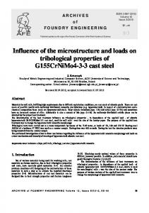
(a)
3. Results 3.1. Removal of bone soft tissues All the bone samples showed appreciable changes after 5 days maceration in water and enzymatic solution at room temperature. All the marrows were easily removed using a wooden rod and a brush. The soft tissues of the bone samples macerated in water were relatively difficult to remove. Cleaning was conducted carefully by scratching the soft tissues using a dull knife. The soft tissues of the bone samples in enzymatic solution became completely separated and could easily be removed with a scouring pad without any damage to the bone surfaces.
(b)
Figure 1. Scanning electron micrographs of the microstructures of the bones macerated in (a) water and (b) enzymatic solution.
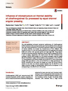
3.2. Microstructure The SEM micrographs demonstrating the microstructures of the bone samples prepared in water and enzymatic solution are shown in figure 1. There are no visible differences among these microstructures. All microstructures show the typical osteonal structure of compact (cortical) bone. Figure 2 shows typical osteons, which were formed rather like plywood, from sheets of alternating lamellae that can be laid flat, or curved around in circles to protect blood vessels. The osteons are elliptical. One has a major axis of length of approximately 100 μm and a minor axis of length of approximately 50 μm. Larger pores with diameters larger than 10 μm were formed from Haversian canals. Smaller pores were formed from osteocytic lacunae and lacuna canaliculi. Details of woven bone and interstitial lamellae are also observed in figure 2. Figure 3(a) demonstrates one example of identification, analysis and calculation of the porosity in a region of interest. Figure 3(b) shows the pore size distribution in the region, indicating that 4% pores had diameters of 10–20 μm and 96% of the pores had diameters smaller than 10 μm. It is interesting to observe the small bump at around 5 μm in figure 3(b), which is probably due to the osteocytes. The porosities measured for the bones prepared under different macerations are plotted in figure 4. Each datum is the average with one standard deviation of three measurements at random locations of the bone sample. The mean porosities
of bones macerated in water and enzymatic solution were approximately 5%. There is no significant difference between the porosities of bones macerated in enzyme solution and water (ANOVA, p > 0.05). 3.3. Microhardness Figure 5 demonstrates the Vickers hardness against the applied load for the bones macerated in water and enzymatic solution. The dots represent the measurement means and the error bars represent the 95% confidence intervals from 18 indentation measurements in three samples. The hardness values were found to increase with the applied load. They are significantly different for the bones macerated in water and enzymatic solution (ANOVA, p < 0.05). The bone tissue macerated in water has higher hardness than that in enzyme solution.
4. Discussion Soft tissues of bone mainly contain organic protein, fat and collagen [17]. Bone marrow is the tissue comprising the center of the medullary cavity and contains a rich vascular network and cells that may produce blood cells, i.e. red bone marrow, 3
Biomed. Mater. 5 (2010) 015006
L Yin et al Pore formed from lacuna canaliculi
Osteons
Pore formed from Haversian canal Concentric lamellae
Pores formed from osteocytic lacunae Woven structure Interstitial Lamellae
Figure 2. High-magnification SEM micrograph of the microstructure of the compact bone macerated in water.
(a) 70
(b)
60 Pore Count
50 40 30 20 10 0
5
10
15
20
Pore Diamater (µm)
Figure 3. (a) Porosity identification and (b) distribution of pore diameters. (This figure is in colour only in the electronic version)
4
Biomed. Mater. 5 (2010) 015006
L Yin et al
canaliculi) [26]. Figure 2 demonstrates that pores in bone were mainly formed from Haversian canals of larger diameters, and osteocytic lacunae and canaliculi of much smaller diameters. Figure 3 shows that the pores formed from Haversian canals with diameters of 10 μm or larger constituted less than 5% of pores. 95% of pores were due to osteocytic lacunae and canaliculi, the tiny channels which run through the laminae that comprise the osteon. The primary mineral material in bone is in the form of crystals of nano-scale inorganic carbonated hydroxyapatite, a hard, brittle mineral material based on calcium phosphate [27–29]. Given that enzymatic solution and water applied in this study had pH values of 7.8 and 7, respectively, it is unlikely that maceration with these agents could have any chemical reactions with the mineral structure of bone. On the other hand, collagens, constituting the major structural proteins in the extracellular matrix, provide mechanical strength and structural integrity to the various connective tissues in bone [10, 11, 30]. Trypsin could have weakened the collagens in bone structure. This explains why maceration in water and enzyme solution had a significant effect on the yielded hardness values shown in figure 5. The applied load also influenced the hardness as shown in figure 5, in which hardness values increased with the applied load. For engineering materials such as ceramics or metals, the hardness–load curve is often referred to as the indentation size effect, which follows Meyer’s law [31]. Hardness versus load is either constant or decreases with load or the hardness has an abrupt transition to a constant value [31]. For bone, however, hardness versus load shows an increase with load (figure 5). Unlike single crystals, polycrystals or amorphous structures in engineering materials, bone is a composite of a fibrous polymer (organic collagen fibers) matrix reinforced by ceramic nanoparticles (inorganic carbonated hydroxyapatite) [9]. The collagen fiber matrix exhibits high toughness and elasticity, while the crystallized hydroxyapatite is very brittle. In indentation, bone materials exhibit viscoelastic behavior [32].
7
Porosity (%)
6
5
4
3 Water
Trypsin
Solution
Figure 4. Porosity of bone macerated in each solution. Each point is the average value from three repeated measurements and the error bar is ±1 standard deviation of the repeated measurements.
Vickers Hardness (GPa)
0.45
0.40
0.35
0.30
Water Trypsin
0
2
4 6 8 Indentation Force (N)
10
12
Figure 5. Vickers hardness for the bones macerated in water and enzymatic solution. Each dot represents the average measurement value obtained from 18 repeated indentation measurements in three samples and the error bar is the 95% confidence interval of the 18 repeated indentation measurements in three samples.
or be transformed into fat, i.e. yellow bone marrow. All these soft tissues are essentially macromolecular substrates [22]. Water maceration at room temperature is traditionally considered the safest method for bone cleaning. This is because no heat or chemicals are applied that may disrupt bone integrity. However, the process of protein degradation to soften the soft tissue is notoriously malodorous. Enzymes, on the other hand, digest a macromolecular substrate and exemplify an important category of hydrolytic reactions [23]. Trypsin is a proteolytic enzyme that can attack the peptide molecule, whereas the exopeptidases hydrolyze the terminal peptide bonds [24]. Compact bone is essentially a solid material, in which spaces for blood vessels and living cells give it a porosity of about 5%. It makes up the majority of long bones [9]. There are three major anatomic cavities: Haversian and Volkmann’s canals, osteocytic lacunae and canaliculi [25]. It has been reported that Haversian/Volkmann’s canals are the major contributions to the total porosity, with much less porosity contributed by the other cavities (lacunae and
5. Conclusions We cleaned fresh lamb femurs using enzymatic maceration in comparison with water at room temperature. The microstructures of the bone samples were examined using SEM. The Vickers hardness was tested using a microhardness tester for mechanical properties. It is found that there were no significant differences between the microstructures in terms of the porosity of bones macerated in water and enzymatic solution. However, maceration in enzymatic solution significantly influenced the yielded bone hardness. This is particularly valuable for micro- and nano-scale mechanical testing of bone tissues, in which caution should be taken to avoid the weakening of bone tissues in sample preparation. Further, applied loads also affected bone hardness. The results indicate that bone mechanical properties depend on sample preparation and applied loads. 5
Biomed. Mater. 5 (2010) 015006
L Yin et al
Acknowledgments This work was supported by the Australian Research Council Discovery Project grant no DP0665941. The authors would like to thank Drs Anthony Flynn, Zbigniew Stachurski and Anthony Jones of the Australian National University (ANU) for valuable discussions. They are grateful to Drs Frank Brink, Cheng Huang and Mr Geoffrey Hunter of the ANU Electron Microscopy Unit for experimental assistance. Mr Fabian Ehrich of Swiss Federal Institute of Technology, Switzerland, is also acknowledged for assistance in sample preparation.
[13]
References
[17]
[14] [15] [16]
[1] Mairs S, Swift B and Rutty G N 2004 Detergent—an alternative approach to traditional bone cleaning methods for forensic practice Am. J. Forensic Med. Pathol. 25 276–84 [2] Belfie D and Clark J 1992 Enzymatic preparation of allograft bone Clin. Res. 40 134A [3] Mooney M P, Kraus E M, Bardach J and Snodgass J I 1982 Skull preparation using the enzyme-active detergent technique Anatomical Record 202 125–9 [4] Rennick S L, Fenton T W and Foran D R 2005 The effects of skeletal preparation techniques on DNA from human and non-human bone J. Forensic Sci. 50 1–4 [5] Steadman D W, DiAntonio L L, Wilson J J, Sheridan K E and Tammariello S P 2006 The effects of chemical and heat maceration techniques on the recovery of nuclear and mitochondrial DNA from bone J. Forensic Sci. 51 11–7 [6] Ho S P, Goodis H, Balooch M, Nonomura G, Marshall S J and Marshall G 2004 The effect of sample preparation technique on determination of structure and nanomechanical properties of human cementum hard tissue Biomaterials 25 4847–57 [7] Guidoni G, Denkmayr J, Sch¨oberl T and J¨ager I 2006 Nanoindentation in teeth: influence of experimental conditions on local mechanical properties Phil. Mag. 86 5705–14 [8] Craig R G 1993 Restorative Dental Materials (St Louis: Mosby) [9] Currey J D 2002 Bones: Structure and Mechanics 2nd edn (Princeton, NJ: Princeton University Press) [10] Fratzl P, Gupta H S, Paschalis E P and Roschger P 2004 Structural and mechanical quality of the collagen–mineral nano-composite in bone J. Mater. Chem. 14 2115–23 [11] Gupta H S, Seto J, Wagermaier W, Zaslansky P, Boesecke P and Fratzl P 2006 Cooperative deformation of mineral and collagen in bone at the nanoscale Proc. Natl Acad. Sci. 21 17741–6 [12] Gupta H S, Wagermaier W, Zickler G A, Aroush D R B, Funari S S, Roschger P, Wagner H D and Fratzl P 2005
[18]
[19]
[20] [21] [22] [23] [24] [25] [26] [27] [28]
[29] [30] [31] [32]
6
Nanoscale deformation mechanisms in bone Nano Lett 5 2108–11 Mackie I G, Green M, Clarke H and Isaac D H 1989 Human bone microstructure studied by collagenase etching J. Bone Jt. Surg. 71-B 509–13 Mackie I G, Green M, Clarke H and Isaac D H 1989 Osteoporotic bone microstructure by collagenase etching Ann. Rheum. Dis. 48 464–9 Taylor D, Hazenberg J G and Lee T C 2007 Living with cracks: damage and repair in human bone Nat. Mater 6 263–8 Gupta H S, Schratter S, Tesch W, Roschger P, Berzlanovich A, Schoeberl T, Klaushofer K and Fratzl P 2005 Two different correlations between nanoindentation modulus and mineral content in the bone–cartilage interface J. Struct. Biol. 149 138–48 Mahoney E, Holt A, Swain M and Kilpatrick N 2000 The hardness and modulus of elasticity of primary molar teeth: an ultra-micro-indentation study J. Dent. 28 589–94 Xu H H K, Smith D T, Jahanmir S, Romberg E, Kelly J R, Thompson V P and Rekow E D 1998 Indentation damage and mechanical properties of human enamel and dentin J. Dent. Res. 77 472–80 Aerssens J, Boonen S, Lowet G and Dequeker J 1998 Interspecies differences in bone composition, density, and quality: potential implications for in vivo bone Endocrinology 139 663–70 Holliday L 1966 Composite Materials (London: Elsevier) Wang X and Ni Q 2003 Determination of cortical bone porosity and pore size distribution using a low field pulsed NMR approach J. Orthop. Res. 21 312–9 Vigue-Martin J 2004 Atlas of the Human Body (Lisse: Rebo) Stryer L 1988 Biochemistry 3rd edn (New York: Freeman) Beck I T 1973 The role of pancreatic enzymes in digestion Am. J. Clin. Nutr. 26 311–25 Tortora G J 2002 Principles of Human Anatomy (New York: Wiley) Martin R B 1984 Porosity and specific surface of bone Crit. Rev. Biomed. Eng. 3 179–222 Peterlik H, Roschger P, Klaushofer K and Fratzl P 2006 From brittle to ductile fracture of bone Nat. Mater 5 52–5 Hassenkam T, Fantner G E, Cutroni J A, Weaver J C, Morse D E and Hansma P K 2004 High-resolution AFM imaging of intact and fracture trabecular bone Bone 35 4–11 Gao H, Ji B, J¨ager I L, Arzt E and Fratzl P 2003 Materials become insensitive to flaws at nanoscale: lessons from nature Proc. Natl Acad. Sci. 13 5597–600 Liebschner M A K 2004 Biomechanical consideration of animal models used in tissue engineering of bone Biomaterials 25 1697–714 Quinn J B and Quinn G D 1997 Indentation of brittle ceramics: a fresh approach J. Mater. Sci. 32 4331–46 Everitt N M, Rajah S and McNally D S 2006 Bone recovery following indentation J. Bone Jt. Surg. (Br.) B 88 398