BIOLOGY OF REPRODUCTION 54, 1317-1325 (1996)
Inhibitory Effect of Peritoneal Macrophages on Progesterone Release from Cocultured Rat Granulosa Cells Is Reversed by Dexamethasone: Evidence for an Action Independent of Nitric Oxide and Distal to Cyclic Adenosine 3',5'-Monophosphate Generation' Saffron Whitehead
2
and Michael Lacey
Departmentof Physiology, St George's HospitalMedical School, London, SWI 7 ORE, United Kingdom ABSTRACT We investigated the effects of cocultured macrophages on progesterone release from granulosa cells obtained at different stages of the rat estrous cycle and determined whether the immunosuppressive actions of the synthetic glucocorticoid can modulate the effects of cocultured macrophages. Basal and hCG-stimulated progesterone release from granulosa cell-only cultures varied according to the stage of the estrous cycle at which granulosa cells were obtained, but macrophages exerted profound inhibitory effects on both basal and hCGstimulated steroidogenesis at all stages. These inhibitory effects were partially reversed by the addition of dexamethasone (5x 10- 7to 5 x 10⁻⁵ M)to the culture medium. Measurement of nitrite accumulation inthe media, as an index of nitric oxide production, showed that granulosa cells produce very low levels of this metabolite compared with macrophages, although granulosa/luteal cells from freshly ruptured follicles of estrous rats produced significantly higher concentrations of nitrite compared with granulosa cells obtained from diestrous and proestrous rats. Dexamethasone had only limited effects in suppressing the accumulation of nitrite, showing that the macrophages were synthesizing nitric oxide synthase and suggesting that the action of dexamethasone was independent of nitric oxide. Macrophages also inhibited the steroidogenic response of granulosa cells to forskolin (5x 10⁻⁶ Mto 10 4M)and dibutyryl cAMP (1 mM and 0.5 mM) and the potentiation of the response by the addition of hCG. However, this inhibition was reversed by the presence of 5 x 10⁻⁶ M dexamethasone, and responses were comparable with those measured from granulosa cell-only cultures. Overall, the results suggest that macrophage inhibition of progesterone synthesis acts at a site distal to the generation of cAMP and that dexamethasone reverses this inhibition. INTRODUCTION Numerous experiments have shown that the two major cytokine secretions of macrophages, namely interleukin (IL)-1j3 and tumor necrosis factor a (TNFa), can affect steroidogenesis in both thecal and granulosa cells. However the data have not been consistent, with stimulatory, inhibitory, or no effects having been reported [1-4]. Norman and Brannstrom [5] suggest that the modulatory action of these cytokines varies according to the stage of the ovarian cycle, and thus the hormonal environment to which follicular cells are exposed before experimentation may affect their response to cytokines. In this context, it is worth noting that the majority of experiments concerned with immune modulation of follicular steroidogenesis have been carried out either on cells obtained from immature rats treated with estrogen antagonists/gonadotropins to induce follicular development or on human granulosa/luteal cells obtained from in vitro fertilization programs. Since there are resident macrophages in the ovary [61 and an increasing infiltration of these leukocytes into the ovary as ovulation approaches [71, we previously investigated the effects of cocultured macrophages on steroidogenesis in granulosa cells obtained from untreated proestrous rats. The Accepted January 30, 1996. Received July 11. 1995. 'This work was supported by The Wellcome Trust. 2 Correspondence: Dr. S.A. Whitehead, Department of Physiology, St. George's Hospital Medical School, Cranmer Terrace, London, SW 17 ORE, UK. FAX: 44 181 725 2993; e-mail:
[email protected]
macrophages exerted powerful inhibitory effects on progesterone secretion. This was dose-dependent on the number of cocultured macrophages and was not associated with any reduction in granulosa cell viability or morphological changes during the 48-h culture period [4]. While macrophages do release high concentrations of both IL-13 and TNFa [8], no effects of recombinant cytokines could be observed in our system, suggesting that macrophages were acting via another pathway. One possibility is nitric oxide (NO), which is produced by activated macrophages through the catalytic action of the inducible isoform of nitric oxide synthase (iNOS) on L-arginine [9]. In pancreatic islet cells, IL-10-induced NO production has been shown to have cytotoxic effects and to suppress insulin synthesis [10]; while in whole ovarian dispersates, the cytotoxic effects of IL-1p, thought to reflect tissue remodeling events associated with ovulation, have also been shown to be mediated through NO production [11]. However, both of these findings have been challenged in more recent reports [12, 13] that demonstrate that the cytotoxic actions of IL-10 are independent of NO production. Finally, it has been shown that endothelial NOS is present in human granulosa-luteal cells and that NO donors inhibit estradiol and progesterone release while NO synthase inhibitors increase estradiol release [14]. The synthesis of iNOS can be inhibited by glucocorticoids (e.g., dexamethasone and hydrocortisone) [15], which is of particular interest with regard to the evidence for a direct action of glucocorticoids at the ovarian level. Adashi 1317
1318
WHITEHEAD AND LACEY
et al. [16] showed that glucocorticoids could potentiate FSHinduced progesterone production in granulosa cells obtained from immature rats. In contrast, Michael et al. [17] reported that glucocorticoids inhibited progesterone secretion in human granulosa cells obtained from in vitro fertilization programs. Furthermore, the enzyme that inactivates glucocorticoids is present in granulosa cells, and the activity of the enzyme has been correlated with the success or failure of in vitro fertilization procedures [18]. While the physiological role of circulating glucocorticoids has not yet been established, it is known that excess secretion of cortisol is associated with several syndromes of ovarian dysfunction, and these steroids may also play some role in the infertility associated with stress [19]. Alternatively, their immunosuppressant properties may modulate immune/ovarian interactions, and such effects may vary according to the stage of the estrous/menstrual cycle. Thus ovarian macrophages, cytokines, nitric oxide, and the immunosuppressant glucocorticoids are all reported to have effects on ovarian function. Against this backdrop, experiments were undertaken to answer the following questions. 1) Does the action of cocultured macrophages on basal and gonadotropin-induced progesterone secretion from granulosa cells vary according to the stage of the rat estrous cycle at which the granulosa cells were obtained? 2) What are the effects of dexamethasone on granulosa cell-only cultures and cocultures? 3) Do the effects of macrophages involve NO production? 4) Do macrophages alter steroidogenesis by an action on hCG receptors or intracellular signal transduction mechanisms? MATERIALS AND METHODS Female Porton Wistar rats weighing 200-250 g were housed under controlled conditions of light (lights-on 0600-1800 h) and temperature (20°C) and had free access to food and water. Daily vaginal smears were taken, and only those rats exhibiting at least two consecutive regular 4-day estrous cycles were used for experimentation. Granulosa cells were obtained from rats between 0800 and 0900 h at diestrous Day 2, proestrus, or estrus.
pension through a 25-gauge needle and recovered by centrifugation. After two washes and further passages through a syringe needle, the cells were cultured in McCoy's 5A culture medium containing 25 mM HEPES, 2 mM glutamate, 0.1% BSA (all from Sigma, Poole, UK), 100 U/ml penicillin, and 100 1pg/ml streptomycin sulphate (Life Sciences, Paisley, Scotland). They were plated out in 1-ml aliquots at a concentration of 5 X 10⁵ cells/ml. Macrophages were obtained by injecting 10 ml sterilized saline into the peritoneal cavity of rats at different stages of the estrous cycle and, after gentle agitation of the abdomen, exposing the abdominal cavity and recovering the fluid. After centrifugation, the macrophages were washed twice in culture medium (as described above). Viewed (unstained) under the light microscope, the cell suspension contained large cells with serrated-type cell membranes and smaller cells with a smooth outer membrane. Only the larger cells were counted and plated at a concentration of 105 cells/ml in culture medium containing 5% fetal calf serum (Gibco). During a 2-h incubation, the macrophages adhere to the base of the culture well, and after this period nonadherent leukocytes can be washed off. This technique for separating macrophages of > 95% purity has previously been validated [4]. After 2 h and two washes, 1 ml granulosa cell suspension in serum-free medium was added to the wells, or macrophages were cultured on their own, again in the same serum-free medium as used for the granulosa cells. All cells were cultured for a period of 48 h with or without the addition of the following drugs: 5 IU hCG, dexamethasone, dibutyryl adenosine 3',5' cAMP (dbcAMP), and/or forskolin (all from Sigma). Drugs were added in 10-1l volumes at appropriate concentrations. Dexamethasone and forskolin were initially dissolved in ethanol, and stock solutions were subsequently diluted with culture medium. Controls were carried out to ensure that the maximum volume of the ethanol diluent (5 ytl) added to the cultures had no effect on progesterone release. Samples of media were taken at the end of the culture period for analysis of progesterone and nitrite release, and subsequently an assessment of cell viability was made. Assays of Cell Viability
Granulosa Cell Cultures and Cocultures The methods for granulosa cell cultures and cocultures have previously been described in detail [4]. Briefly, the ovaries from 4-5 animals were teased apart in ice-cold saline, and the follicles (> 300 lm) were dissected out. In the case of estrous rats, only the freshly ovulated follicles were dissected; these could easily be distinguished by their fragile, semi-translucent appearance and (usually) a surface network of fine blood vessels, not observed in developing follicles. After rupture of the follicles in culture medium, granulosa cells were dispersed by passing the cellular sus-
Cell viability was measured either by the trypan blue dye exclusion test, as determined by microscopy, or quantitatively by the ability of the cells to convert 3-[4,5-dimethylthrazol-2-yl]-2,5-diphenyltetrazolium bromide (MTT; 0.5 mg/ml) into formazan during a 4-h incubation at 370 C. The resulting blue precipitate (an index of various hydrogenase enzymes) was dissolved by adding 20% SDS in 0.02 N HCI at a 1:4 (v:v) ratio to the cultures. Samples were read on an Anthos 2001 scanning microplate reader (Anthos Lab Tec Instruments, Salzberg, Austria) at a wave length of 570 nm and a reference wavelength of 630 nm.
1319
MACROPHAGES, STEROIDOGENESIS, AND DEXAMETHASONE
Nitrite and ProgesteroneAssays nr
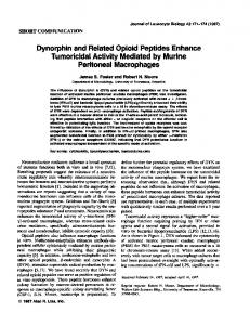
Production of NO was assayed by measuring (in triplicate) the accumulation of nitrite in the culture medium. Fifty microliters of conditioned medium was mixed with 50 tilof Griess reagent, which was prepared fresh by mixing equal volumes of stock A (10% sulfanilamide, 40% phosphoric acid) and stock B (1% N-[1-naphthyl] ethylenediamine dihydrochloride). After a 10-min incubation at room temperature, the absorbency was read at 550 nm in a scanning microplate reader, and concentrations were determined against a standard curve with sodium nitrite used as a standard. Interassay variation was < 10% as determined by assaying an aliquot of pooled samples from macrophage cultures with each assay. Progesterone concentrations in the media were measured in duplicate by a direct RIA (Immunodiagnostic Systems Ltd., Tyne and Wear, UK) that has been validated for measuring steroids in both plasma and culture medium. The cross-reactivity of the progesterone antiserum with 17a-hydroxyprogesterone and pregnenolone was 2% and 0.3%, respectively, and inter- and intraassay variations were 7% and 3.5%, respectively. Possible cross-reactivity of dexamethasone in the RIA was tested by adding a range of concentrations of the steroid (5 x 10- 7 to 5 X 10 -3 M) to culture media and assaying as for the samples. No crossreactivity was observed even at the highest concentration. StatisticalAnalysis The data points represent the mean SEM of triplicate (occasionally duplicate) cultures from at least three independent experiments, and for each experiment controls (granulosa cells with or without hCG) were routinely run to ensure normal responsiveness for each batch of granulosa cells processed. When two groups of data were compared, statistical significance was determined with a Student's ttest; when three or more groups were compared, an analysis of variance followed by Gabriel's test was used. The latter test is suitable for groups of unequal size. RESULTS Basal and hCG-stimulated progesterone secretion from cultured granulosa cells was dependent on the stage of the estrous cycle at which the cells were obtained (Fig. 1). Granulosa cells obtained from diestrous rats showed a low level of basal progesterone release and were apparently insensitive to the action of hCG. Granulosa cells from proestrous rats similarly showed a low level of basal progesterone release but a marked increase (p < 0.001) in response to 5 IU hCG. In contrast, cells obtained from freshly ruptured follicles of estrous rats showed a high basal secretion of progesterone that could not be further stimulated by the addition of hCG. At all stages of the estrous cycle, cocultured macrophages inhibited both basal and hCG-stimu-
ou
=Con MhCG
60 E ro § 40 -
:*
0
o 20 0 nv
M e D2
I
.
_
P
G cells only
E
l
D2
P
//
E
Co-cultures
FIG. 1. Comparison of mean (+ SEM) progesterone release from granulosa cellonly cultures (Gcells only) and granulosa cells cocultured with peritoneal macrophages (Co-cultures). Granulosa cells were obtained from dissected follicles obtained at diestrous Day 2 (D2), proestrus (P), or estrus (E)and were cultured at a concentration of 3 x 10⁵ cells/ml for 48 hwith or without macrophages (105 cells/ ml) and with (hatched bars) or without (open bars) 5 IUhCG. For each observation, n = 8-12; *, p < 0.001 compared with corresponding basal value (Student's t-test). Con, control.
lated progesterone release, although there was a small but detectable release of progesterone from granulosa/luteal cells obtained from estrous rats (Fig. 1). The effects of dexamethasone (5 X 10 - 7 to 5 X 10 5 M) on basal and hCG-stimulated progesterone release from granulosa cells also showed variations according the estrous cycle, but the most marked effect of this synthetic glucocorticoid was seen in the cocultures. In granulosa cell-only cultures obtained from diestrous and estrous rats, there was no significant effect of dexamethasone on basal progesterone release, or indeed on progesterone release in the presence of hCG (Fig. 2, a and c). The latter would be expected since these cells were found to be insensitive to hCG. However, in granulosa cells obtained from proestrous rats, the two higher doses of dexamethasone significantly enhanced the progesterone response to hCG (Fig. 2b). With regard to the cocultures, dexamethasone partially reversed the inhibitory effects of macrophages at all stages of the estrous cycle. Progesterone release from granulosa cells obtained from diestrous rats was significantly increased by 5 X 10 - 6 and 5 x 10- 5 M dexamethasone (Fig. 2a), and the same was observed in granulosa/luteal cells from estrous rats, although in this case the lowest dose of 5 x 10- 7 M exerted significant effects (Fig. 2c). In granulosa cells from proestrous animals, dexamethasone suppressed the inhibitory action of macrophages not only on basal release but also on hCG-stimulated release (Fig. 2b). Figure 3 shows the concentrations of nitrites measured from macrophages, cocultures, and granulosa cells at dif-
1320
WHITEHEAD AND LACEY ,1 1a
. 1 -
o Con
DIOESTRUS
+ hCG
I 2 a)
10 -I
b I
G-Cells only 50 -
0 0 0 P 0 (L 0
E
60 -
I
Co-Cultures
;
L 40 -
5-
.
a Z
30 -
..
o5x10-7
0
5x10
-
Dexamethasone o Con
(b) 60-
5x10
z
- s
M]
20 -
10 -
PROESTRUS
* + hCG
04
*
only FIG. 3.
E
.
u
-
Macrophages
.
_
u
Co-Cultures
Granulosa cells
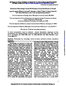
Mean concentrations (+ SEM) of nitrites measured in macrophages, mac-
rophages cocultured with granulosa cells, and granulosa cell-only cultures after 48h culture. Cells were obtained at different stages of the rat estrous cycle as indicated. In each group of results (n = 6-12 for each observation), statistically significant results are indicated by different letters; p < 0.01, analysis of variance followed by Gabriel's test. D2,diestrous Day 2; P, proestrus; E,estrus.
40
0 O 20o 0.
1
5- - ,1, x
,,
x
00
z Co-Cultures _
.j ..-
5x10-
510-
e G-Cells only -
5x10 5
Dexamethasone [M] o Control
(C) 120
OESTRUS
* + hCG
b b*~
100
-
E
b
b
G-Cells only
b
a
80
C
e
o
60
y _/===iCo-Cultures
/.
40 -
0o O. 20
-
,
'
,
/" 0
O
5x10
-7
5x10
-s
-
5x10
Dexamethasone IM] FIG. 2. Effects of increasing concentrations of dexamethasone on basal (open circles) and hCG-induced (closed circles) release of progesterone from granulosa cell-
only cultures (G-cells, solid lines) and granulosa cells cocultured with peritoneal macrophages (Co-Cultures, broken lines). Granulosa cells were obtained from rats at diestrous Day 2 (a), proestrus (b), or estrus (c); data show mean concentrations (i SEM) of progesterone in media after 48-h culture from 8-12 observations. Note different scales of y-axes in the panels. Different letters on each dose-response curve represent significant differences (p < 0.01) according to analysis of variance followed by Gabriel's test. Significant differences (*, p < 0.01) between basal and hCG-stimulated progesterone release in different groups were analyzed with Student's t-test. Con, control.
ferent stages of the estrous cycle. While the concentrations of nitrites were comparable in macrophage-only cultures and cocultures, levels of nitrites measured from granulosa cells were low. However, worthy of note is the fact that levels of nitrites measured in granulosa/luteal cells obtained from the ruptured follicles of estrous animals were significantly higher that those measured in granulosa cells obtained from proestrous and diestrous rats. In the presence of increasing concentrations of dexamethasone there was a small inhibition of nitrite formation both in macrophageonly cultures (Fig. 4a) and in cocultures (Fig. 4b). While some statistically significant differences were observed in these results, there was no clear dose-response relationship in either case. Determination of cell viability by measurement of cellular dehydrogenases showed that granulosa/luteal cells from newly formed corpora lutea had a significantly higher (p < 0.01) number of viable cells after 2-day culture (OI) 0.052 + 0.004, n = 12) compared with granulosa cells from proestrous follicles (OD 0.027 + 0.005, n = 13). The corresponding figures for cocultures were OD 0.201 + 0.048 (estrus) and 0.205 ± 0.06 (proestrus), while in macrophageonly cultures the OD was 0.128 + 0.014. Dexamethasone had no effect on cell viability (Fig. 5, a and b), nor did it have any effect on cell viability in cocultures, for which representative data obtained from estrous animals are shown in Figure 5c. Preliminary studies on granulosa cells obtained from proestrous animals showed that forskolin stimulated pro-
1321
MACROPHAGES, STEROIDOGENESIS, AND DEXAMETHASONE 0
(.1
-
O
PROESTRUS
D Macrophages P Macrophages E Macrophages
(al 0.06 -
+ hCG
b 50 -
C
b
H, 0.
0.04 -
0 C, CO
go I f-.
40-
T
10
*
I
0.02 -
6
6 I.
30o-
_
20 -
z
Con
=
_
_
-
I IY 1 1111
_ _
_
5x10
O
'7
Dexamethasone 10 -
=
Con
=
+ hCG
-
5x10 '
5x10O
M]
OESTRUS
(bi 0.06 -
0-
O
5x10 -
7
5x10 '-
5x10'-
Dexamethasone [M1
(b) 60
EJ
40
T
0.02 -
O
I
a o -
i
5x1o
i
-
-7
5x10o
5x10lo
Dexamethasone [M]
a : Con
a'
"
30-
(c) 0.25
4-
Z
T
T
o to 0 oI
o D Macrophages P Macrophages E Macrophages
50
T
T
'1
0.04-
20 -
0.20 *
E
OESTRUS - Co-cultures
+hCG
[
I
I
c
0 a -
10 -
M
0.15
0
0.1
00
5x10⁻⁷
5x10-
d
5x10⁻⁵
Dexamethasone [M] FIG. 4. Effect of increasing concentrations of dexamethasone on nitrite accumulation in media of macrophage-only (a) and macrophages cocultured with granulosa cells (b)over 48-h period. Cells for cultures were obtained at different stages of the rat estrous cycle as indicated. Data represent mean + SEM of 6-10 observations. For each dose response curve, significant differences (p < 0.005-0.01) are indicated by different letters (analysis of variance followed by Gabriel's test); comparisons between groups at each dose level were analyzed with Student's t-test: *, p < 0.001. D,diestrous Day 2; P,proestrus; E, estrus.
o 0.05
n u
-
_
5x10
_
7
_
1
5x10o
Dexamethasone
-
5x10
[M]
FIG. 5. Viable cell mass of granulosa cells (as assessed by ability of cultures to reduce MTT) after 48-h culture with increasing doses of dexamethasone, with (hatched bars) and without (open bars) 5 IU hCG. Data represent mean + SEM from duplicate/triplicate observations of 3 independent experiments with granulosa cells obtained from proestrous (a) or estrous (b,c) rats and cultured on their own (a,b) or with granulosa cells (c). *, p < 0.01 compared with corresponding value without hCG-Student's t-test. Con, control.
WHITEHEAD AND LACEY
1322
TABLE 1. Comparison of effects of dbcAMP, hCG, and dexamethasone (DEX) on progesterone release from granulosa cells obtained at proestrus and cultured 48 h with or without peritoneal macrophages.
PROESTRUS (a) 40 -
Progesterone concentrations (nmols/ ml)a
+ DEX
Dose added to cultures 30-
E
E C
+ DEX *
c o
calls only
20-
0
o
10'
0
Forskolin M) .
ID)
14
^
-
OESTRUS 4+DEX
120 -
0
E loo
.It-
E-100
C 0 c.
4) c 0
Cocultures 3.62 ± 1.08 0 2.98 + 0.72 36.19 2.04**
Granulosa cells 29.66 18.54 56.98 64.70
± ± ± ±
4.4 0.88 10.92* 7.18*
aData represent mean + SEM of 6-9 observations. *p < 0.01, compared with values measured in the presence of dbcAMP only (Student's t-test). **p < 0.001, compared with all other values of cocultures (Gabriel's test).
(
0.1
1 mM dbcAMP 0.5 mM dbcAMP 1 mM dbcAMP + 51U's hCG 1 mM dbcAMP + 5 x 10 6 M DEX
40 -
a-
b
0.
b
20-
aa n-
0
5x10
10-'
5x10- ' 10-
4
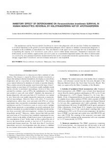
Forskolin (M) FIG. 6. Progesterone release from granulosa cell-only cultures (open circles) or cocultures (closed circles) cultured for 48 h in presence of increasing doses of forskolin. Granulosa cells were obtained from proestrous (a) or estrous (b) rats. Hatched bars show progesterone response when cells were cultured with forskolin (at dose indicated) and 5 x 10 6 M dexamethasone (DEX). Data represent mean + SEM from 8-10 observations. *, p < 0.001 compared to response observed in absence of dexamethasone; +, p < 0.05 compared with progesterone release in absence of forskolin (Student's t-tests).
gesterone secretion in a dose-dependent manner (Fig. 6a) and, at the highest dose investigated, progesterone release was comparable to that achieved with stimulation by 5 IU hCG (see Fig. 2b). In the presence of macrophages, the effects of forskolin were virtually abolished. However, when a submaximal dose of forskolin (10-5 M) plus 5 X 10 6 M
dexamethasone was added to the cocultures, there was a large progesterone response, far exceeding that measured when a similar dose of dexamethasone alone was added to 0.96 nmol/l prococultures (i.e., 21.3 + 2.12 vs. 4.98 gesterone, respectively). In the presence of 10 - ' M forskolin, the potentiating effect of dexamethasone was even greater (Fig. 6a). The addition of 10 5 M forskolin plus 5 IU hCG to the cocultures did not reverse the inhibitory effect of the macrophages, with the same minimal concentrations of progesterone being measured in the medium-0.86 0.50 nmol/l. This compares with granulosa cell-only cultures, in which the effects of hCG were additive with the response to 10 - 5 M forskolin. Thus, in the presence of 10 M forskolin only, mean progesterone concentrations in the media were 3.71 + 0.99 nmol/l compared with 16.7 ± 1.02 nmol/l when 5 IU hCG plus 10-' forskolin was added to the cultures. In granulosa/luteal cells obtained from estrous rats and cocultured with macrophages, there was a small but significant increase in progesterone release in response to increasing concentrations of forskolin (Fig. 6b); and at a dose of 10 5 M forskolin this response was enhanced by the presence of 5 IU hCG. Thus the mean progesterone concentrations were increased from 17.05 2.0 to 45.34 0.58 by the addition of hCG. However, with the same concentration of forskolin plus 5 x 10 6 M dexamethasone, progesterone release was increased to 121.35 6.07 nmol/l (Fig. 6b). In parallel, experiments were undertaken to assess the effects of dbcAMP on progesterone release from granulosa cells obtained from proestrous animals. The same picture emerged as that seen with forskolin. Granulosa cells cultured on their own showed a dose-related increase in progesterone secretion with increasing doses of this stable analogue of cAMP, and the effect was potentiated by addition of hCG (Table 1). In contrast, only the highest dose of dbcAMP showed a weak stimulation of progesterone secretion in cocultures, and this could not be further stimulated by the addition of hCG (Table 1). However, in the presence of 5 X 10- 6 M dexamethasone, the response was restoredcl
MACROPHAGES, STEROIDOGENESIS, AND DEXAMETHASONE
to that observed in granulosa cell-only cultures incubated with 1 mM dbcAMP. DISCUSSION Granulosa cells obtained from untreated adult female rats at different stages of the estrous cycle showed a marked variation in both their basal and hCG-stimulated steroidogenic responses. Granulosa cells obtained from diestrous animals failed to respond to hCG during a 48-h culture period; this is consistent with the fact that the number of LH receptors increases progressively during the final stages of follicular maturation as does the steroidogenic response to LH [20]. In contrast, granulosa cells from proestrous animals showed a large and significant response to hCG, whereas the high steroidogenic capacity of granulosa-luteal cells from freshly ruptured follicles could not be further stimulated by the addition of hCG. The inhibitory action of macrophages on steroidogenesis showed no cyclic variation, with virtually complete inhibition of both basal and hCG-stimulated progesterone release observed in granulosa cells obtained at all stages of the cycle. However, it should be noted that the number of macrophages within the ovary does change according to the stage of the estrous cycle [7]; this variable was not addressed in the present experiments. The inhibitory effects of macrophages on steroidogenesis are consistent with studies reported by Sun et al. [21], who showed that cocultured macrophages inhibited testosterone secretion from cultured Leydig cells; but they contrast with findings in two previous reports [22, 23] that showed that macrophages stimulated progesterone release from both mouse and human granulosa-luteal cells. However, in these latter studies, granulosa cells had been obtained from patients or immature mice previously treated with either human menopausal gonadotropin or eCG, respectively, followed by hCG. Other differences that might explain the discrepancies with the present report are the periods of granulosa cell culture prior to the addition of macrophages, the use of serum-supplemented medium, a higher concentration of macrophages, species differences, and whether or not the macrophages were activated. In the studies cited, there was no indication as to whether or not the macrophages were activated. However, it is clear from the measurements of nitrite in the media reported here, coupled with our previous findings that peritoneal macrophages produce high levels of both IL-I P and TNFa [8], that macrophages obtained in our experiments are activated at the time of culture. While the inhibitory effects of peritoneal macrophages are both dramatic and consistent, there remains the problem of the biological relevance of these observations to both normal and abnormal ovarian physiology. We tried to isolate ovarian macrophages (by adherence, as described above) from whole ovarian dispersates or cells from freshly
1323
ruptured follicles, which contain the highest number of macrophages [7]. However, with use of this technique, the yield of macrophages is extremely low and also contaminated with granulosa/luteal cells, as evidenced by the ability of the cultures to produce low levels of progesterone. Perhaps the use of Dynabeads coated with the macrophage ED2 antibody, as has been described for the isolation of testicular macrophages [241, might provide a satisfactory yield of relatively pure ovarian macrophages. Thus, while the experimental model used in these experiments may not be ideal for extrapolating to the biological role of resident and infiltrating ovarian macrophages, it is worth noting that Sun et al. [21] have shown that testicular macrophages produce a similar inhibition of testosterone secretion compared with peritoneal macrophages. Furthermore, in vivo, the ovaries are in contact with peritoneal macrophages, and there is evidence that infertility associated with endometriosis, for example, is associated with a higher number of peritoneal macrophages and higher concentrations of IL-1 3 and TNFa in peritoneal fluid compated with control subjects without endometriosis [251. With regard to the ratio of macrophages to granulosa cells, immunohistochemical studies have shown that the ratio of macrophages to granulosa cells ranges from 0.008 to 0.002 depending on the stage of follicular maturation 26]. In these studies, a higher ratio was used (0.33), but in our previous experiments we showed that even when the ratio was reduced to 0.01 the peritoneal macrophages still produced a significant inhibitory effect on steroidogenesis [4]. Thus, it may be that when macrophages infiltrate the ovary during the periovulatory period, they inhibit progesterone synthesis from granulosa cells by inhibiting responses to gonadotropins and thus prevent premature luteinization of the follicles and ovulatory failure. There is also evidence that macrophages may be involved in luteolysis and the decline in progesterone secretion [5]. Dexamethasone had little effect on basal progesterone release from granulosa cells obtained at diestrus and proestrus, but there was a small dose-related increase in basal progesterone release from granulosa/luteal cells obtained from estrous animals. It may be that the dexamethasonestimulated increases observed in granulosa/luteal cells of freshly ruptured follicles reach statistical significance only because the relative increases are amplified by the already high basal secretory rates observed in these cells. In contrast, the very weak and statistically insignificant stimulatory effects of dexamethasone seen in granulosa cells from diestrous and proestrous rats reflect the low levels of basal progesterone release; thus any increment is proportionately smaller and does not reach statistical significance. In granulosa cells from proestrous animals, the response to hCG was markedly enhanced by the presence of the two higher doses of dexamethasone. In contrast to the small effects on granulosa cell-only cultures, dexamethasone partially re-
1324
WHITEHEAD AND LACEY
versed the inhibitory effects of macrophages on both basal and hCG-stimulated release. While it is recognized that the doses of dexamethasone were relatively high, these doses were chosen to span the range of concentrations used by both endocrinologists and immunologists. Adashi et al. [16] showed that maximal glucocorticoid stimulation of progesterone release in granulosa cells obtained from diethylstilbestrol-treated rats was observed between 10 7 and 10 -6 M, while in studies reported by Michael et al. [17] on human granulosa cells, doses of glucocorticoids up to 10- 4 M were used. They reported that the EDso for the inhibitory action of dexamethasone on pregnenolone release was in the range of 0.4-2.0 X 10- 7 M, although the dose for maximal inhibition was not provided in their report. In contrast, experiments concerned with the inhibitory effects of dexamethasone on macrophage activity typically use doses ranging from 10 - 6 to 10 - 4 M [27-29]. Thus the doses used in these experiments ranged from 5 x 10 - 7 to 5 x 10 - 5 after preliminary experiments had revealed that lower doses had no effect either in granulosa cell-only cultures or in cocultures. The present findings agree with those reported by Hsueh and Erickson [30] in that dexamethasone potentiated the gonadotropin-induced release of progesterone but had little effect on basal release. They are in contrast to the findings of Michael et al. [17], who reported an inhibitory action of dexamethasone on pregnenolone release from human granulosa-luteal cells. We have also found that dexamethasone potently inhibits progesterone release in human granulosa cells (unpublished observations), and thus these discrepancies between stimulatory and inhibitory effects of glucocorticoids in rat and human granulosa cells may lie in species differences or prior exposure to gonadotropins during in vitro fertilization treatment. The failure of dexamethasone to reduce nitrite accumulation to concentrations similar to those measured in granulosa cell-only cultures suggests that the effects of this synthetic steroid were independent of NO. While high levels of nitrite were measured in both macrophage-only and cocultures, with or without the presence of dexamethasone, only very low levels were measured in cultures of granulosa cells. However, a significantly higher level of nitrite was measured in granulosa/luteal cells obtained from freshly ruptured follicles of estrous rats compared with cells obtained at other stages of the cycle. This is consistent with the evidence that there is an increasing infiltration of macrophages into the follicle during the periovulatory period [7]. However, in this context, it should be noted that Voorhis et al. [14], using the polymerase chain reaction, demonstrated that mRNA for endothelial NO synthase is present in human granulosa /luteal cells and that such cells are capable of converting L-arginine to citrulline. A comparison of the effects of forskolin and dbcAMP on progesterone release in granulosa cell-only cultures and co-
cultures suggests that macrophages do not primarily inhibit the hCG-induced increase in cAMP that ultimately leads to an increased steroidogenesis [31]. What the results do suggest is that macrophages inhibit steroidogenesis at a site distal to the generation of cAMP and that this inhibition can be reversed by dexamethasone. Whether this is at the level of a cAMP-dependent kinase, a steroidogenic enzyme, or some genomic expression needs to be determined. Interestingly, recent studies [32] have also shown that the inhibitory effects of conditioned medium from testicular macrophages on testosterone release from cultured Leydig cells are exerted beyond cAMP production and act at a site prior to the formation of pregnenolone. In the same context, Adashi et al. [16] showed that dbcAMP potentiated the effects of dexamethasone on FSHinduced progesterone secretion, suggesting that glucocorticoids also act at a point distal to cAMP generation. More recently, Zachow et al. [33] reported that the inhibitory effects of TNFa on LH-stimulated androstenedione production by theca-interstitial cells of immature rats involved multiple sites in the protein kinase A pathway, including a site distal to the generation of cAMP. The current experiments do not allow an explanation as to why granulosa/luteal cells, from estrous rats, that are cocultured with macrophages still retain a limited ability to respond to forskolin compared with cocultures of granulosa cells obtained from proestrous animals. It may be that steroids or the LH surge up-regulates signal transduction pathways, leaving some spare capacity, or that other signal transduction pathways are activated. Furthermore, activation of LH receptors stimulates not only adenyl cyclase but also phospholipase C. Thus both the production of cAMP and activation of protein kinase A and the production of diacylglycerol and activation of protein kinase C are second messenger pathways for LH [31], and both are potential sites for steroid modulation. Overall, the results show that dexamethasone can partially reverse the inhibitory effects of cocultured activated macrophages by an action apparently independent of nitric oxide. Raising intracellular levels of cAMP with forskolin or dbcAMP failed to revoke this inhibitory effect of cocultured macrophages, but, in the presence of dexamethasone, these drugs were able to stimulate progesterone release similar to that observed in granulosa cell-only cultures. This provides evidence that the effects of dexamethasone on macrophage inhibition of steroidogenesis are exerted on the signal transduction pathway distal to the generation of cAMP. REFERENCES 1. Andreani CL, Payne DW, Packamn JN, Resnick CE, Hurwitz A. Adashi EY. Cytokine mediated regulation of ovarian function. J Biol Chem 1991; 266:6761-6766. 2. Yan Z, Hunter V, Week J, Hutchison S. Lyles R., Terranova P. Tumor necrosis factor-a alters steroidogenesis and stimulates proliferation of human ovarian granulosa cells in vitro. Fertil Steril 1993; 59:332-338.
MACROPHAGES, STEROIDOGENESIS, AND DEXAMETHASONE 3. Wang LJ, Brannstrom M, Robertson SA, Norman RJ.Tumor necrosis factor in the human ovary: presence in follicular fluid and effects on cell proliferation and prostaglandin production. Fertil Steril 1992; 58:934-940. 4. Shakil T, Whitehead SA. Inhibitory action of peritoneal macrophages on progesterone secretion from cocultured rat granulosa cells. Biol Reprod 1994; 50:1183-1189. 5. Norman RJ, Brannstr6m M. White cells in the ovary-incidental invaders or essential effectors? J Endocrinol 1994; 140:333-336. 6. Hume DA, Halpin D, Charlton H, Gordon S. The mononuclear phagocyte system of the mouse defined by immunohistochemical localization of antigen F4/80: macrophages of endocrine organs. Proc Natl Acad Sci USA 1984; 81:4147-4177. 7. Brannstrom M, Mayrhofer G, Robertson SA. The localization of leucocyte subsets in the rat ovary during the periovulatory period. Biol Reprod 1993; 48:277-286. 8. Shakil T, Whitehead SA, Wilson BMG. Granulosa cells enhance the release ofinterleukin1 (IL-I) and tumor necrosis factor-a (TNF-t) from co-cultured macrophages. J Endocrinol 1993; 137(suppl):176. 9. Hibbs JB, Taintor RR, Vavrin Z. Macrophage cytotoxicity: role for L-arginine deiminase and imino nitrogen oxidation to nitrite. Science 1987; 235:473-476. 10. Corbett JA, McDaniel nML. Reversibility of interleukin-l3-induced islet destruction and dysfunction by the inhibition of nitric oxide synthase. Biochem J 1994; 299:719-724. 11. Ellman C, Corbett JA, Misko TP, McDaniel M, Beckerman KP. Nitric oxide mediates interleukin-l-induced cellular cytotoxicity in the rat ovary. J Clin Invest 1993; 92:30533056. 12. Eizirik DL, Sandler S, Welsh N, Cetkovic-Cvrtje M, Nieman A, Geller DA, Pipeleers DG, Bendtsen K,Hellerstrom C. Cytokines suppress human islet function irrespective of their effect on nitric oxide generation. J Clin Invest 1994; 93:1968-1974. 13. Ben-Shlomo I, Adashi EY, Payne DW. The morphogenic/cytotoxic and prostaglandinstimulating activities of interleukin-li in the rat ovary are nitric oxide independent. J Clin Invest 1994; 94:1463-1469. 14. Van Voorhis BJ, Dunn MS, Snyder GD, Weiner CP Nitric oxide: an autocrine regulator of human granulosa-luteal cell steroidogenesis. Endocrinology 1994; 135:1799-1806. 15. Di Rosa M, Radomski M, Carnuccio R, Moncada S. Glucocorticoids inhibit the induction of nitric oxide synthase in macrophages. Biochem Biophys Res Commun 1990; 172:1246-1252. 16. Adashi EY, Jones PBC. Hsueh AJW. Synergistic effects of glucocorticoids on the stimulation of progesterone production by follicle-stimulating hormone in cultured rat granulosa cells. Endocrinology 1981; 109:1888-1894. 17. Michael AE, Pester LA,Curtis P, Shaw RW, Edwards CRW, Cooke BA. Direct inhibition of ovarian steroidogenesis by cortisol and the modulatory role of Ii-hydroxysteroid dehydrogenase. Clin Endocrinol 1993; 38:641-644. 18. Michael AE, Gregory L, Walker SM, Antoniw JW, Shaw RW, Edwards CRW, Cooke BA.
19. 20.
21.
22. 23.
24. 25. 26.
27.
28. 29.
30. 31. 32. 33.
1325
Ovarian 11 3-hydroxysteroid dehydrogenase: potential predictor of conception by invitro fertilization and embryo transfer. Lancet 1993; 342:711-712. Brann DW, Mahesh VB. Role of corticosteroids in female reproduction. FASEB J 1991; 5:2691-2698. Fortune JE, Hilbert JL. Oestradiol secretion by granulosa cells from rats with four- or five-day estrous cycles: the development of responses to follicle-stimulating hormone versus luteinizing hormone. Endocrinology 1986; 118:2395-2401. Sun XR, Hedger MP, Risbridger CP. The effect of testicular macrophages and interleukinI on testosterone production by purified adult rat Leydig cells cultured under in vitro maintenance conditions. Endocrinology 1993; 132:186-192. Kirsch TM, Friedman AC, Vogel RL, Flickinger GL. Macrophages in corpora lutea of mice: characterization and effects on steroid secretion. Biol Reprod 1981; 25:629-638. Halme J, Hammond MG, Syrop CH, Talbert LM.Peritoneal macrophages modulate human granulosa-luteal cell progesterone production. J Clin Endocrinol & Metab 1985; 61:912-916. Dirami G, Poulter LW, Cooke BA. Separation and characterization of Leydig cells and macrophages from rat testes. J Endocrinol 1993; 130:357-365. Ramey JW, Archer DE Peritoneal fluid: its relevance to the development of endometriosis. Fertil Steril 1993; 60:1-14. Fukumatsu Y, Katabuchi H, Naito M, Takeya M, Takahashi K, Okamura H. Effect of macrophages on proliferation of granulosa cells in the ovary in rats. J Reprod Fertil 1992; 96:241-249. Baydoun AR, Bogle RG, Pearson JD, Mann GE. Selective inhibition by dexamethasone of induction of NO synthase, but not of induction of L-arginine transport, in activated murine macrophageJ774 cells. BrJ Pharmacol 1993; 110:1401-1406. Sowa G, Przewlocki R. cAMP analogues and cholera toxin stimulate the accumulation of nitrite in rat peritoneal macrophage cultures. EurJ Pharmacol 1994; 266:125-129. Bogle RG, Whitley GStJ, Soo S-C, Johnstone AP, ValIance P. Effects of anti-fungal imidazoles on mRNA levels and enzyme activity of inducible synthase. BrJ Pharmacol 1994: 111:1257-1261. Hsueh AJW, Erickson GE Glucocorticoid inhibition of FSH-induced estrogen production in cultured rat granulosa cells. Steroids 1978; 32:639-648. Davis JS. Mechanisms of hormone action: luteinizing hormone receptors and secondmessenger pathways. Curr Opin Obstet Gynecol 1994; 6:254-261. Sun XR, Risbridger GP Site of macrophage inhibition of luteinizing hormone-stimulated testosterone production by purified Leydig cells. Biol Reprod 1994; 50:363-367. Zachow RJ, TashJS, Terranova PF Tumor necrosis factor-alpha attenuation of luteinizing hormone-stimulated androstenedione production by ovarian thecal-interstitial cells: inhibition at loci within the adenosine 3',5'-monophosphate-dependent signalling pathway. Endocrinology 1993; 133:2269-2276.