for example during the budding process. To exclude the .... supported by grant no. 2o2o8 of the Fonds voor Wetenschappelijk Geneeskundig Onderzoek.
J. gen. Virol. 0976), 34, 353-36I
353
Printed in Great Britain
Interaction of Mouse Peritoneal Macrophages with Different Arboviruses in vitro By G. VAN DER GROEN,* D. A. R. V A N D E N BERGHE]" AND S. R. P A T T Y N t * Institute for Tropical Medicine, B-2000 Antwerp, and University of Antwerp, B-2610 Wilrijk, Belgium (Accepted 2I September I976) SUMMARY
In vitro cultured mouse peritoneal macrophages are inefficient host cells for both alpha and flaviruses tested. Production of infectious virus ceased 6 to 24 h after infection, except for virulent and avirulent SF strains. This limited growth is unrelated to interferon production. No correlation was found between the LDs0 of different virus strains for mice and the virus yields of in vitro infected macrophages therefrom. When macrophages from SF immunized mice were inoculated in vitro with SF or EEE, a cytopathic effect occurred, while the multiplication of SF, but not of EEE, was decreased. Virus multiplication in proteose peptone induced macrophages was enhanced for SF strains only. These results are discussed in relation to the virulence of several arboviruses for mice.
INTRODUCTION
Macrophages are generally considered important barriers against the spread of infectious agents from the primary site of infection or inoculation to other susceptible cells or organ systems of the host. Conflicting results concerning the relation between the capacity of a virus to multiply in macrophages in vitro and the virulence of the virus for the host have been published (Eustatia et al. I972 ; Allison, I974). Apart from the probability that most viruses are taken up by macrophages, it is not yet possible to generalize about the influence of macrophages on the outcome of virus infections. This is particularly true in the field of arboviruses where most studies have been done with flaviviruses, little being known about the interaction of alphaviruses with macrophages. It appears that the importance of macrophage-virus interaction requires individual assessment with each virus. In the present study we report the in vitro interaction of different alphavkuses and some flaviviruses with mouse peritoneal macrophages.
METHODS
Cells and viruses. Primary chick embryo cells were grown in medium I99 (Wellcome) supplemented with o.o2 % (w/v) glutamine, o'o4 % (w/v) sodium pyruvate, 2 % (v/v) chick embryo extract and 4% inactivated new born calf serum (Flow laboratories). L 929 cells were grown in TC medium I99 supplemented with o.o2 % (w/v) glutamine, 5 % (v/v) chick embryo extract and 30 % inactivated calf serum. Vero cells were cultured in TC medium I99
354
G. VAN DER GROEN AND OTHERS
supplemented with 7 % inactivated calf serum. All media were buffered with 13 raM-sodium bicarbonate. The virulent V 13 strain and the avirulent A 7 strain of Semliki Forest virus (SFV and SFA respectively) supplied by C.J. Bradish (Microbiological Research Establishment, Porton, Salisbury) were used. Middelburg virus (MIB) was supplied by J. S. Porterfield (National Institute for Medical Research, Mill Hill, London). Western equine encephalomyelitis (WEE), Eastern equine encephalomyelitis (EEE) and West Nile (WN) viruses were obtained from the National Institutes of Health (Research Resources Branch). Yellow fever I7 D (YF I7 D) was the vaccine virus. Vesicular stomatitis virus (VSV) was obtained from A. Billiau, Rega Institute, B-Leuven, Belgium. Virus stocks of SFA, SFV, EEE, WEE and VSV were prepared in chick embryo tissue cultures. YF, W N and MIB virus were prepared from infected newborn mouse brain. Virus assays. Alphaviruses were assayed by the plaque method in chick embryo cell monolayers. Serial tenfold dilutions were made in phosphate buffered saline containing 1% (w/v) gelatine and 2.2 mu-tris. Samples (0"5 ml) of each dilution were inoculated into 50 mm diam. plastic Petri dishes (Nunc Denmark) containing 5 x io 7 cells which formed a monolayer within 24 h. After virus adsorption for I h at 37 °C, the infected monolayers were overlayed with minimum essential medium (Flow laboratories) supplemented with 0"0075 % (w/v) neutral red (0"9 %, w/v) agar and 0.o2 M-N-2-hydroxyethylpiperazine-N-2-ethane sulphonic acid (HEPES). Plaques were counted on the 2nd and 3rd day after infection. W N and YF were assayed in Vero cells by the 50 % end point method. Serial tenfold dilutions were made in medium described by Hronovsky, Plaisner & Benda (I975) and 0"25 ml samples of each dilution were inoculated into 4 wells of a microtissue culture plate, each well containing 1.5 x io 4 cells. Titres were calculated from the observed c.p.e, using the Reed & Muench (I938) method. Macrophage cultures. Macrophages were obtained from 6- to 7-week-old female mice strain OF 1 (Iffa-Credo, Lyon, France) by intraperitoneal (i.p.) washings of 2 ml TC medium I99, containing heparin 50 units/ml, penicillin 250 units/mt and streptomycin 250 #g/ml. The cells from Io to 20 mice were pooled, and tissue culture tubes were seeded with 2"3 x io 6 cells. After I h incubation at 37 °C the non-adherent cells were removed and the medium was changed to TC medium I99 supplemented with 3o % inactivated foetal calf serum. Non-adherent cells were counted ; they usually represented IO % of the total number of cells ; thus the tubes contained, in general, 2 x Io 6 adherent cells, more than 9o % of them having the morphology of macrophages. Proteose peptone stimulated macrophages (PPS) were induced by i.p. injection of o.I ml of a Io % (w/v) proteose peptone (Difco) solution, 72 h before preparation of macrophage cultures. SF immune macrophages were harvested I2 to 14 days after i.p. inoculation of 4-week-old mice with 8 × Io 3 p.f.u, of SFA virus. Inoculation and sampling of macrophage cultures. After 24 h in vitro, the macrophage cultures were drained, covered with 0.2 ml virus suspension in macrophage medium and incubated for 2 h at 37 °C. The supernatant fluids were pooled and the infected macrophages were rinsed four times with Hanks' solution, then covered with I ml macrophage culture medium. This was considered as time zero. Final rinsing fluids were pooled and stored. At different time intervals thereafter, supernatant fluids were harvested for virus titration (extracellular virus). The remaining cells were rinsed three times with Hanks' solution, frozen and thawed three times in borate saline, p H 9"o (Clarke & Casals, I958), and titrated
Arbovirus macrophage interaction
355
Table I. Thermal inactivation of different arboviruses in macrophage culture medium at 37 °C Virus strain Slope*
WN
SF
SFA
WEE
SFV
EEE
MIB
- I'O58 (+0'27)
-o'912 (+0'06)
-o'9oz (+0"04)
-0"828 (_+0"03)
-0"790 (+o-oz)
-0'534 (+0"07)
-o'516 (_+0'03)
* Slopes of the linear relationship between log10 p.f.u./ml and time (in days) have been calculated by the least squares method. Mean estimates are shown together with standard error estimates.
for cell-associated virus. All harvested samples were stored at - 70 °C, until they were titrated for virus. The same procedure was followed for L 929 cell cultures. Neutralization tests. Neutralization activity inthe serum of vaccinated mice wasdetermined by the 5o % plaque reduction test. Interferon assay. Culture fluids to be assayed for interferon were acidified with 6 N-HC1 to p H 2 and held for 24 h at room temperature. The pH was then adjusted to 7"2 to 7"4 with 5 N-NaOH. Interferon activity was assayed by the TCDs0/ml reduction method. Monolayers of L 929 cells in microtitre plates (I 5 ooo cells/well) were pre-treated for 24 h with o. I ml of the interferon (IF) or control preparations. Fluids were then removed and plates inoculated with Ioo TCD~o]ml of VSV. TCDso]ml were determined 3 days after incubation. The highest dilution of IF preparation inhibiting the c.p.e, of too TCDs~ of VSV virus was taken as the titre. RESULTS
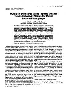
Thermal inactivation of arboviruses at 37 °C in macrophage culture medium In order to interpret extracellular virus titres measured after infection of in vitro cultures of mouse peritoneal macrophages it is necessary to follow the thermal inactivation of these viruses in a cell free system under identical conditions. Virus titres in macrophage culture medium at 37 °C have been determined at appropriate time intervals. In all cases a linear relationship between logao p.f.u./ml and time was found, allowing the calculation of the slope of the inactivation curve (Table I). The viruses studied can be classified according to increasing thermal stability, the flaviviruses being the most thermolabile and MIB the most thermostable. Thermal inactivation rate was the same in the presence of dead macrophages or in macrophage culture medium that had been in contact for 24 h with living macrophages. Multiplication of arboviruses in macrophages Determination of the extracellular virus titres from arbovirus infected mouse macrophages at different times after infection indicated that the production of virus was maximal between 6 and 24 h after infection (Fig. I). However, virus titres measured were always very low. No virus was found in the final rinses at time zero, indicating that all extracellular virus after 2 h adsorption was completely removed. For MIB and WN, the inactivation rate of the virus was higher in the presence of macrophages compared with the cell free system; this result indicates that in some macrophagearbovirus systems inactivation occurs. In contrast, the extracellular virus titres for SFV and SFA remained significantly higher than those predicted from thermal inactivation. For all viruses studied there was a parallel evolution of extra- and intracellular virus titres : the former always being up to tenfold higher than the latter. However with both SF
356
G. VAN DER GROEN AND OTHERS I
I
I
I
6
I
I
I
l
l
l
t
l
EEE
i
i
I
I
II
I
I
I
MIB
%\ x
1
_o
l
r
3
~~%\\ \,
"%"'0 I
e~
i
i
WEE
5 4
i
I
I
6
I
I
I
% O\N
I
I
SFV
I
I
N
SFA
I
YF WN
5 4
2 'x \%
1 I
I
I\~0
1
2
3
\
4
I
I
l
I
5
6
7
1
'o I
I
I
I
I
I
2
3
4
5
6
7
56 7 I
1
2
3
4
I
i
Time after infection(days) Fig. i. Time course of extracellular virus titres ( • - - O ) in arbovirus infected mouse peritoneal macrophages ; O - - - O, calculated virus titres according to thermal inactivation. Each point represents the mean of at least 3 separate experiments. Virus yields of YF ( • - - • ) and WN ( • - - • ) are expressed as loga0 TCDs0/ml; A - - A , calculated WN titre according to thermal inactivation. strains, no intracellular infectious virus could be demonstrated. The explanation could be that infectious SF was formed only during the latest phase of intracellular multiplication, for example during the budding process. To exclude the possibility that SF virus detected in the culture fluid later than 6 h postinfection was input virus gradually released from the cell surface, the following experiment was performed. Cultures of macrophages were inoculated with a multiplicity of 0.08 p.f.u./ cell of SFA virus. After 2 h adsorption the inoculum was removed, cultures were rinsed 3 times with M I99 and then treated with a I/IOO dilution of SFA immune mouse serum for 0"5 h in order to neutralize remaining traces of virus adsorbed to the cell surface. Immune serum was removed, cultures again rinsed 3 times and incubated at 37°C. As a control, macrophages were treated in the same way with normal mouse serum. At varying time intervals individual cultures were harvested and infectious virus was assayed in the supernatant fluids. The same extracellular infectious virus titres were found in both series, indicating that virus detected after 6 h incubation was newly synthesized virus and not the result of elution of virus originally adsorbed to the cell surface.
Arbovirus macrophage interaction
357
I
I
I
I
I
I
I 1
I 2
I 3
I 4
I 5
I 6
~4 .4
o~ 3
7
Time after infection (days) Fig. 2. ExtraceUular SFA virus yields in normal peritoneal mouse macrophages ( 0 - - D ) and L 929 cells ( A - - , ) at 37 °C; /~ - - - A, cell associated infectious virus yields in L 929 cells. Table 2. LDso values of different alphaviruses after intraperitoneal inoculation
of 6- to 7-week-old female OF1 mice Virus EEE WEE SFV SFA
Mouse LDso (p.f.u.)
Yield from maciophages* (log10p.f.u./ml)
88 7o7 2800 Not pathogenic
o'9 o 0-6 3"t
* Two-day titre with macrophages minus z-day titre without macrophages. No obvious cytopathic changes were observed in virus infected macrophages, observed for 6 to 7 days. These observations show that cultured macrophages from adult strain OF ~ mice are poorly susceptible to both alpha- and flaviviruses, only YF and the two strains of SF appearing to replicate in macrophages. Yields were much lower than those obtained in other murine cells, for example, L 929 cells infected with SFA virus, as shown in Fig. 2. Furthermore, in L 929 cells there was a parallel evolution of extracellular and intracellular virus titres, whereas this was not the case in macrophages (see above).
Lack of correlation between infectivity of arboviruses in macrophages and pathogenicity for OF I mice The LDs0 of different arboviruses for the OF I mouse strain by the i.p. route (Table 2) showed no correlation with the virus yields of in vitro infected macrophages.
G. VAN DER GROEN AND OTHERS
358 |
I
I
I
I
I
I
I
I
I
SFA
6
I
SFV
51 A
4 3 2 \
I
t
1
I
\
\ \
N \
N \x
\ i
I
i
i
~+
i
2 o
--
EEE
MIB
6
5' 4 \X
3
xZ
\
2 1
1
2
t
I
I
3
4
5
I
t
I
6 1 2 Time after infection (days)
I
I
1
I
3
4
5
6
Fig. 3. Time course of extracellular virus in arbovirus infected macrophages from normal (O--O), SFA-immunized (A - - - A) and proteose-peptone-stimulated (m--m) mice.
Multiplication of arboviruses in proteose peptone stimulated macrophages A significant increase of extracellular virus titre was obtained when both SF strains were inoculated into PPS macrophages (Fig. 3). The effect was less pronounced with the virulent SF strain. No extracellular infectious MIB virus could be demonstrated in PPS macrophages.
Multiplication of arboviruses in macrophages of vaccinated mice In further experiments virus production was studied in macrophages from animals previously immunized with SFA. In control experiments, macrophages from SFA pretreated mice were tested for the presence of infectious SFA virus, both extra- and intracellular. None was found. Sera from immunized mice had a 5o % neutralization activity at a dilution of ~o-3 to [o-3.5. There was a small but significant decrease in the duration of multiplication and of virus yields of SFA and SFV in ' i m m u n e ' macrophages (Fig. 3). The multiplication of EEE virus in SF immune macrophages was no different from the multiplication in normal cells. A clear c.p.e, was observed in macrophages from SFA immunized mice when infected
Arbovirus m a c r o p h a g e interaction
359
with SFA, SFV and EEE. This contrasts with the absence of an2¢ c.p.e, after normal macrophages were infected with the same viruses. The c.p.e, was characterized by pronounced rounding of the cells and partial lysis, and became manifest from the first day post infection. Interferon production
No interferon was detected in uninfected normal and immune macrophage cultures or in those infected with SFV, SFA or WEE virus after I to 7 days incubation at 37 °C. With EEE virus infected normal macrophages a very low interferon titre (I/2 to I/8) was demonstrated from the 3rd until the 7th day of incubation.
DISCUSSION
In some instances there is a relationship between the virulence of a virus for a given animal and its capacity to multiply in vitro in macrophages of the same host. Goodman & Koprowski (I962) showed that macrophages from susceptible mice support the multiplication of YF and WN very efficiently, whereas macrophages from resistant mice do not. However, this correlation was strongly dependent on the strain of mice used, and not always absolute. For example, in later work Hansen et al. 0969) showed that WN multiplication occurred in macrophages from both resistant (C3 HRv) and susceptible (C3-H) mouse strains. After 48 h incubation virus yields in cultures from resistant animals levelled off, whereas they cominued to rise in macrophage cultures from susceptible ones. Olson, Sithisarn & Djinawi (I975) reported the age-dependent resistance of mice to intraperitoneal infection with Wesselsbron virus as opposed to Germiston virus. This resistance was correlated with the capacity of peritoneal macrophages in vitro to destroy Wesselsbron virus, whereas Germiston virus replicated in these cells. Johnson (1964) concluded from studies with the fluorescent antibody technique that Sindbis virus does not multiply in adult or suckling mouse macrophages in vitro or in vivo. Additional evidence for the inactivation of Sindbis virus by mouse macrophages was given by McFarland (I974) and Lagwinska et al. 0975). The present work has shown that macrophages from adult strain OF I mice are inefficient host cells for both alpha- and flaviviruses, whether these viruses are virulent for mice or not. Extracellular virus yields were low and of the same order of magnitude as those found with Germiston virus (Olsen et al. r975) and other viruses as, for example, Sendal, herpes simplex and mouse hepatitis viruses (Eustatia et al. I972). It is not clear why the SF strains and YF multiply better in macrophages than the other viruses tested. It could be related to the mechanism by which the virus is taken up in the cell, as suggested by Olson et al. 0975) : penetration into the cytoplasm, where multiplication can take place, instead of phagocytosis followed by digestion in phagolysosomes. Different processing of arboviruses by normal macrophages would presumably be related to a different nature of the virus envelopes. Entry of virus into a macrophage can disturb some of its functions, for example, bactericidal activity is inhibited by Germiston virus (Olson et al. I975). One could think of an analogous mechanism whereby the first infecting virus particles might themselves inhibit normal macrophage function, including viricidal activity, sufficiently to allow limited virus replication. Little is known about the interaction of arboviruses and stimulated macrophages, which have been shown to be young macrophages freshly released from the bone marrow (Van Furth, i975). Hirsch, Zisman & Allison (I97O) working with herpes simplex virus demon-
360
G. V A N
DER
GROEN
AND
OTHERS
strated that PPS macrophages produced considerably less virus compared with normal macrophages. We have demonstrated a significant increase of extracellular virus with both SF strains in PPS macrophages, whereas no virus was produced after infection with MIB. Lagwinska et al. 0975) have shown that Sindbis virus replicated in thioglycollate stimulated macrophages but not in normal unstimulated macrophages. Thus, virus multiplication in stimulated macrophages seems dependent both on the virus and on the compound used to mobilize the macrophage. The importance of the latter was clearly demonstrated by Heessen (I975). Avila, Schultz & Tompkins 0972) and Schultz, Woan & Tompkins (I974), working with vaccinia virus, reported that macrophages from animals immunized against a given virus are immune to challenge with the homologous virus but support the replication of antigenically unrelated viruses. We obtained similar results with rnacrophages from SF vaccinated mice, which allow a normal growth cycle of EEE but not of SF virus. The effect ascribed to 'immune' macrophages may be due to the presence of small amounts of antibody (cytophilic antibody; Schultz et aI. I974) or to contaminating lymphoid cells. The latter may be difficult to remove completely from macrophage cultures as shown by Sheagren et aL (I975). Such antigen stimulated lymphocytes or the lymphokines produced by them might activate the killing activity of macrophages. Perhaps this mechanism may be responsible for the suppression of virus multiplication in cultured macrophages from immune animals. Our experiments show that the role of macrophages in the pathogenesis of peripheral arbovirus infections is difficult to establish. The macrophage-arbovirus interaction may vary from virus to virus as exemplified by SF compared with the other viruses. If the in vitro experiments reflect in vivo events, it can be stated that macrophages are not necessarily absolute barriers for arbovirus infection. Our findings support the view of Bradish, Allner & Fitzgeorge (I975) that virulence of an arthropod-borne virus for a certain host is a multifactorial event. The susceptibility or resistance of macrophages to the infection with a given virus in vitro determines only in some instances the resistance of the host to this agent. The authors thank A. Francken and J. Peel for technical assistance. This work was supported by grant no. 2o2o8 of the Fonds voor Wetenschappelijk Geneeskundig Onderzoek.
REFERENCES
ALLISON,A. C. (I 974). On the role of mononuclear phagocytes in immunity against viruses.Progress in Medical Virology xS, ~5-3I. AVILA, F. A., SCHULTZ, R. M. & TOMPKINS, W. A. F. (I972). Specific m a e r o p h a g e i m m u n i t y to vaccinia virus : m a c r o p h a g e virus interaction. Infection and Immunity 6, 9. BRADISH, C. J., ALLNER, K. & FITZGEORGE, R. (1975). I m m u n o m o d i f i c a t i o n a n d the expression of virulence in
mice by defined strains of Semliki Forest virus : the effects of myocrisin and L-asparaginase. Journal of General Virology 28, 239-25o. CLARKE,D. I-I. & CASALS,J. (I958). Techniques for hemagglutination and hemagglutination inhibition with arthropod-borne viruses. American Journal of Tropical Medicine and Hygiene 7, 561-573. ELrSTATIA,J. M., MAA.SE,E., VAN HELDEN, P. & VAN DER VEEN, .L (1972). Viral replication in m o u s e m a c r o p h a g e s Archiv fiir die gesamte Virusforschung 39, 376-38o.
~OODMAN,~. T. e, KOPROWSICI,n. (~962). Study of the mechanism of innate resistance to virus infection. Journal of Cellular and Comparative Physiology 59, 333-373. ttANSEN, B., KOPROWSKI, H., BARON, S. & BUCKLER, Ctf. E. (I969). Interferon m e d i a t e d n a t u r a l resistance o f
mice to arbo B virus infection. Microbios xB, 51-58. HE,SEN,F. W. A. (I975). The role of macrophages in persistent adenovirus infections. Doctors thesis, chapter 4, PP. 64-67. Nijrnegen, Krips Repro Meppel. HIRSCH, M. S.) ZISMAN)1~. & ALLISON,A. C. (I970). M a c r o p h a g e s a n d age d e p e n d e n t resistance to herpes simplex virus in mice. Journal of Immunology xo4, 1 i 6 o - I I65.
Arbovirus macrophage interaction
36I
rmor~ovsKv, v., PLAXSNER,V. & BENDA,R. (I975). A modified plaque method for arboviruses on plastic panels. Acta Virologica I9, I5o-I54. JOHNSON, R. T. (I964). The pathogenesis of herpes virus encephalitis. II. A cellular basis for the development of resistance with age. Journal o f Experimental Medicine x2o, 359-374. LAGW~NSKA,E., STUART, ¢. C., ADLES,¢. & SCnLESINGER,S. 0975)" Replication of lactic dehydrogenase virus and Sindbis virus in mouse peritoneal macrophages. Virology 65, 2o4-214. MCrARLAND, n. F. (X974). In vitro studies of cell-mediated immunity in an acute viral infection. Journal of Immunology xx3, I73-I8o. O~ON, e. ¢., SITHISARN,V. & DJINAWI,N. K. (I975). Role of macrophages in Wesselsbron and Germiston virus infections of mice. Journal of Infectious Diseases x3I, I I9-127. REED, L. J. & MUENCH,rE (t938). A simple method of estimating fifty percent endpoints. American Journal o f Hygiene 27, 493-495. scrrueTz, R. M., WOAN, M. C. & TOrn'IONS, W. A. F. (r974). Maerophage immunity to vaccinia virus : factors affecting macrophage immunity in vitro. Journal of the Retieuloendothelial Society x6, 37-47. S~EAGREN, J. H., SIMON,H. B., TUAZON, C. V. & MEHROTA,I'. (I975)- In Mononuelear Phagocytes in Immunity, Infection and Pathology, 1st ed., chapter 42, pp. 654-658. Edited by R. van Furth. Oxford, London : Blackwell Scientific Publications. van FORTH, R. (I975). Modulation of monocyte production. In Mononuelear Phagocytes in Immunity, Infection and Pathology, Ist ed., chapter io, pp. I6I-I74. Edited by R. van Furth. Oxford, L o n d o n : Blackwell Scientific Publications. van FURTH, R., COHN, Z. A., HIRSCH, J. G., HUMPHREY,J. H., SPECTOR,W. O. & LANGEVOORT,H. L. (I972). The mononuelear phagocyte system : a new classification of macrophages, monocytes and their precursor cells. Bulletin of the Worm Health Organization 46, 845-852.
(Received 6 August I976)