www.nature.com/scientificreports
OPEN
received: 18 April 2016 accepted: 10 August 2016 Published: 02 September 2016
Integration of transcriptomic and genomic data suggests candidate mechanisms for APOE4-mediated pathogenic action in Alzheimer’s disease Laura Caberlotto1, Luca Marchetti1, Mario Lauria1, Marco Scotti1,2 & Silvia Parolo1 Among the genetic factors known to increase the risk of late onset Alzheimer’s diseases (AD), the presence of the apolipoproteine e4 (APOE4) allele has been recognized as the one with the strongest effect. However, despite decades of research, the pathogenic role of APOE4 in Alzheimer’s disease has not been clearly elucidated yet. In order to investigate the pathogenic action of APOE4, we applied a systems biology approach to the analysis of transcriptomic and genomic data of APOE44 vs. APOE33 allele carriers affected by Alzheimer’s disease. Network analysis combined with a novel technique for biomarker computation allowed the identification of an alteration in aging-associated processes such as inflammation, oxidative stress and metabolic pathways, indicating that APOE4 possibly accelerates pathological processes physiologically induced by aging. Subsequent integration with genomic data indicates that the Notch pathway could be the nodal molecular mechanism altered in APOE44 allele carriers with Alzheimer’s disease. Interestingly, PSEN1 and APP, genes whose mutation are known to be linked to early onset Alzheimer’s disease, are closely linked to this pathway. In conclusion, APOE4 role on inflammation and oxidation through the Notch signaling pathway could be crucial in elucidating the risk factors of Alzheimer’s disease. Alzheimer’s disease (AD) is the most common cause of dementia, characterized clinically by a decline in cognitive function and by distinctive brain pathology with neuronal loss and the formation of amyloid plaques and neurofibrillary tangles. Early onset AD is rare and is caused by mutations in specific genes such as amyloid precursor protein (APP), presenilin 1 (PSEN1) and presenilin 2 (PSEN2). Late onset AD is the most common form but, although several putative susceptibility genes have been reported, APOE, coding for the Apolipoprotein E, is the most robust susceptibility gene known to date. Three common isoforms of APOE have been recognized: APOE2 (cys112, cys158), APOE3 (cys112, arg158) and APOE4 (arg112, arg158) and the presence of the alleles coding for the APOE4 isoform are associated with an increased risk (up to tenfold in homozygous cases1) of late onset AD when compared to the most common APOE3 allele or APOE2, a rarer allele, that appears to have, instead, a protective effect2,3. APOE is a multifunctional glycosylated protein with a major role in lipid transport and atherosclerosis pathogenesis and it is expressed in several organs, with the highest expression in the liver and brain. In the central nervous system, although neurons can produce APOE under certain conditions, non-neuronal cells, mainly astrocytes and to some extent microglia, are the major cell types that express APOE in the brain4,5. Numerous mechanisms by which APOE influences AD pathogenesis have been proposed, including a role in the clearance of Amyloid β6,7, but how this influences the pathogenic molecular processes remains to be clarified.
1
The Microsoft Research, University of Trento Centre for Computational Systems Biology (COSBI), Piazza Manifattura 1, 38068, Rovereto, Italy. 2GEOMAR Helmholtz Centre for Ocean Research Kiel, Düsternbrooker Weg 20, 24105 Kiel, Germany. Correspondence and requests for materials should be addressed to L.C. (email: Laura.
[email protected])
Scientific Reports | 6:32583 | DOI: 10.1038/srep32583
1
www.nature.com/scientificreports/
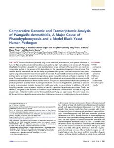
Figure 1. Schematic representation of the network analysis workflow. The molecular biomarker associated to APOE44-AD was extracted from transcriptomic data of post-mortem cerebral cortices of AD affected individuals carrying the APOE33 or APOE44 alleles. Network analysis was then performed using NetWalker technique with the reference protein-protein interaction (PPI) network derived from HPRD data. The functionality of the network was then performed by testing over-represented Gene Ontology biological process terms and pathways. Genomic data analysis was then performed with the analysis of genes in the network displaying a genetic interaction with APOE.
Study
Type of data/Technology
#Subjects in the original study
#Subjects selected for the study and APOE genotype
Brain Region
Data source
Temporal, Parietal, Frontal Cortex
NCBI GEO GSE15222
1-Mayer’s lab
Transcriptomic/Illumina ref-seq 8
364
77 (43APOE33 34 APOE44)
2-Mayo
Transcriptomic/Illumina HiSeq 2000
278
39 (30 APOE33 9 APOE44)
Temporal Cortex
AMP AD knowledge Portal syn3163039
3- Astrocyte study Transcriptomic/2.0 Affymetrix gene arrays
18
18 (9 APOE4-9 APOE4+)
Frontal Cortex (Astrocyte)
NCBI GEO GSE29652
4-ADNI
Genomic/ILLUMINA Human610-Quad BeadChip
757
139 (APOE4: 0 = 45;1 = 65;2 = 29)
NA
adni.loni.usc.edu
5-Mayo LOAD GWAS
Genomic/ILLUMINA HumanHap300
2099
580 (APOE4: 0 = 175; 1 = 289; 2 = 98)
NA
AMP AD Knowledge Portal syn3157238
Table 1. Datasets used in the study. Table describing the datasets used in the study, type of data and technology platform used, total number of subjects in the original study and number of subjects selected for the present study including APOE alleles composition. Brain regions analyzed in the study (when applicable) and data source are also described.
To gain some insight into the molecular mechanisms responsible for the pathogenic impact of APOE, we used a systems biology approach to integrate pre-existing genomic and transcriptomic data in an unbiased fashion. A network-based analysis combined with a novel technique for biomarker identification revealed specific biological processes altered in APOE44 carriers affected by AD. Molecular processes related to inflammation, oxidative stress and metabolism were shown to be altered, demonstrating that APOE4 possibly quickens pathological alterations naturally induced by aging. The integration of genomic data evidenced that components of the Notch pathway could be among the upstream effectors involved in this action. Overall, this study provides a contribution to the long-standing APOE4 debate by suggesting an evidence driven hypothesis on the mechanism by which APOE4 confers risk for the development of Alzheimer’s disease.
Material and Methods
A schematic representation of the study workflow is shown in Fig. 1.
Transcriptomic and Genomic data. The microarray dataset (study number 1, Table 1) was downloaded from the Myers laboratory website (http://labs.med.miami.edu/myers/LFuN/data.html, GEO reference: GSE15222). The complete dataset consists of transcriptome-wide gene expression data from post-mortem cerebral cortical area (temporal, parietal and frontal cortices), all cortical areas involved in the disease, of Alzheimer’s
Scientific Reports | 6:32583 | DOI: 10.1038/srep32583
2
www.nature.com/scientificreports/ disease affected subjects at all stages of the disease (Braak stages ≥3) and matched controls (188 controls and 176 AD), stratified by their APOE genotype8. In the study, data from 77 subjects affected by AD (43 APOE33 and 34 APOE44) was used (Table 1) matched for age (average age APOE33-AD 83.58 years [72–102 years]; APOE44-AD 83.62 years [68–92 years]) and post-mortem interval (average PMI APOE33-AD 10.54 hours [1.3–26.9 hours]; APOE44-AD 10.39 hours [1.2–17.7 hours]). For the validation studies, an additional dataset was evaluated: the Mayo RNA sequencing study (study number 2 Table 1) of temporal cortex accessed through the AMP AD Knowledge Portal (syn3163039; https://www. synapse.org/#!Synapse:syn2580853/wiki/77552). The original study consisted of RNA sequencing data of 278 samples from post-mortem temporal cortical tissue of AD affected individuals at all stages of the disease (Braak stages ≥4) and matched controls. In the present study a sub-population of 39 subjects was used (30 APOE33 and 9 APOE44 carriers) all subjects were affected by AD (average age APOE33-AD 76.5 years [65–80 years]; APOE44-AD 72.3 years [66–78 years] (Table 1). No information on post-mortem interval was provided. In order to evaluate the role of the specific pathways highlighted in our network analysis in astrocytes of APOE4 individuals, an additional dataset was used (GEO accession: GSE29652; study number 3, Table 1). This dataset includes microarray profiles of laser-captured microdissected astrocytes of post-mortem human temporal cortex of AD affected individuals APOE4- and APOE4+carriers at different Braak stages. NCBI GEO’s tool GEO2R was used to compare expression between the APOE4- and APOE4+groups, each group including subjects at all stages of the disease. GEO2R analyzes gene expression using GEOquery and the Linear Models of Microarray Analysis R package (limma)9,10. Genes were considered differentially expressed if they showed a minimum 1.5-fold change at p-value