Research Article
2031
Integrin clustering induces kinectin accumulation Huan Tran1, Roumen Pankov1, Simon D. Tran1, Brian Hampton2, Wilson H. Burgess2 and Kenneth M. Yamada1,* 1Craniofacial
Developmental Biology and Regeneration Branch, National Institute of Dental and Craniofacial Research, National Institutes of Health, Bethesda, MD 20892-4370, USA 2Department of Tissue Biology, Jerome H. Holland Laboratory, American Red Cross, Rockville, MD 20855, USA *Author for correspondence (e-mail:
[email protected])
Accepted 19 February 2002 Journal of Cell Science 115, 2031-2040 (2002) © The Company of Biologists Ltd
Summary Integrin receptors mediate the formation of adhesion complexes and play important roles in signal transduction from the extracellular matrix. Integrin-based adhesion complexes (IAC) contain proteins that link integrins to the cytoskeleton and recruit signaling molecules, including vinculin, paxillin, focal adhesion kinase, talin and αactinin. In this study, we describe a ~160 kDa protein that is markedly enriched at IAC induced by clustering integrins with fibronectin-coated beads. Protein sequence analysis reveals that this ~160 kDa protein is kinectin. Kinectin is an integral membrane protein found in endoplasmic reticulum, and it serves as a receptor for the Introduction Integrin transmembrane receptors play critical structural roles in linking the actin skeleton to the extracellular matrix (ECM). The integrin family is also involved in cellular processes such as adhesion, signal transduction, migration, growth and differentiation (Hynes, 1992; Sonnenberg, 1993; Clark and Brugge, 1995; Schwartz et al., 1995; Sastry and Horwitz, 1996; Aplin et al., 1999; Giancotti and Ruoslahti, 1999; Humphries, 2000; Hynes and Zhao, 2000; Yamada and Danen, 2000). When cells attach to ECM proteins such as fibronectin, laminin and collagen via integrins, these ligands crosslink or cluster integrins into cell surface complexes. Ligand-binding and clustering by integrins are critically important for activation of intracellular responses. They lead to the recruitment and organization of a number of cytoskeletal and signaling proteins into specialized cell-substrate adhesions, such as focal and fibrillar adhesions, where integrins serve as a complex link to the ECM (Burridge et al., 1988; Jockusch et al., 1995; Geiger et al., 2001; Zamir and Geiger, 2001). These multimolecular complexes are known to play important roles in modulating cell signaling and cytoskeletal dynamics (Sastry and Burridge, 2000; Geiger et al., 2001), but their constituents are not yet fully characterized. To analyze cellular responses to integrin clustering, an experimental approach was developed using beads coated with integrin ligands to mimic cell-to-ECM adhesion formation (Grinnell and Geiger, 1986; Plopper and Ingber, 1993). For example, fibronectin-coated beads can rapidly induce the accumulation of vinculin, paxillin, α-actinin, talin and F-actin (Grinnell and Geiger, 1986; Mueller et al., 1989; Plopper and Ingber, 1993; Lewis and Schwartz, 1995; Miyamoto et al., 1995b). This procedure also permits the rapid isolation of
motor protein kinesin. Fibronectin-induced IAC sequestered over half of the total cellular content of kinectin within 20 minutes. In addition, two other ERresident proteins, RAP [low-density lipoprotein receptorrelated protein (LRP) receptor-associated protein] and calreticulin, were found to be clustered at IAC, whereas kinesin was not. Our results identify a novel class of constituents of IAC.
Key words: Adhesion, Integrin, Kinectin, Kinesin-binding proteins, Endoplasmic reticulum, Fibronectin
intact integrin-based adhesion complexes (IAC) after integrin crosslinking and allows searches for cytoskeletal and other components that associate with IAC (Plopper and Ingber, 1993). Previous studies using beads coated with integrin ligands or integrin antibodies have shown that both integrin occupancy and clustering into aggregates play important roles in IAC formation, and that these two signals can synergize; a ‘hierarchy’ of molecules requiring either one or both of these signals to accumulate was identified (Miyamoto et al., 1995a; Miyamoto et al., 1995b). Numerous cytoskeletal proteins have been localized to the IAC by morphological and biochemical approaches, but integrin clustering can also induce the aggregation of growth factor receptors, stimulate the phosphorylation of FGF, EGF and PDGF receptors, and activate downstream signal transduction (Plopper et al., 1995; Schwartz et al., 1995; Miyamoto et al., 1996; Sundberg and Rubin, 1996). In addition, recent studies have shown that mRNA and ribosomes also localize to IAC when integrins are clustered with FN-coated beads (Chicurel et al., 1998). Furthermore, cell adhesion induces increased translation of pre-existing mRNAs (Benecke et al., 1978; Farmer et al., 1978). Taken together, these studies indicate that IAC are important for redistribution of cytoplasmic components and a variety of integrin functions. In the present study, we have used beads coated with a specific integrin ligand to isolate intact IAC in order to identify new proteins that associate with these complex cellular structures. We searched for novel component(s) that might be particularly markedly sequestered from the cytoplasm into IAC compared with other components to potentially establish the concept of differential recruitment.
2032
Journal of Cell Science 115 (10)
Unexpectedly, we found that kinectin, an integral membrane protein prominent in endoplasmic reticulum (ER) that binds to kinesin and is involved in vesicle transport, becomes strikingly localized to intracellular sites at regions where cells are in contact with FN ligand. Over 50% of total cellular kinectin becomes associated with fibronectin-integrin adhesion sites, an enrichment many times higher than the classical components vinculin and paxillin. Furthermore, two ER-resident proteins, RAP and calreticulin, accumulate at these sites. Our results demonstrate that IAC formation can trigger integrin-mediated differential recruitment of cytoplasmic components, and suggest control of the localization of a portion of the ER, thereby potentially influencing sites of ER-based protein synthesis and secretion. Materials and Methods Antibodies and reagents Mouse monoclonal antibodies against fibronectin, PKCδ and paxillin were purchased from Transduction Laboratories. The kinectinspecific monoclonal antibody KR160.9 has been described previously (Toyoshima et al., 1992) and was kindly provided by Michael Sheetz (Columbia University). Talin rabbit polyclonal antibody was generously provided by Keizo Takenaga [National Institute of Dental and Craniofacial Research (NIDCR), NIH]. Monoclonal actin, α-actinin, dynein, tubulin, vinculin and VSV glycoprotein (P5D4) antibodies were obtained from Sigma. Polyclonal anti-α5β1 integrin rabbit antibody (Rab 3847) was described previously (Roberts et al., 1988), and a polyclonal anti-β1 integrin rabbit antibody (Rab 4080) was produced against a synthetic peptide representing the β1 C-terminal cytoplasmic domain. Function-blocking anti-β1 rat mAb13 monoclonal antibody and noninhibitory ES66 control antibody (same IgG isotype that binds without affecting function to chicken but not human β1) were characterized previously (Duband et al., 1988; Akiyama et al., 1989; Miyamoto et al., 1995a). Mouse mAb 12G10 against human β1 integrin was described previously (Mould et al., 1995). Calreticulinand tensin-specific antibodies were purchased from Upstate Biotechnology. Kinesin-specific monoclonal antibody (MAB 1614) was obtained from Chemicon, and rhodamine-phalloidin was purchased from Molecular Probes. Monoclonal antibody (mAb 7F1) against LDL receptor-related protein (LRP) receptor-associated protein (RAP) was generously provided by Dudley Strickland, (American Red Cross, Rockville, MD). Non-conjugated, Cy3-, or FITC-conjugated goat anti-mouse, -rabbit, or -rat IgG antibodies were purchased from Jackson Immunoresearch Laboratories. Polystyrene microbeads (mean diameter 10 µm) were obtained from Polysciences, Inc. Tosyl-activated magnetic microbeads (mean diameter 4.5 µm) were purchased from Dynal. Human plasma fibronectin (FN) and vitronectin (VN) were purified as previously described (Miekka et al., 1982; Yatohgo et al., 1988). Collagen I (Vitrogen 100) was purchased from Collagen Corp. Concanavalin A (ConA) and poly-DL-lysine were obtained from Sigma. FN fragments containing type III repeats 5-10 (III5-10) and type III repeats 5-10 with the RGD sequence mutated to KGE (III5-10KGE) were described previously (Danen et al., 1995) and were generously provided by Shinichi Aota (CDBRB, NIDCR, NIH). Plasmid DNAs for pRK-VSV-tensin and pRK-VSV were kindly provided by Kazue Matsumoto (CDBRB, NIDCR, NIH). The pRKVSV plasmid was prepared by inserting two continuous VSV sequences with a Kozak consensus sequence in a cytomegalovirus promoter-based expression system. The puromycin resistance plasmid pHA262pur was generously provided by Hein te Riele (Netherlands Cancer Institute, Amsterdam, The Netherlands) (Lacalle et al., 1989).
Preparation of cDNAs encoding kinectin and its fragments Plasmid constructs designed to express five portions of kinectin and full-length kinectin are depicted in Fig. 7A. The eukaryotic expression vector pRK-VSV containing a VSV tag was used to place the kinectin cDNAs under the transcriptional control of the cytomegalovirus promoter. The cDNAs encoding various portions of kinectin shown in Fig. 7A were assembled from restriction fragments of PCR-generated kinectin cDNAs, which were generated using as template the full-length chick kinectin cDNA plasmid pGinKNT (Yu et al., 1995) kindly provided by Michael Sheetz (Columbia University). The fragment I construct (designated PCR fragment 1; Table 1) encoding residues 1-326 of kinectin was generated by PCR using primers 1 and 2 (Table 2). PCR fragment 1 (1007 bp) was cleaved by EcoRI and SalI and ligated into EcoRI-SalI linearized vector pRK-VSV. Similarly, constructs for fragments II, III, IV and V (Table 1) encoding various portions (residues 327-629, 630-935, 935-1365 and 327-1365) of kinectin were prepared as described for fragment I. For construction of full-length kinectin, pGinKNT (Yu et al., 1995) plasmid DNA was digested with BglI and BglII to liberate a 2759 bp kinectin fragment encoding kinectin nucleotides 1015 to 3774. Kinectin contains both BglI and BglII sites (1015 and 3774 in the kinectin sequence). PCR-generated fragment 1 was subjected to EcoRI and BglI digestion to obtain an EcoRI-BglI fragment encoding kinectin nucleotides 58-1015. PCR-generated fragment 4 was digested with BglII and SalI restriction enzymes to generate a BglIISalI fragment encoding kinectin nucleotides 3774 to 4176. Full-length 4126bp kinectin cDNA was prepared by ligation of EcoRI-BglI fragment 1, the 2759bp BglI-BglII kinectin fragment, and BglII-SalI fragment 4 together into a linearized EcoRI-SalI pRK-VSV vector. Plasmid DNAs of all PCR-generated fragments were subjected to complete DNA sequencing analysis to confirm the integrity of each construct. Cell culture and transfection Primary human foreskin fibroblasts (HFF) and 293 cells were maintained in Dulbecco’s modified Eagle (DME) medium (Gibco/BRL Life Technologies) supplemented with 10% fetal bovine serum (Hyclone), 100 units/ml penicillin and 100 µg/ml streptomycin. The cells were a gift from Susan Yamada (NIDCR, NIH) and were used at cell passages 9-22. The NIH3T3 cell line was obtained from American Type Culture Collection. These cells were maintained in DME medium supplemented with 10% bovine calf serum, 100 units/ml penicillin and 100 µg/ml streptomycin. Transient transfections of kinectin and its fragments into NIH3T3 cells were performed using Lipofectamine Plus (Gibco/BRL Life Technologies) according to the protocol provided by the manufacturer. In other experiments, kinectin plasmid DNAs were introduced into HFF by electroporation. Briefly, pRK-VSV containing either no insert or fulllength kinectin, kinectin fragments or tensin (30 µg each) was transfected into 1.5×10⁶ cells by electroporation. Cells were then cultured in serum-containing medium for 24 hours with 5 mM sodium butyrate to enhance expression of transfected genes. In some experiments, plasmid DNAs encoding kinectin or its fragments were transfected into HFF together with 6 µg of the puromycin selection vector pHA262pur. Cells were subcultured at a 1:3 dilution for 24 hours after transfection and were selected for 2 days in 1 µg/ml puromycin-containing medium. Isolation of integrin-based adhesion complexes (IAC) IAC were isolated as described previously (Plopper and Ingber, 1993) with some minor modifications. Tosyl-activated magnetic beads (4.5 µm; Dynal) were coated with either anti-integrin antibodies, FN, FN fragment III5-10, III5-10KGE, or ConA at 5 µg per 107 beads in 0.1 M phosphate buffer, pH 7.4, according to the manufacturer’s protocol.
Integrin-induced clustering of kinectin Table 1. Description of PCR-generated kinectin fragments Fragment Nucleotide Amino acid designation residues residues 1 2 3 4 5
58-1062 1035-1971 1944-2889 2856-4176 1035-4176
1-326 327-629 630-935 935-1365 327-1365
Upstream Downstream Construct primer* primer* name 1 3 5 7 3
2 4 6 8 8
I II III IV V
PCR fragments 1-5 were generated using the indicated primers with chick kinectin as template. *The primer designations refer to those described in Table 2.
Table 2. Oligonucleotide primers used in generation of kinectin cDNAs Primer no. 1 2 3 4 5
Cells were removed from flasks by trypsin-EDTA and allowed to recover in DME medium containing 10% fetal bovine serum. Cells were then washed twice with DME medium containing 1% BSA and 20 mM Hepes, pH 7.6 and resuspended at 1×10⁶ cells/ml. Cells were mixed with FN-, ConA-, III5-10-, III5-10KGE-, mAb13-, or ES66coated beads (10 beads/cell) and rotated gently for 20 minutes using a rotator at 37°C. The complex of beads and bound cells was collected by a magnetic particle concentrator (Dynal) and was washed with 1 ml cold CSK buffer [50 mM NaCl, 300 mM sucrose, 3 mM MgCl2, protease inhibitor mixture (Boehringer Mannheim), and 1 mM PMSF in 10 mM PIPES, pH 6.8] without detergent. The pellet containing cell-bead complexes was extracted with 0.5 ml of cold CSK buffer containing 0.5% Triton X-100 and sonicated for 10 seconds with a 50 watt Ultrasonic Processor (Aldrich, model GE 50, set at amplitude 20). The beads-protein complexes were washed five times with 1 ml cold CSK buffer containing detergent. In experiments to test for nonspecific binding, beads were incubated with cell homogenates. Cells were homogenized in CSK buffer containing detergent and clarified by centrifugation in a microfuge at 19,000 g for 15 minutes at 4°C. The supernatants were incubated with FN- or ConA-coated beads for 15 minutes at 4°C; the beads were then washed and processed as described above. Proteins were extracted from beads and subjected to SDS-PAGE and immunoblot analyses. The ratio of bead-bound to non-bound (soluble) protein was determined by quantitative densitometry and correction for the proportion of sample used for immunoblotting. For example, if gels were loaded with all of the material extracted from beads and 1/10 of the sonicated supernatant containing non-bound material, we calculated the ratio of bead-bound protein as R=B/10S, where R is the ratio, B is the absorbance of immunoblotted bound material, and S is the absorbance of immunoblotted supernatant fraction (1/10 volume). Alternatively, we calculated P=[B/(B+10S)]×100, where P is the percentage of a protein that was bead-bound. Immunoprecipitation and immunoblot analyses Confluent 293 cells (15 cm diameter dish) were lysed with 1 ml of RIPA lysis buffer [50 mM N-2-hydroxyethylpiperazine-N′-2ethanesulfonic acid (Hepes; pH 7.5), 150 mM NaCl, 10% glycerol, 1.5 mM MgCl2, 1 mM EGTA, 1 mM sodium vanadate, 10 mM sodium pyrophosphate, 100 mM NaF, 1% Triton X-100, 1% sodium deoxycholate, 0.1% sodium dodecyl sulfate (SDS), protease inhibitor mixture (Boehringer Mannheim) and 1 mM phenylmethylsulfonyl fluoride]. The cell extracts were clarified by centrifugation at 20,000 g for 15 minutes at 4°C. The supernatants were absorbed with protein G-Sepharose for 1 hour at 4°C and centrifuged at 5000 g for 5 minutes at 4°C. Antibodies (4 µg/ml) and a fresh aliquot of protein GSepharose (30 µl/1 ml of lysate) were added to the supernatants and incubated for 1 hour or overnight at 4°C. The immunoprecipitated proteins were washed five times with RIPA buffer at 4°C. Protein complexes bound to beads were solubilized in reducing SDS-PAGE sample buffer, boiled for 5 minutes, separated by SDS-PAGE, and transferred to nitrocellulose membranes (Novex) for 1.5 hours at 30 V.
2033
6 7 8
Oligonucleotide sequence AGAAAAgaattcATGGAGTTTTACGAGTCT EcoRI CTTGTCgTcgAcCTaATGTATAGCTGCAGCCGA SalI GCAGTCgaAttcCAGCTGCAAGACAAGGAG EcoRI CAAGACgTCgAcCTaTTCTTCCTTGCTTGTTAA SalI AGCAAGGAAttcGAGCTCAAGGTCTTGCAG EcoRI CCGGCTgTcGAccTaCTGGACTTGGGATGCAGC SalI GCTGCAgaattcGTCCAGGACCTGGAGAGC EcoRI TCCCCCgtcgACTCATTCTACTAGCTGGTAATG SalI
Positions* 58-88 1030-1063 1048-1078 1939-1972 1957-1987 2856-2889 2869-2899 4164-4197
Mismatches with template are indicated in lowercase letters. Stop codons are shown in boldface. Restriction site recognition sequences are underlined. *The numbers correspond to residues within chick kinectin cDNA (Yu et al., 1995).
Unoccupied protein binding sites were blocked by incubating the membranes with blocking buffer [5% nonfat dried milk in TBS-T (150 mM NaCl, 50 mM Tris-HCl, 0.1% Tween-20, pH 7.4)]. Antibodies were diluted in blocking buffer and incubated with membranes for 1 hour at room temperature. The membranes were washed with TBS-T and incubated with horseradish peroxidase-conjugated goat antimouse or -rabbit IgG (Bio-Rad). Bound antibodies were visualized using a SuperSignal kit (Pierce) and Hyperfilm (Amersham). Partial amino acid sequence analysis Partially immuno-affinity purified ~160 kDa polypeptide was
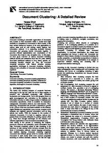
Fig. 1. Immunoblot identification of accumulation of a 160 kDa protein with FN-coated beads. Unbound fractions (lane 1), beadbound protein complexes (lane 2) from either ConA- or FN-coated beads, and rat brain extract (10 µg) were electrophoresed on an SDScontaining 4-12% polyacrylamide gel under reducing conditions and transferred to nitrocellulose. The membrane was probed with a putative PKCδ monoclonal antibody. Indicated on the right are the molecular masses (kDa) of marker proteins.
2034
Journal of Cell Science 115 (10)
Kinectin Peptide 1
191
Q E A L P 195 QEALP
Kinectin
394
H V S Y Q E T Q Q M Q 404
Peptide 2
HVSYQETQ- MQ
Kinectin Peptide 3
544
Q L E Q R L M Q L M 553 QLEQRLMQLM
Kinectin
746
V E E L L E T G L I Q V A T K 760
Peptide 4 Kinectin Peptide 5
V- - LL- TGL I Q V A T K 810
K S V E E L L E A E L L K 822 KSVEELLEAELLK
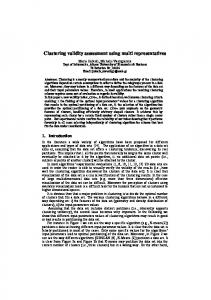
Fig. 2. Partial amino acid sequence alignment of 160 kDa polypeptides and human kinectin. The sequence of Lys-C-digested peptides of 160 kDa protein were aligned with human kinectin (GenBank accession no. Z22551). The numbers represent the amino acid positions of kinectin.
separated by SDS-PAGE and transferred to nitrocellulose. The polypeptide band migrating at ~160 kDa was visualized by staining with Ponceau S (Sigma) and excised from the membrane. The nitrocellulose strip containing the 160 kDa polypeptide was digested with endoproteinase Lys-C (Boehringer Mannheim) in 0.1 M NH4HCO3 as described (Aebersold et al., 1987). The liberated digest was fractionated on a Vydac C18 column using a microbore HPLC (Model 130; Applied Biosystems). Peptides from individual peaks were then subjected to Edman degradation using a protein sequencer (Model 477A; Applied Biosystems). Indirect immunofluorescence microscopy and bead-binding assay Polystyrene beads (mean diameter 10 µm, or 4.5 µm for comparisons) were coated with 50 µg/ml FN, 100 µg/ml ConA, or 100 µg/ml polylysine in 0.1 M carbonate buffer, pH 9.5, at 4°C overnight. The ligand-coated beads were blocked with 1% BSA in Dulbecco’s PBS (D-PBS) for 1 hour at room temperature. Glass coverslips (12 mm; Carolina Biological Supply Company) were coated overnight with 20
µg/ml type I collagen (Vitrogen 100, Collagen Corp.) in D-PBS and then blocked with 1% heat-denatured BSA for 1 hour at room temperature. To inhibit FN synthesis so as to retain a diffuse distribution of FN receptors, HFF cells were incubated with DME medium containing 25 µg/ml of cycloheximide and 10% FN-depleted fetal bovine serum for 2 hours at 37°C. Cells were detached with trypsin-EDTA and allowed to recover in DME medium containing cycloheximide and FN-depleted serum for 45 minutes. Cells (2×10⁴) were plated on collagen-coated coverslips for 30 minutes at 37°C. Ligand-coated beads were then added to the cells at a ratio of 10 beads per cell and incubated for 20 minutes at 37°C. Subsequently, the cells were fixed with 4% paraformaldehyde with 5% sucrose in D-PBS for 20 minutes and permeabilized with 0.4% Triton X-100 in PBS for 5 minutes at room temperature. For kinectin staining, HFF were plated on FN-coated coverslips (10 µg/ml) for time periods ranging from 20 minutes to 2 hours, fixedpermeabilized for 3 minutes with the above fixative containing 0.5% Triton X-100, and post-fixed for an additional 20 minutes with the fixative alone. The coverslips were incubated with an appropriate dilution of different primary antibodies in D-PBS for 1 hour at room temperature. The coverslips were then washed with D-PBS three times and incubated with Cy3-conjugated secondary IgG in D-PBS. Double staining with two primary mouse antibodies was achieved as follows: The fixed samples were blocked with 20% donkey serum in D-PBS for 30 minutes, washed, and stained with mouse anti-kinectin antibody for another 30 minutes. After three 5-minute washes with D-PBS containing 0.05% Tween 20 (D-PBST), the samples were incubated for 30 minutes with goat anti-mouse IgG (25 µg/ml in D-PBST, 0.5% donkey serum) and washed as above. To prevent possible redistribution of bound antibodies, the samples were fixed briefly again (10 minutes), washed, and blocked again with 20% donkey serum in D-PBS for 15 minutes. The coverslips were then stained with the appropriate dilution of the second primary mouse antibody (anti-α-actinin) for 30 minutes. After three washes with D-PBST, the samples were incubated for 30 minutes with a mixture of labeled secondary antibodies consisting of FITC-conjugated donkey anti-goat and Cy3-conjugated donkey antimouse IgGs to visualize kinectin and α-actinin, respectively. Specificity was confirmed by appropriate controls. After three washes with D-PBST, each coverslip was mounted on a glass slide with mounting medium (Biomeda) containing 1 mg/ml 1,4 phenylenediamine to minimize photobleaching. Immunofluorescent samples were examined using a Zeiss Axiophot fluorescence microscope. Accumulation of a particular protein at a bead was assayed as described previously (Miyamoto et al., 1995b) and scored as positive if it localized as aggregates or thick zones around at least half of the bead.
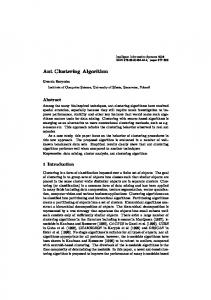
Fig. 3. Kinectin is specifically associated with adhesion complexes induced by FN-coated beads and α5β1 integrin. (A) Bead-associated protein complexes induced by FN, fibronectin fragment III510, or mutant fragment III5-10KGE. (B) Beadassociated protein complexes were induced by mAb13 against the β1 integrin or control antibody ES66. (C) Concanavalin A-coated (ConA) or fibronectin-coated (FN) beads were either mixed directly with HFF homogenates (Lysate) or incubated with intact, living HFF (Cells). Beadbound protein complexes were then isolated, separated on 4-12% SDS-containing polyacrylamide gels, transferred to nitrocellulose, and probed with kinectin KR160.9 monoclonal antibody. Note that kinectin is present only in the integrin-based adhesion complexes.
Integrin-induced clustering of kinectin
2035
1). Although subsequent studies showed that PKCδ itself could not be detected in association with IAC (data not shown), this ~160 kDa protein was exceptionally enriched in bead-induced IAC. Crossreactivity of a particular monoclonal antibody with epitopes on two proteins is not unusual (Marchalonis et al., 2001) and, in this case, it permitted further studies of this fortuitously identified integrin-redistributed protein. Partial amino acid sequence analysis of 160 kDa protein Internal proteolytic peptides were generated from the 160 kDa protein by Lys-C digestion. These peptides were purified by HPLC and subjected to microsequencing. The amino acid sequence of these peptides was used to search protein databases. As shown in Fig. 2, the sequence of these peptides matched exactly with the human CG1 or kinectin sequence (Toyoshima et al., 1992; Futterer et al., 1995; Print et al., 1996).
Fig. 4. Kinectin is markedly enriched in adhesion complexes induced by FN and FN III5-10 fragment-coated beads. Beads coated with fibronectin (FN), a fibronectin fragment containing type III domains 5-10 (III5-10RGD), or the same fragment with an inactivated RGD site (III5-10KGE) were used to obtain bead-associated protein complexes (Beads) or unbound fractions (Sup). The samples were separated on a 4-12% SDS-containing polyacrylamide gel, transferred to nitrocellulose, and probed with the indicated antibodies. Note that in the beads fraction, kinectin is remarkably enriched, while kinesin and tubulin are virtually absent. In this particular experiment, 91% of total cellular kinectin was found in the beads fraction, while the corresponding percentages of total α-actinin and actin in the same fraction were 22% and 23%, respectively. Also note that no proteins are observed in the unbound fractions using the functionally inactivated fibronectin fragment (III5-10KGE), because the absence of integrin binding and adhesion to this control polypeptide resulted in no cells being isolated at the first step of the procedure where beads and bound cells are isolated using a magnetic concentrator.
Results Accumulation of 160 kDa protein with FN-coated beads In order to identify new proteins involved in integrin-mediated responses that might meet our hypothesized criterion of marked enrichment at integrin-based adhesion complexes (IAC), we used magnetic beads coated with integrin ligands to specifically induce formation of IAC. This system is known to induce transmembrane aggregation of a variety of cytoskeletal proteins involved in adhesion and signaling. This bead system for rapid experimental induction of IAC was initially used to explore whether protein kinase C family members physically associate with IAC. The proteins that bound to FN- or ConAcoated magnetic beads were electrophoresed on SDScontaining polyacrylamide gels, transferred to nitrocellulose, and probed with a putative PKCδ monoclonal antibody. We detected the prominent accumulation of a ~160 kDa protein with FN-coated beads but not with ConA-coated beads (Fig.
Kinectin accumulation with FN-coated beads Immunoblot analysis using the monoclonal KR160 antibody against kinectin confirmed that the 160 kDa protein associated with FN was kinectin (Fig. 3A). In order to test the specificity of kinectin association with IAC induced by integrin clustering, we used beads coated with an FN fragment containing the cellbinding domain (III5-10) or the adhesion-blocking/mimicking β1 integrin monoclonal antibody mAb13 to induce formation of IAC. As shown in Fig. 3, kinectin accumulated with wildtype fragment III5-10RGD (Fig. 3A) or mAb13-coated beads (Fig. 3B) but not with a mutated version of III5-10 that had its RGD site substituted by KGE (III5-10KGE, Fig. 3A), nor with class-matched control monoclonal antibody ES66-coated beads (Fig. 3B). We next examined whether kinectin specifically accumulates with IAC or merely binds directly to FN-coated beads. As shown in Fig. 3C, kinectin did not bind to FN- or ConA-coated beads that were mixed with cell homogenates (Fig. 3C, Lysate). In contrast, we again detected kinectin accumulation with IAC after integrin clustering induced by the same FN-coated beads interacting with the exterior of living cells (Fig. 3C, Cells). Taken together, these results indicate that kinectin is specifically recruited to IAC after integrins are clustered. The proportion of total cellular kinectin bound to beads was determined by densitometry of western immunoblots (e.g. Fig. 4). The ratio of bead-bound to soluble kinectin was 1.3±0.31 (s.e.m.), indicating that 57% of total cellular kinectin became associated with bead-bound complexes. In this bead-binding system, cytoskeletal molecules such as actin, vinculin, paxillin, talin, tensin and α-actinin also accumulated with beads coated with FN or FN fragment III5-10 (Fig. 4, Beads). None of these proteins was clustered and isolated with FN mutant fragment III5-10KGE-coated beads (Fig. 4) or with control ConA-coated beads (data not shown). Interestingly, the bead-binding ratios for the well-known IAC constituents vinculin and paxillin were only 1.6% and 0.2%, respectively, of the total cellular content of these proteins. Since kinectin is a kinesin-binding protein and an ER integral membrane protein, we examined whether other proteins involved in vesicular transport could accumulate with FN-coated beads. As shown in Fig. 4, in comparison to kinectin, relatively little dynein accumulated after binding of
2036
Journal of Cell Science 115 (10)
Cellular distribution of kinectin Immunofluorescence analysis of human fibroblasts with anti-kinectin antibodies at early times after adhesion to tissue culture substrates revealed a linear staining pattern in addition to the expected endoplasmic reticulum staining. This staining was less intense than within the ER, but it was highly reproducible and particularly prominent at early times of cell spreading (Fig. 5). This kinectin localization was organized as fibrils (Fig. 5A,B) that were parallel to actin stress fibers (Fig. 5B,b,b’,c,c’). Kinectin staining was absent from α-actinin-positive focal adhesions (Fig. 5A,c,c’). After 1 hour of spreading, only occasional cells demonstrated this fibrillar pattern of staining, indicating that this kinectin accumulation was transient. In order to test further for the accumulation of kinectin after integrin crosslinking, we used immunofluorescence staining of human foreskin fibroblasts after binding of FN-coated beads in a system for visualizing rapid experimental induction of adhesive complexes. As shown in Fig. 6A, kinectin showed marked accumulation in a zone around the FN-coated beads. In the experiment shown in Fig. 6, semi-quantitative analysis showed that 53% of the FN-coated beads induced clear kinectin complex formation, while polylysine-coated beads were only 3% positive (significant at the P