JOURNAL OF BACTERIOLOGY, Feb. 2008, p. 798–806 0021-9193/08/$08.00⫹0 doi:10.1128/JB.01115-07 Copyright © 2008, American Society for Microbiology. All Rights Reserved.
Vol. 190, No. 3
Interaction of Bacillus subtilis CodY with GTP䌤† Luke D. Handke,1 Robert P. Shivers,1,2‡ and Abraham L. Sonenshein1,2* Department of Molecular Biology and Microbiology, School of Medicine,1 and Program in Molecular Microbiology, Sackler School of Graduate Biomedical Sciences,2 Tufts University, Boston, Massachusetts 02111 Received 16 July 2007/Accepted 5 November 2007
Many of the adaptive mechanisms that allow Bacillus subtilis to adjust to changes in nutrient availability are controlled by CodY. Binding of CodY to its target genes is stimulated by interaction with its effectors, GTP and the branched-chain amino acids (BCAAs). Upon nutrient limitation, intracellular pools of these effectors are depleted and CodY can no longer repress genes required for adaptation. In vitro studies reported here explored in more detail the interaction of CodY with GTP. DNase I footprinting experiments indicated that CodY has an affinity for GTP in the millimolar range. Further, CodY was shown to interact specifically with GTP and dGTP; no other naturally occurring nucleotides that were tested, including ppGpp and pppGpp, resulted in DNA protection. Two nonhydrolyzable analogs of GTP were fully able to activate CodY binding to target DNA, demonstrating that GTP hydrolysis is not necessary for CodY-dependent regulation. GTP and the BCAAs were shown to act additively to increase the affinity of CodY for DNA; increased protection was observed in DNase I footprinting experiments when both effectors were present, compared to either effector alone, and in in vitro transcription reactions, transcriptional repression by CodY was stronger in the presence of both GTP and BCAAs than of BCAAs alone. Thus, interaction of CodY with GTP is specific and results in increased affinity for its target genes. This increase in affinity is independent of GTP hydrolysis and is augmented in the presence of BCAAs.
B. subtilis codY mutant strains sporulate readily in a nutrientrich medium (12, 15, 31). CodY exists primarily as a dimer of 29-kDa monomers (2, 20; K. Matsuno and A. L. Sonenshein, unpublished observation) and senses nutrient availability through interaction with two effectors, GTP and the branched-chain amino acids (BCAAs) isoleucine, leucine, and valine (31, 36, 37). Binding of either GTP or BCAAs stimulates the DNA-binding activity of B. subtilis CodY (17, 27, 31, 35, 36). Conformational changes that occur in CodY upon corepressor binding are thought to alter the C-terminal helix-turn-helix motif such that CodY can bind more tightly to DNA (17, 20). Limitation of nutrients at the onset of stationary phase is associated with a transient drop in intracellular GTP and BCAA pools (11, 21, 22, 26, 28, 41), coincident with derepression of CodY targets. The first direct evidence that GTP is an effector of CodY was presented by Ratnayake-Lecamwasam et al. (31). CodY was found to bind GTP in UV-induced cross-linking experiments. In competition assays, ATP could compete with radioactively labeled GTP for binding to CodY while CTP and UTP could not. These authors showed that the ability of CodY to inhibit in vitro transcription from the dpp promoter is greatly stimulated by a physiological concentration of GTP (2 mM), while addition of ATP did not induce repression by CodY. As the competition experiments were not performed under equilibrium conditions, they left open the possibility that CodY can interact with other nucleotides as effectors. In addition, these nucleotides could promote binding to DNA apart from any effect on transcription. We report here an analysis of the interaction of CodY with GTP. DNase I footprinting experiments were used to demonstrate that the affinity of CodY for GTP is in the millimolar range. In addition, we demonstrate that interaction with GTP results in a fourfold increase in CodY’s affinity for DNA. CodY
The ability to adapt to changes in nutrient availability is central to bacterial colonization of diverse habitats. When nutrients are limiting, the gram-positive bacterium Bacillus subtilis employs several adaptive strategies. It may migrate to a more favorable environment; secrete enzymes to degrade macromolecules; induce the expression of transport systems for amino acids, peptides, DNA, and other potential nutrients; and activate intracellular catabolic pathways. In addition, the cell may synthesize antibiotics to eliminate nearby competitors. If these adaptive strategies fail to restore conditions suitable for growth, B. subtilis can differentiate to form a dormant spore. CodY, a highly conserved regulatory protein in gram-positive bacteria (8, 20, 23, 29, 43, 44), is one of the factors that control the expression of these adaptive mechanisms. CodY was initially identified in B. subtilis as a regulator of the dipeptide permease (dpp) operon (25, 34, 38–40), but we now know that the B. subtilis CodY regulon is large, encompassing nearly 200 genes (1, 7, 10, 16, 27, 46). Many of these genes encode proteins that mediate the adaptive responses cited above. In addition, CodY has been proved to repress branched-chain amino acid biosynthesis (36) and activate the expression of the acetate kinase gene (35). CodY also participates in repression of the initiation of sporulation; unlike wild-type cells, those of
* Corresponding author. Mailing address: Department of Molecular Biology and Microbiology, Tufts University School of Medicine, 136 Harrison Avenue, Boston, MA 02111. Phone: (617) 636-6761. Fax: (617) 636-0337. E-mail:
[email protected]. † Supplemental material for this article may be found at http://jb .asm.org/. ‡ Present address: Department of Biology, Massachusetts Institute of Technology, Cambridge, MA 02139. 䌤 Published ahead of print on 9 November 2007. 798
B. SUBTILIS CodY AND GTP
VOL. 190, 2008
was found to interact specifically with GTP (or dGTP); this interaction was independent of GTP hydrolysis. Finally, GTP and the BCAAs were shown to act additively to increase the affinity of CodY for DNA and repress transcription. MATERIALS AND METHODS Expression and purification of CodY. A C-terminally six-histidine-tagged version of the CodY protein was expressed from the arabinose-inducible promoter of plasmid pKT1 in Escherichia coli host strain KS272 (18). CodY, purified with TALON metal affinity resin (Clontech, Mountain View, CA) by a previously described method (36), was free of contaminating proteins by sodium dodecyl sulfate-polyacrylamide gel electrophoresis analysis and was quantified by the Bio-Rad protein assay (Bio-Rad, Hercules, CA). Concentrations given here refer to the monomer form of CodY. DNase I footprinting. A 453-bp segment of the ilvB promoter region was amplified from plasmid pRPS5 (36) by PCR with Platinum Taq High Fidelity DNA polymerase (Invitrogen, Carlsbad, CA) and primers ORPS7 (36) and ORPS6 (36). The latter had been labeled with [␥-32P]ATP by treatment with T4 polynucleotide kinase (Invitrogen) (19). DNA sequencing of pRPS5 revealed that the ilvB promoter region in this plasmid differs from the corresponding sequence from B. subtilis strain 168 at four positions. None of these alterations resides in the region (I, II, III, or IV) known to be bound by CodY (36, 37), and purified CodY protein was able to bind to the 168 and pRPS5 versions of the sequence equally well in the presence of GTP and BCAAs (data not shown). Ten-microliter binding reaction mixtures were assembled with 5 fmol radioactively labeled ilvB promoter DNA (approximately 60,000 cpm), purified CodY protein, and the following components: 20 mM Tris (pH 8.0), 50 mM sodium glutamate, 10 mM MgCl2, 0.5 mM EDTA, 0.05% Nonidet P-40 (Igepal; SigmaAldrich, St. Louis, MO), 5% (vol/vol) glycerol, and 250 ng calf thymus DNA. CodY protein was diluted in 10% glycerol containing 125 mM KCl and 20 mM Tris, pH 8. pppGpp was synthesized and provided by Jade Wang (Baylor College of Medicine), and ppGpp was purchased from TriLink Biotechnologies (San Diego, CA). All other nucleotides were purchased from Sigma-Aldrich. Additional components were added to binding reaction mixtures as indicated. Following 15 min of incubation at room temperature, 1 l of a solution of 120 mM CaCl2 and 115 mM MgCl2 was added to the binding reaction mixtures. Digestions were then performed for 1 min with 1 l (0.16 U) RQ1 DNase (Promega, Madison, WI). The reactions were stopped by addition of 48 l stop solution (0.5 M ammonium acetate, 0.125% sodium dodecyl sulfate, 12.5 mM EDTA) and incubation on ice. Twenty micrograms of glycogen was added to each reaction mixture, and samples were purified by phenol-chloroform extraction. Digestion products were precipitated by addition of cold 100% ethanol, and the pellet was washed with cold 70% ethanol. Samples were resuspended in 4 l sequencing gel loading buffer (95% formamide, 0.1% bromphenol blue, 0.1% xylene cyanol FF) (32). Sequencing reactions were performed on pRPS5 plasmid DNA with primer ORPS6 and a Sequenase kit (USB, Cleveland, OH); [␣-35S]dATP was used to label these reaction mixtures. Digestion products and sequencing reaction products were separated on an 8 M urea–6% polyacrylamide gel by electrophoresis at 1,200 V for 5 h. Gels were dried under vacuum and exposed to a PhosphorImager screen. Screens were scanned with a Molecular Dynamics STORM 860 imager (GE Healthcare BioSciences, Piscataway, NJ), and densitometry was performed with ImageQuant TL software (GE Healthcare Bio-Sciences). To compare extents of protection within and among experiments, the number of pixels for the appropriate region in a sample without added CodY protein was assigned a protection value of 0%. The number of pixels for that region in samples with saturating CodY or saturating effector was assigned a protection value of 100%. All other densitometry values were scaled as a fraction of 100% protection. In vitro transcription. A 453-bp ilvB template that gives rise to a 150-nucleotide RNA product was amplified from plasmid pRPS5 with primers ORPS6 and ORPS7 (36). A 396-bp DNA segment that includes the veg promoter was used to produce a 138-nucleotide RNA product (35). In vitro transcription reaction mixtures were assembled and incubated as described previously (35). GTP and BCAAs were added to reaction mixtures as indicated.
RESULTS The concentration of GTP needed to activate CodY is in the millimolar range. Attempts to measure directly the affinity of
799
CodY for GTP by equilibrium dialysis or filter binding assays had to be abandoned because of the low affinity of CodY for GTP (P. Joseph and A. L. Sonenshein, unpublished observation). Fluorescence-quenching experiments also were unsuccessful because of the absence of tryptophan residues in CodY and the high fluorescence of GTP. We therefore sought to determine the affinity indirectly by a DNase I footprinting approach. In this assay, the Kd of CodY for GTP was assumed to be reflected in the concentration of ligand at which 50% of the target DNA is protected from cleavage by DNase I when CodY is in excess over DNA, but at a concentration far below the Kd for CodY binding in the absence of ligand. For instance, at 125 nM CodY, only 12% protection of a labeled DNA fragment containing the ilvB promoter region, a known target of CodY, was seen in the absence of GTP (Fig. 1A). When GTP was present in the binding reaction mixture, however, the same concentration of CodY gave 50% protection at 1.1 mM GTP (Fig. 1B). Full protection was observed at 4 mM GTP. The affinity of CodY for target DNA is increased with GTP addition. Previous gel shift studies performed with the ilvB promoter region and purified CodY demonstrated that addition of GTP to the binding reaction mixtures increased CodY’s affinity for this DNA (27, 36). This approach, however, may have resulted in an underestimation of the effect of GTP due to loss of the effector and consequent dissociation of proteinDNA complexes during electrophoresis. Footprinting experiments avoid this complication but have only been reported in the presence of both GTP and BCAAs (17, 36, 37). To determine precisely the increase in CodY’s affinity for the ilvB promoter region that is conferred by GTP, DNase I footprinting was performed with increasing concentrations of CodY protein in the absence of GTP or in the presence of 4 mM GTP (Fig. 2). In the absence of GTP, 50% of the ilvB promoter DNA molecules were protected at 540 nM CodY (see Fig. S1 in the supplemental material). When 4 mM GTP was added to the binding reaction mixtures, 50% protection was seen at lower CodY levels, at 130 nM protein (see Fig. S1 in the supplemental material). Thus, addition of 4 mM GTP resulted in a fourfold increase in the affinity of CodY for the ilvB promoter. CodY is specifically activated by GTP. To determine whether nucleotides other than GTP can induce CodY binding to target DNA, DNase I footprinting reactions were performed with 125 nM CodY and various concentrations of GTP, GDP, ATP, CTP, and UTP (Fig. 3A). As expected, GTP addition resulted in the protection of promoter DNA while addition of GDP, CTP, or UTP did not. Interestingly, ATP, which was able to compete with GTP for binding to CodY in UV cross-linking experiments (31), did not induce protection of the ilvB promoter. We conclude that CodY is activated specifically with GTP. Further, the ␥ phosphate of GTP must be important in the interaction because GDP did not promote CodY binding to ilvB. Three additional compounds (dGTP, ITP, and cyclic GMP [cGMP]) related to GTP were also tested by DNase I footprinting (Fig. 3B). ITP and cGMP failed to promote binding of CodY to the ilvB promoter fragment, but dGTP did enhance binding, indicating that CodY does not discriminate between the ribose and deoxyribose forms of GTP. We also tested whether the highly phosphorylated guanine
800
HANDKE ET AL.
J. BACTERIOL.
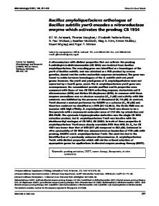
FIG. 1. (A) DNase I footprinting analysis of CodY binding to the ilvB promoter region. Lane 1, no CodY protein or GTP; lane 2, 125 nM CodY and no GTP; lanes 3 to 6, 125 nM CodY and increasing amounts of GTP. The bracket on the left shows the regions compared by densitometric analysis and corresponds to a portion of high-affinity region II of reference 36. The values under the lanes indicate the percent protected ilvB promoter DNA, compared to the leftmost lane, as assayed by densitometry. A sequencing ladder is shown at the right. (B) Plot of percent protection of ilvB promoter DNA versus GTP concentration in the presence of 125 nM CodY. We have assumed a linear relationship between GTP concentration and CodY binding but have not ruled out other potential kinetic relationships.
nucleotide GDP 3⬘-diphosphate (ppGpp), GTP 3⬘-diphosphate (pppGpp), or guanosine 5⬘-tetraphosphate (ppppG) could activate CodY (Fig. 3C). ppGpp and pppGpp did not promote binding of CodY to the ilvB promoter fragment. Addition of the synthetic nucleotide ppppG to the binding reaction mixtures resulted in better protection than was observed with corresponding concentrations of GTP.
GTP hydrolysis is not required for CodY binding to target DNA. Sequence analysis has indicated that putative GTP-binding motifs reminiscent of those found in small GTPase proteins (4) are present within CodY (31). To determine whether GTP hydrolysis is required for DNA binding by CodY, two nonhydrolyzable analogs of GTP, guanosine 5⬘-[␥-thio]triphosphate (GTP-␥-S) and guanosine 5⬘-[␥-imido]triphosphate (GMP-
VOL. 190, 2008
B. SUBTILIS CodY AND GTP
801
FIG. 2. DNase I footprinting analysis of CodY binding to the ilvB promoter in the presence of GTP. Leftmost lane, no CodY, no GTP; other lanes, various concentrations of CodY (0.1 to 3.2 M) with or without GTP (4 mM). The bracket on the left shows the regions compared by densitometric analysis and corresponds to a portion of high-affinity region II of reference 36. The values under the lanes indicate the percent protected ilvB promoter DNA, compared to the leftmost lane, as assayed by densitometry.
PNP), were used in footprinting reactions (Fig. 4). Both of these GTP analogs increased CodY binding to the ilvB promoter at concentrations similar to those at which GTP was active. Thus, we conclude that GTP hydrolysis is not necessary for CodY binding. This result is in accord with previous experiments in which CodY failed to cleave GTP (30). GTP and BCAAs act additively to induce CodY binding to target DNA. Further DNase I footprint analysis has demonstrated that GTP and isoleucine act additively to increase the affinity of CodY for DNA (Fig. 5). In this experiment, increasing concentrations of GTP were added to DNase I footprinting reaction mixtures containing 20 nM CodY. At this concentration of CodY, even 4 mM GTP was insufficient to induce full protection of the ilvB promoter DNA (Fig. 5, lanes 3 to 6). While addition of 10 mM isoleucine alone resulted in an increase in protection by CodY (Fig. 5, lane 8), addition of 10 mM isoleucine plus concentrations of GTP as low as 0.5 mM resulted in further protection of labeled DNA (Fig. 5, lanes 9 to 12). The reverse experiment was also performed, in which 20 nM CodY failed to fully protect ilvB promoter DNA in the presence of isoleucine alone (Fig. 5, lanes 13 to 15), but full protection was observed when 2 mM GTP was added simultaneously with isoleucine (Fig. 5, lanes 16 to 18). We conclude that isoleucine increases the affinity of CodY for GTP, in that protection is observed at lower GTP concentrations (⬃50% protection at 0.5 mM GTP) in the presence of isoleucine than in its absence. GTP also increases CodY’s affinity for isoleucine, as ⬃50% protection was observed at 2.5 mM isoleucine in the presence of GTP, compared to 5 to 10 mM in its absence. It can be inferred from these data that GTP and isoleucine probably bind to different sites on CodY since a saturating concentration of one effector does not preclude stimulation by the other effector. The observation that both effectors together confer increased protection of the ilvB promoter was confirmed by additional DNase I footprinting studies (Fig. 6). In this exper-
iment, increasing concentrations of the CodY protein were tested in the absence of effectors or in the presence of 2 mM GTP, the BCAAs at 10 mM each, or both effectors together. While high concentrations of CodY protein were sufficient to protect the highest-affinity binding site, region II, in the absence of effectors, lower protein concentrations were sufficient for protection upon the addition of either GTP or BCAAs to the binding reaction mixtures. While the BCAAs alone promoted CodY binding to the lower-affinity region III binding site (36), supplementation with both effectors further enhanced binding to this region, as well as to another low-affinity CodY binding site, region IV (36). These results confirm that the effectors work together to enhance the binding of the protein to its target sites. GTP and BCAAs act additively in runoff transcription assays. To further demonstrate that GTP and BCAAs act additively to induce the binding of CodY to its targets, runoff in vitro transcription assays were performed with a DNA fragment containing the ilvB promoter (Fig. 7). In reaction mixtures containing CodY protein and increasing concentrations of BCAAs, nearly complete repression of ilvB transcription was observed at concentrations of 1 to 4 mM BCAAs. In contrast, in the presence of 2 mM GTP, ilvB transcription was completely repressed by CodY at a lower concentration of BCAAs; under these conditions, only 0.2 to 0.4 mM BCAAs was necessary to shut off the expression of ilvB. Control reactions were performed simultaneously with the veg promoter, which has been shown not to be regulated by CodY (27, 36). As expected, addition of CodY, BCAAs, and GTP to reaction mixtures containing veg as the template did not appreciably affect expression. DISCUSSION The affinity of CodY for GTP, as determined indirectly with a DNase I footprinting assay, is in the millimolar range. This
802
HANDKE ET AL.
J. BACTERIOL.
FIG. 3. (A) DNase I footprinting analysis of ilvB promoter binding by CodY in the presence of various nucleotides. Lane 1, 125 nM CodY without nucleotides; other lanes, 125 nM CodY with 2, 5, or 10 mM nucleotides, as represented by the increasing size of the wedge. (B) DNase I footprinting analysis of ilvB promoter binding by CodY in the presence of various nucleotides. Lane 1, 125 nM CodY without nucleotides; other lanes, 125 nM CodY with 2, 5, or 10 mM nucleotides, as represented by the increasing size of the wedge. (C) DNase I footprinting analysis of ilvB promoter binding by CodY in the presence of various nucleotides. Lane 1, 150 nM CodY without nucleotides; other lanes, 150 nM CodY with 1, 2, or 4 mM nucleotides, as represented by the increasing size of the wedge.
result stands in contrast to findings for other GTP-binding proteins, which have affinities for GTP in the nanomolar range (4), but is consistent with the role of CodY in the cell. CodY’s relatively low affinity for GTP allows the protein to distinguish between 2 to 3 mM GTP, the concentration in rapidly growing cells, and 300 M GTP, the concentration in early-stationaryphase cells (42; T. Blais, T. Conway, T. Henkin, and A. L. Sonenshein, unpublished observation). The drop in intracellular GTP during stationary phase can be partly attributed to activation of the stringent response (6), which reduces the pool of GTP both directly and indirectly. The stringent response is initiated in the cell when an uncharged tRNA enters into the
ribosome acceptor site. As a result, the ribosome-associated RelA protein is activated and catalyzes a reaction between GTP and ATP that produces pppGpp. This nucleotide is further changed into ppGpp by multiple mechanisms (6). In addition, the accumulation of ppGpp lowers the intracellular GTP indirectly by inhibition of IMP dehydrogenase (28), an enzyme required for de novo guanine nucleotide biosynthesis. Inaoka and Ochi have recently shown that a relA strain of B. subtilis is impaired in competence and sporulation (15). It was demonstrated that GTP pools remain high in this mutant, likely resulting in sustained CodY activation. In agreement with this notion, when both codY and relA were deleted, com-
VOL. 190, 2008
FIG. 4. DNase I footprinting analysis of ilvB promoter binding by CodY in the presence of nonhydrolyzable analogs of GTP. Lane 1, no CodY or nucleotides; lane 2, 125 nM CodY without effectors; other lanes, 125 nM CodY with 2, 5, or 10 mM nucleotides, as represented by the increasing size of the wedge.
petence and sporulation efficiencies were restored. Our observation that pppGpp and ppGpp do not activate CodY is consistent with the current model that depletion of GTP to low levels by stringent-response activation renders CodY unable to interact with this effector and results in inactivation of CodY. Recent X-ray crystallography studies performed by Levdikov et al. (20) have given clues about the residues of CodY important in BCAA binding. A hydrophobic pocket in the N-terminal half of CodY appears to envelop the isobutyl side chain of Ile, while other nearby residues appear to participate in polar interactions with the amino and carboxyl groups of Ile. In contrast, the means by which CodY binds GTP remains unclear. Results presented here indicate that this interaction is specific; several other nucleotides tested, even at a 10 mM
B. SUBTILIS CodY AND GTP
803
concentration, did not promote CodY binding to its target site. Since GDP is not an effector of CodY, the ␥ phosphate of GTP is essential for activation of CodY. However, the GTP-binding region of CodY must be able to accommodate an additional bulky phosphate group, as ppppG was able to activate CodY. Further, the two-amino group of the guanine base appears to be important because ITP, which lacks this group, is not an effector of CodY. In contrast, dGTP enhanced CodY binding to the ilvB promoter, indicating that CodY does not distinguish between deoxyribose and ribose forms of GTP. We assume that GTP is the primary nucleotide effector of CodY in cells since the intracellular dGTP concentration is much lower than that of GTP (3). The presence of GTP increases the affinity of CodY for the ilvB target site fourfold. Two possibilities have been put forward to explain how CodY interacts with GTP (20, 31). First, three putative GTPbinding motifs common to the GTPase superfamily of proteins, termed G1, G3, and G4, have been found in CodY (31). Despite the strong conservation of the primary sequences of these motifs, several lines of evidence cast doubt on their roles in the interaction of CodY with GTP. First, these motifs are typically found within a single domain in small GTPase proteins (20). In CodY, the G1 motif is found in the N-terminal half of the protein while the G3 and G4 motifs are in the C-terminal half and on different faces of this domain (20). As a result, a large conformational change would be necessary to form a site for GTP binding. However, partial proteolysis, a technique used to detect changes in protein structure (5), with five different proteases has failed to reveal any major conformational change in CodY induced by interaction with GTP (P. Joseph, L. D. Handke, and A. L. Sonenshein, unpublished results). In addition, in members of the GTPase superfamily of proteins, the side chain from a highly conserved aspartic acid residue in the G4 motif (NXXD) forms hydrogen bonds with the guanine base (4, 33, 45). In CodY, this position is occupied by a leucine residue (31).
FIG. 5. DNase I footprinting analysis of the additive effects of GTP and isoleucine on CodY binding to the ilvB promoter. Lane 1, no CodY or effectors; lane 2, 20 nM CodY without effectors; other lanes, 20 nM CodY and effectors as indicated. When the GTP concentration was varied, it was at 0.5, 1, 2, or 4 mM, as represented by the increasing size of the wedge. When the isoleucine concentration was varied, it was at 2.5, 5, or 10 mM, as represented by the increasing size of the wedge. The bracket on the left shows the regions compared by densitometric analysis and corresponds to a portion of high-affinity region II of reference 36. The values under the lanes indicate the percent protected ilvB promoter DNA, compared to the leftmost lane, as assayed by densitometry.
804
HANDKE ET AL.
J. BACTERIOL.
FIG. 6. DNase I footprinting analysis of the additive effects of GTP and BCAAs on CodY binding to the ilvB promoter. First set of lanes, various CodY concentrations (0, 50, 100, 300, and 500 nM) in the absence of effectors (⫺); second set of lanes, various CodY concentrations in the presence of 2 mM GTP (⫹G); third set of lanes, various CodY concentrations in the presence of 10 mM BCAAs (⫹B); fourth set of lanes, various CodY concentrations in the presence of 2 mM GTP and 10 mM BCAAs (⫹G ⫹B). A sequencing ladder is shown at the right.
An alternative view is that CodY binds GTP through a GAF domain that overlaps the site of Ile interaction (20). These domains, which have been described in phosphodiesterases and adenylyl cyclases, have been shown to bind nucleotides, including cGMP, as effectors (13, 14). Binding of CodY to the ilvB promoter region was not stimulated by cGMP. Genetic experiments are in progress to assess the roles of the three putative GTP-binding motifs, the GAF domain, and other regions of CodY in GTP binding. Partial-proteolysis studies have demonstrated that CodY un-
dergoes a major change in conformation in the presence of BCAAs (20). Further, it has been shown that upon binding of BCAAs to the N-terminal domain of CodY, there is a shift in the position of the C-terminal domains of the CodY dimer (V. Levdikov and A. J. Wilkinson, personal communication). By contrast, any conformational change induced by GTP must be subtle since no conformational change induced by GTP has been detected. DNase I footprinting and in vitro transcription assays demonstrated that CodY binds to the ilvB promoter more tightly in
FIG. 7. In vitro transcription analysis of ilvB expression in the presence of either BCAAs alone or BCAAs and GTP as effectors. In vitro transcription reactions were performed with 200 nM CodY when indicated and a 453-bp fragment of the ilvB promoter. Various concentrations of BCAAs were added as indicated, in the presence or absence of 2 mM GTP. Each tube additionally contained 150 M GTP as part of the transcription reaction mixture. A promoter known not to be CodY regulated, veg, was used as a control in these assays.
B. SUBTILIS CodY AND GTP
VOL. 190, 2008
the presence of both effectors than of either effector alone. Whether binding of both effectors is required for repression of CodY targets in vivo is unclear, but recent in vitro evidence indicates that for at least one CodY-regulated gene, ackA, BCAAs are sufficient for CodY activity (35). This observation indicates that binding of CodY to a given target may depend on which effector is bound. In fact, the observation that partial proteolysis reveals only the conformational change induced by BCAAs implies that binding of the two effectors changes the protein structure in different ways. Accumulating evidence suggests that CodY has a role in the regulation of virulence gene expression in pathogenic grampositive bacteria (43). Two virulence-regulatory systems in Streptococcus pyogenes, pel/sagA and mga, have recently been shown to be positively regulated by CodY (23). In Clostridium difficile, a codY null mutation causes strong derepression of all of the genes of the pathogenicity locus and CodY binds in vitro to the promoter region of tcdR, the gene for the locus-specific regulator (9, 24). This binding is synergistically activated by GTP and BCAAs. Disruption of the codY gene in Staphylococcus aureus strains SA564 and UAMS-1 has recently been shown to derepress the expression of several virulence factors (C. Majerczyk, M. Sadykov, T. T. Luong, C. Lee, G. Somerville, P. Dunman, and A. L. Sonenshein, submitted for publication); Tu Quoc et al. (44) found that CodY is needed for biofilm formation in S. aureus strain S30. The activity of S. aureus CodY in vitro is strongly increased by the combination of GTP and BCAAs (C. Majerczyk and A. L. Sonenshein, unpublished results). Not all CodY-like proteins are activated by GTP, however. In Lactococcus lactis, addition of decoyinine, an inhibitor of guanine nucleotide synthesis, does not result in derepression of opp, a CodY-regulated gene in this species (29). Further, GTP does not enhance the binding of purified CodY protein from L. lactis (8) or from Streptococcus pneumoniae (R. P. Shivers and A. L. Sonenshein, unpublished observation) in gel shift assays. Both of these CodY proteins are stimulated by BCAAs, however. The notion that CodY regulates virulence factor expression, and the strong conservation of CodY among the low-G⫹C gram-positive bacteria, suggests a novel treatment for certain infections. Analogs of guanine nucleotides or BCAAs might alter CodY activity in such a way as to limit the expression of virulence genes. Ongoing studies seek to test the feasibility of this approach. ACKNOWLEDGMENTS We thank Boris Belitsky for critical review of the manuscript, Silvia Picossi-Gon ˜i for expert advice on footprinting assays, and Boris Belitsky, Sean Dineen, Anuradha Villapakkam, and AJ Wilkinson for helpful discussions. We thank Jade Wang for the gift of pppGpp. This work was supported by a research grant (GM042219 to A.L.S.) and a National Research Service award (GM082155 to L.D.H.) from the U.S. Public Health Service. REFERENCES 1. Bergara, F., C. Ibarra, J. Iwamasa, J. C. Patarroyo, R. Aguilera, and L. M. Marquez-Magana. 2003. CodY is a nutritional repressor of flagellar gene expression in Bacillus subtilis. J. Bacteriol. 185:3118–3126. 2. Blagova, E. V., V. M. Levdikov, K. Tachikawa, A. L. Sonenshein, and A. J. Wilkinson. 2003. Crystallization of the GTP-dependent transcriptional regulator CodY from Bacillus subtilis. Acta Crystallogr. D Biol. Crystallogr. 59:155–157.
805
3. Bochner, B. R., and B. N. Ames. 1982. Complete analysis of cellular nucleotides by two-dimensional thin layer chromatography. J. Biol. Chem. 257: 9759–9769. 4. Bourne, H. R., D. A. Sanders, and F. McCormick. 1991. The GTPase superfamily: conserved structure and molecular mechanism. Nature 349:117–127. 5. Carey, J. 2000. A systematic and general proteolytic method for defining structural and functional domains of proteins. Methods Enzymol. 328:499– 514. 6. Cashel, M., D. Gentry, V. Hernandez, and D. Vinella. 1996. The stringent response, p. 1458–1496. In F. Neidhardt and R. Curtiss (ed.), Escherichia coli and Salmonella: cellular and molecular biology, 2nd ed. ASM Press, Washington, DC. 7. Debarbouille, M., R. Gardan, M. Arnaud, and G. Rapoport. 1999. Role of bkdR, a transcriptional activator of the SigL-dependent isoleucine and valine degradation pathway in Bacillus subtilis. J. Bacteriol. 181:2059–2066. 8. den Hengst, C. D., P. Curley, R. Larsen, G. Buist, A. Nauta, D. van Sinderen, O. P. Kuipers, and J. Kok. 2005. Probing direct interactions between CodY and the oppD promoter of Lactococcus lactis. J. Bacteriol. 187:512–521. 9. Dineen, S. S., A. C. Villapakkam, J. T. Nordman, and A. L. Sonenshein. 2007. Repression of Clostridium difficile toxin gene expression by CodY. Mol. Microbiol. 66:206–219. 10. Ferson, A. E., L. V. Wray, Jr., and S. H. Fisher. 1996. Expression of the Bacillus subtilis gabP gene is regulated independently in response to nitrogen and amino acid availability. Mol. Microbiol. 22:693–701. 11. Freese, E., J. E. Heinze, and E. M. Galliers. 1979. Partial purine deprivation causes sporulation of Bacillus subtilis in the presence of excess ammonia, glucose and phosphate. J. Gen. Microbiol. 115:193–205. 12. Fujita, M., and R. Losick. 2005. Evidence that entry into sporulation in Bacillus subtilis is governed by a gradual increase in the level and activity of the master regulator Spo0A. Genes Dev. 19:2236–2244. 13. Ho, Y. S., L. M. Burden, and J. H. Hurley. 2000. Structure of the GAF domain, a ubiquitous signaling motif and a new class of cyclic GMP receptor. EMBO J. 19:5288–5299. 14. Hurley, J. H. 2003. GAF domains: cyclic nucleotides come full circle. Sci. STKE 2003:PE1. doi:10.1126/stke.2003.164.pe1. 15. Inaoka, T., and K. Ochi. 2002. RelA protein is involved in induction of genetic competence in certain Bacillus subtilis strains by moderating the level of intracellular GTP. J. Bacteriol. 184:3923–3930. 16. Inaoka, T., K. Takahashi, M. Ohnishi-Kameyama, M. Yoshida, and K. Ochi. 2003. Guanine nucleotides guanosine 5⬘-diphosphate 3⬘-diphosphate and GTP co-operatively regulate the production of an antibiotic bacilysin in Bacillus subtilis. J. Biol. Chem. 278:2169–2176. 17. Joseph, P., M. Ratnayake-Lecamwasam, and A. L. Sonenshein. 2005. A region of Bacillus subtilis CodY protein required for interaction with DNA. J. Bacteriol. 187:4127–4139. 18. Kim, H. J., S. I. Kim, M. Ratnayake-Lecamwasam, K. Tachikawa, A. L. Sonenshein, and M. Strauch. 2003. Complex regulation of the Bacillus subtilis aconitase gene. J. Bacteriol. 185:1672–1680. 19. Kim, H. J., A. Roux, and A. L. Sonenshein. 2002. Direct and indirect roles of CcpA in regulation of Bacillus subtilis Krebs cycle genes. Mol. Microbiol. 45:179–190. 20. Levdikov, V. M., E. Blagova, P. Joseph, A. L. Sonenshein, and A. J. Wilkinson. 2006. The structure of CodY, a GTP- and isoleucine-responsive regulator of stationary phase and virulence in gram-positive bacteria. J. Biol. Chem. 281: 11366–11373. 21. Lopez, J. M., A. Dromerick, and E. Freese. 1981. Response of guanosine 5⬘-triphosphate concentration to nutritional changes and its significance for Bacillus subtilis sporulation. J. Bacteriol. 146:605–613. 22. Lopez, J. M., C. L. Marks, and E. Freese. 1979. The decrease of guanine nucleotides initiates sporulation of Bacillus subtilis. Biochim. Biophys. Acta 587:238–252. 23. Malke, H., K. Steiner, W. M. McShan, and J. J. Ferretti. 2006. Linking the nutritional status of Streptococcus pyogenes to alteration of transcriptional gene expression: the action of CodY and RelA. Int. J. Med. Microbiol. 296:259–275. 24. Mani, N., D. Lyras, L. Barroso, P. Howarth, T. Wilkins, J. I. Rood, A. L. Sonenshein, and B. Dupuy. 2002. Environmental response and autoregulation of Clostridium difficile TxeR, a sigma factor for toxin gene expression. J. Bacteriol. 184:5971–5978. 25. Mathiopoulos, C., J. P. Mueller, F. J. Slack, C. G. Murphy, S. Patankar, G. Bukusoglu, and A. L. Sonenshein. 1991. A Bacillus subtilis dipeptide transport system expressed early during sporulation. Mol. Microbiol. 5:1903– 1913. 26. Mitani, T., J. E. Heinze, and E. Freese. 1977. Induction of sporulation in Bacillus subtilis by decoyinine or hadacidin. Biochem. Biophys. Res. Commun. 77:1118–1125. 27. Molle, V., Y. Nakaura, R. P. Shivers, H. Yamaguchi, R. Losick, Y. Fujita, and A. L. Sonenshein. 2003. Additional targets of the Bacillus subtilis global regulator CodY identified by chromatin immunoprecipitation and genomewide transcript analysis. J. Bacteriol. 185:1911–1922. 28. Ochi, K., J. Kandala, and E. Freese. 1982. Evidence that Bacillus subtilis
806
29.
30. 31. 32. 33. 34. 35. 36. 37. 38.
HANDKE ET AL. sporulation induced by the stringent response is caused by the decrease in GTP or GDP. J. Bacteriol. 151:1062–1065. Petranovic, D., E. Guedon, B. Sperandio, C. Delorme, D. Ehrlich, and P. Renault. 2004. Intracellular effectors regulating the activity of the Lactococcus lactis CodY pleiotropic transcription regulator. Mol. Microbiol. 53:613– 621. Ratnayake-Lecamwasam, M. 2001. Ph.D. thesis. Tufts University, Boston, MA. Ratnayake-Lecamwasam, M., P. Serror, K. W. Wong, and A. L. Sonenshein. 2001. Bacillus subtilis CodY represses early-stationary-phase genes by sensing GTP levels. Genes Dev. 15:1093–1103. Sambrook, J., E. F. Fritsch, and T. Maniatis. 1989. Molecular cloning: a laboratory manual, 2nd ed. Cold Spring Harbor Laboratory Press, Cold Spring Harbor, NY. Scheffzek, K., C. Klebe, K. Fritz-Wolf, W. Kabsch, and A. Wittinghofer. 1995. Crystal structure of the nuclear Ras-related protein Ran in its GDP-bound form. Nature 374:378–381. Serror, P., and A. L. Sonenshein. 1996. CodY is required for nutritional repression of Bacillus subtilis genetic competence. J. Bacteriol. 178:5910– 5915. Shivers, R. P., S. S. Dineen, and A. L. Sonenshein. 2006. Positive regulation of Bacillus subtilis ackA by CodY and CcpA: establishing a potential hierarchy in carbon flow. Mol. Microbiol. 62:811–822. Shivers, R. P., and A. L. Sonenshein. 2004. Activation of the Bacillus subtilis global regulator CodY by direct interaction with branched-chain amino acids. Mol. Microbiol. 53:599–611. Shivers, R. P., and A. L. Sonenshein. 2005. Bacillus subtilis ilvB operon: an intersection of global regulons. Mol. Microbiol. 56:1549–1559. Slack, F. J., J. P. Mueller, and A. L. Sonenshein. 1993. Mutations that relieve
J. BACTERIOL.
39.
40.
41.
42.
43. 44.
45.
46.
nutritional repression of the Bacillus subtilis dipeptide permease operon. J. Bacteriol. 175:4605–4614. Slack, F. J., J. P. Mueller, M. A. Strauch, C. Mathiopoulos, and A. L. Sonenshein. 1991. Transcriptional regulation of a Bacillus subtilis dipeptide transport operon. Mol. Microbiol. 5:1915–1925. Slack, F. J., P. Serror, E. Joyce, and A. L. Sonenshein. 1995. A gene required for nutritional repression of the Bacillus subtilis dipeptide permease operon. Mol. Microbiol. 15:689–702. Soga, T., Y. Ohashi, Y. Ueno, H. Naraoka, M. Tomita, and T. Nishioka. 2003. Quantitative metabolome analysis using capillary electrophoresis mass spectrometry. J. Proteome Res. 2:488–494. Soga, T., Y. Ueno, H. Naraoka, K. Matsuda, M. Tomita, and T. Nishioka. 2002. Pressure-assisted capillary electrophoresis electrospray ionization mass spectrometry for analysis of multivalent anions. Anal. Chem. 74:6224–6229. Sonenshein, A. L. 2005. CodY, a global regulator of stationary phase and virulence in gram-positive bacteria. Curr. Opin. Microbiol. 8:203–207. Tu Quoc, P. H., P. Genevaux, M. Pajunen, H. Savilahti, C. Georgopoulos, J. Schrenzel, and W. L. Kelley. 2007. Isolation and characterization of biofilm formation-defective mutants of Staphylococcus aureus. Infect. Immun. 75: 1079–1088. Valencia, A., M. Kjeldgaard, E. F. Pai, and C. Sander. 1991. GTPase domains of ras p21 oncogene protein and elongation factor Tu: analysis of three-dimensional structures, sequence families, and functional sites. Proc. Natl. Acad. Sci. USA 88:5443–5447. Wray, L. V., Jr., A. E. Ferson, and S. H. Fisher. 1997. Expression of the Bacillus subtilis ureABC operon is controlled by multiple regulatory factors including CodY, GlnR, TnrA, and Spo0H. J. Bacteriol. 179:5494–5501.