18 Wild, J., Rossmeissl, P., Walter, W. A. and Gross, C. A. (1996) J. Bacteriol. 178,. 3608â ... 34 Scho$n, U. and Schumann, W. (1995) FEMS Microbiol. Lett. 134 ...
367
Biochem. J. (2000) 348, 367–373 (Printed in Great Britain)
Interaction of Bacillus subtilis CsaA with SecA and precursor proteins Jo$ rg P. MU$ LLER*1, Jo$ rg OZEGOWSKI†, Stefan VETTERMANN‡, Jelto SWAVING§, Karel H. M. VAN WELY§ and Arnold J. M. DRIESSEN§ *Institute for Molecular Biology, Jena University, Winzerlaer Strasse 10, D-07745 Jena, Germany, †Institute of Experimental Microbiology, Jena University, Winzerlaer Str. 10, D-07745 Jena, Germany, ‡ProThera GmbH, Winzerlaer Str. 10,D-07745 Jena, Germany, and §Department of Microbiology, Groningen Biomolecular Sciences and Biotechnology Institute, University of Groningen, Kerklaan 30, NL-9751 Haren, The Netherlands
CsaA from the Gram-positive bacterium Bacillus subtilis has been identified previously as a suppressor of the growth and protein-export defect of Escherichia coli secA(Ts) mutants. CsaA has chaperone-like activities in io and in itro. To examine the role of CsaA in protein export in B. subtilis, expression of the csaA gene was repressed. While export of most proteins remained unaffected, export of at least two proteins was significantly reduced upon CsaA depletion. CsaA co-immunoprecipitates and co-purifies with the SecA proteins of E. coli and B. subtilis, and binds the B. subtilis preprotein prePhoB. Purified CsaA
stimulates the translocation of prePhoB into E. coli membrane vesicles bearing the B. subtilis translocase, whereas it interferes with the SecB-mediated translocation of proOmpA into membrane vesicles of E. coli. The specific interaction with the SecA translocation ATPase and preproteins suggests that CsaA acts as a chaperone that promotes the export of a subset of preproteins in B. subtilis.
INTRODUCTION
dependent manner [2]. A second targeting factor, consisting of the Ffh protein and the 4.5 S RNA, assists the export of SecBindependent exported proteins [16]. Other molecular chaperones, such as the heat-shock proteins DnaK\DnaJ\GrpE [17–19] and GroEL\GroES [20], appear also to be involved under specific conditions. The central components of the protein-translocation system of B. subtilis are similar to those of E. coli. So far, homologues of Ffh, 4.5 S RNA, SecA, SecY, SecE, SecG, SecDF and several type-I signal peptidases have been identified (for review see [21]). So far, it is unknown if chaperones are involved in protein translocation in B. subtilis. B. subtilis lacks a SecB homologue [22] and this has raised the question of whether other chaperones are involved in protein translocation. Depletion of signalrecognition-peptide (SRP) components impairs protein translocation [23,24], and the B. subtilis Ffh interacts directly with SecA and promotes the formation of soluble SecA–preprotein complexes [25]. This has led to the suggestion that SRP of B. subtilis not only acts as a targeting factor in co-translational translocation, but also stimulates post-translational translocation of preproteins [25]. The B. subtilis CsaA protein specifies chaperone-like activities possibly related to protein translocation [26]. The csaA gene was identified as a suppressor of growth and secretion defects of E. coli secA(Ts) strains [27]. Presence of CsaA stimulated precursor processing in secA, secB, groEL and dnaJ mutant strains of E. coli and it suppressed the growth defects of dnaK, dnaJ and grpE mutants of E. coli. CsaA stimulates the reactivation of heat-denatured firefly luciferase in groEL, groES, dnaK and grpE mutant strains of E. coli, and prevents the aggregation of heat-denatured luciferase in itro. The exact mechanism by which CsaA suppresses the growth and secretion defects of E. coli secA(Ts) strains is unknown. CsaA may either improve the translocation-competence of exported preproteins, thereby making them better substrates for mutant SecA proteins, or stimulate the translocation activity of the
In Eubacteria, proteins can be translocated across the cytoplasmic membrane and secreted into the medium. These proteins are usually synthesized as precursors with an N-terminal extension, the signal peptide (for review, see [1]), which is essential for directing them into the export pathway (for review, see [2]). Genetically and biochemically the best-characterized bacterial export system is that of Escherichia coli (for reviews, see [2,3]). This system, termed preprotein translocase, consists of the peripheral membrane protein SecA [4] and a multi-subunit membrane-protein complex with SecY, SecE and SecG as subunits [5]. The SecYEG complex acts as a receptor for SecA, and functions as a preprotein-conducting channel [6,7]. During or shortly after the translocation of the preprotein across the membrane, the signal peptide is removed by signal peptidase(s) (for review, see [8]), a prerequisite for the release of the mature protein from the membrane [9]. The integral membrane proteins SecD and SecF [10] are not essential for precursor protein translocation but, when overproduced, stabilize the SecYEGbound SecA in a membrane-inserted state [11]. Whereas bacterial protein translocation is largely uncoupled from ongoing translation [12], efficient export requires chaperones to maintain preproteins in a translocation-competent state and to target them to the membrane-associated part of the translocase. In E. coli, SecB is an export-dedicated cytosolic chaperone that is required for efficient export of a subset of preproteins [13]. SecB stabilizes preproteins in an unfolded, nonaggregated state [14], and binds post-translationally or at the late co-translational stage to the mature region of these proteins [15]. The SecB–preprotein complex is then targeted to the SecYEGbound SecA. The preprotein is subsequently transferred from SecB to SecA, and, upon the ATP-dependent initiation of translocation, the SecB is released into the cytosol [3]. In io, only a subset of preproteins appears to be translocated in a SecB-
Key words : chaperone, co-immunoprecipitation, co-purification, in itro translocation, protein translocation.
Abbreviations used : NaPi, inorganic phosphate, NaH2PO4 ; His6-CsaA, hexa-histidine-tagged CsaA ; His6-SecB, hexa-histidine-tagged SecB ; His6prePhoB, hexa-histidine-tagged prePhoB ; IPTG, isopropyl β-D-thiogalactoside ; Ni-NTA, Ni2+-nitrilotriacetate ; SRP, signal recognition peptide. 1 To whom correspondence should be addressed (e-mail jmueller!imb-jena.de). # 2000 Biochemical Society
J. P. Mu$ ller and others
368
mutant SecA proteins. Expression of CsaA in E. coli suppresses synthesis of SecB [26], whereas an impaired function of general chaperones causes an elevation in SecB expression [28]. These data further suggest a chaperone function of CsaA that could possibly be related to protein translocation. In this paper we have further examined the role of CsaA in protein translocation in B. subtilis. CsaA affects the efficient secretion of some preproteins, and interacts specifically with SecA and preproteins. The data suggest that CsaA acts as a chaperone that promotes the export of a subset of preproteins in B. subtilis.
EXPERIMENTAL Bacterial strains, plasmids and media Bacterial strains and plasmids used in this study are listed in Table 1. Strains were grown in TY medium (Bacto Tryptone\ Bacto Yeast extract) or on TY plates [28]. Pulse labelling of B. subtilis was carried out in defined HPDM medium [29], casamino acids were replaced by amino acids (0.02 mg\ml) excluding methionine and cysteine. If required, ampicillin (80 µg\ml), erythromycin (5 µg\ml) or kanamycin (20 µg\ml) were added.
Biochemicals B. subtilis SecA [32], E. coli SecA [33] and the B. subtilis GroELS complex [34] were isolated from overproducing strains as described. proOmpA [7] and hexa-histidine-tagged prePhoB (His -prePhoB) [35] were purified as described. Purified pro' OmpA, prePhoB and SecA proteins were labelled with carrierfree "#&I according to van Wely et al. [35]. Labelled preproteins were stored frozen in 6 M urea. Polyclonal antiserum was raised
Table 1
Bacterial strains and plasmids, and their genotype/phenotypes
IPTG, isopropyl β-D-thiogalactoside ; His6-CsaA, hexa-histidine-tagged CsaA ; His6-SecB, hexahistidine-tagged SecB. Strain/plasmid
Relevant genotype/phenotype
Strains B. subtilis DB104 his, nprE, aprE DB104 : : pMUTINcsaAh DB104, allows IPTG-inducible expression of csaA E. coli TG1 hsdD5/F traD36, proA+B+, ∆(lac-pro) lacIQ, lacZ ∆M15 TG1(pREP4) Host for overexpression of His-tagged proteins TG1(pREP4, pQE9csaA ) TG1 derivative strain, Kmr, Apr, allows IPTGinducible synthesis of His6-CsaA TG1(pREP4, pQE9secB ) TG1 derivative strain, Kmr, Apr, allows IPTGinducible synthesis of His6-SecB Plasmids pMUTIN2 pBR322-based integration vector for B. subtilis ; containing IPTG-inducible Pspac promoter, Apr, Emr pMUTINcsaAh pMUTIN2 derivative ; carries the 5h part of csaA pREP4 plasmid, containing lacI q repressor gene, Kmr pQE9 pBR322-based vector for IPTG-inducible synthesis of His6-tagged proteins, Apr pQE9csaA pQE9 derivative plasmid, Apr, allows IPTGinducible synthesis of His6-CsaA pQE9secB TG1 derivative plasmid, Apr, allows IPTGinducible synthesis of His6-SecB
# 2000 Biochemical Society
Reference
[30] This study
[31] This study This study This study
[40]
Qiagen Qiagen This study This study
against purified hexa-histidine-tagged CsaA (His -CsaA). Anti' sera against SecA from B. subtilis were from R. Freudl (Forschungszentrum Ju$ lich, Ju$ lich, Germany). Membrane vesicles were prepared from E. coli strain SF100 [36] containing plasmid pET605 overproducing SecYEG from E. coli [37] or plasmid pET822 overproducing SecYEG from B. subtilis DB104 as described in [38]. Membrane vesicles were treated with polyclonal antibodies against E. coli SecA to deplete and inactivate the endogenous SecA [38].
DNA manipulation/techniques Procedures for DNA purification, restriction, ligation, transformation of E. coli and agarose-gel electrophoresis were carried out as described by Sambrook et al. [39]. The nucleotide sequences of cloned PCR fragments were confirmed by DNA sequencing. To amplify csaAh, oligonucleotides 5h-TCTCGAATTCAATAGGAGAAAAAGGAGTT&(%-3h, localized 5h to the ribosomebinding site of csaA and incorporating an EcoRI restriction site, and 5h-GGACCAAGGGATCCGATTTAAATCCGGCG)!&-3h, localized within csaA and incorporating a BamHI restriction site, were used (the numbers refer to those in Mu$ ller et al. [27]). The csaAh gene was amplified by PCR from B. subtilis chromosomal DNA. The amplified fragment was digested with EcoRI and BamHI and cloned into pMUTIN2 [40] digested with the same enzymes. The resulting plasmid pMUTIN csaAh was integrated into the chromosome of B. subtilis DB104 via Campbell-type integration. For synthesis of His-tagged proteins secB and csaA genes were amplified excluding their ATG start codons and were inserted 3h to the His-coding region of pQE9. Genes were amplified by PCR using oligonucleotides 5h-CACGGATCCGAACAAAACAACACTG"!$-3h, incorporating a BamHI restriction site, and 5hCCGACTGCAGTCATTGAAGCATTACG&&)-3h, with a PstI restriction site, for secB (the numbers refer to those used in [41]), and oligonucleotides 5h-GGAGTTATTGGATCCGCAGTTATTGATGAC&*)-3h, with a BamHI restriction site, and 5h-GCCGATCTCTGCAGGCCTTTACGGCACACACG*)"-3h, incorporating a PstI restriction site, for csaA (the numbers refer to those used in [27]). PCR fragments were purified, digested with BamHI and PstI and inserted into pQE9 digested with the same enzymes. The resulting plasmids pQE9secB and pQE9csaA were transformed into E. coli TG1(pREP4).
Pulse–chase protein labelling and analysis of protein secretion Pulse–chase protein-labelling experiments were performed as described by van Dijl et al. [42]. B. subtilis was grown in minimal medium to exponential growth in the absence or presence of 1 mM isopropyl β--thiogalactoside (IPTG), pulse labelled with [$&S]methionine for 1 min and subsequently chased. Samples were taken 2 and 5 min post chase time and proteins secreted into the growth medium were collected as described in [43]. SDS\ PAGE was performed according to Laemmli [44]. "%C-Methylated proteins (Amersham International, Amersham, Bucks, U.K.) were used as molecular-size markers. Relative amounts of radioactivity were estimated by using a PhosphoImager (Fuji) and associated image-analysis software.
Purification of proteins His -CsaA and hexa-histidine-tagged SecB (His -SecB) were ' ' prepared from IPTG-induced E. coli TG1(pREP4, pQE9csaA) and TG1(pREP4, pQE9secB) cultures as abundant proteins and purified by Ni#+-nitrilotriacetate (Ni-NTA) agarose affinity chromatography. Purification was carried out under native
Bacillus chaperone CsaA conditions following standard protocols (Qiagen, Hilden, Germany). Cell lysates prepared in 50 mM NaPi (inorganic phosphate, NaH PO ), pH 8.0, and 300 mM NaCl, were supple# % mented with 10 mM imidazole and applied on to the Ni-NTA column. The column was washed three times with several column volumes of NaPi buffer containing 20 mM imidazole. His-tagged proteins were eluted with NaPi buffer containing 500 mM imidazole. CsaA from B. subtilis was purified immunologically. Affinitypurified rabbit antibodies against CsaA were covalently linked to CNBr-activated Sepharose. Exponentially grown B. subtilis DB104 cells were harvested and extracts were prepared from lysozyme-treated cells via sonication. Cleared cell extracts were loaded on to the anti-CsaA–Sepharose column at room temperature. Anti-CsaA–Sepharose was washed with 30 ml of phosphate buffer (50 mM, pH 7.0) containing 0.5 M NaCl until no detectable protein was eluted from the column. CsaA was eluted with 0.1 M acetic acid, pH 3.0, and subsequently lyophilized. To obtain SecA-free CsaA B. subtilis cell extracts were purified by preparative isoelectric focusing in Sephadex gel with a pH gradient from pH 4.0 to 9.0 for 18 h. After focusing, the Sephadex gel was cleaved to fractions and CsaA-containing fractions (as determined immunologically) were analysed by thin-layer polyacrylamide gel isoelectric focusing with an ampholyte solution (pH 3.5–10.0) and SDS\PAGE. The CsaA was precipitated with ammonium sulphate to a final concentration of 80 % saturation. The precipitated protein was dissolved, dialysed against H O and # lyophilized.
369
experiments, mixtures were incubated for 30 min at 37 mC without membrane vesicles prior to the translocation reactions. After 30 min of translocation at 37 mC samples were chilled on ice, treated with proteinase K (0.5 mg\ml) for 15 min, precipitated with 7.5 % trichloroacetic acid, washed with acetone, dissolved in SDS\PAGE sample buffer, and analysed by SDS\PAGE (10 % gel) and autoradiography.
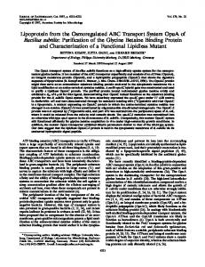
RESULTS Repression of CsaA expression results in alteration of protein secretion In B. subtilis strain DB104 : : pMUTIN2csaAh, csaA is localized downstream of the Pspac promoter, allowing the IPTG-inducible expression of the CsaA protein. Cells grown in the absence of IPTG were normally viable but Western-blotting analysis showed that they contained a dramatically reduced cellular level of CsaA. Induction of the Pspac promoter resulted in a production level of CsaA that was about 2-fold higher than in the wild type (Figure 1A). To study the function of CsaA in protein secretion, the pattern of proteins secreted by B. subtilis DB104 : : pMUTIN2csaAh grown in the absence or presence of IPTG was determined. While the pattern of secreted proteins of the strain grown in presence of IPTG was indistinguishable from that of the wild type, repression of csaA expression resulted in a decrease in at least two proteins with apparent molecular masses of 19 and 36 kDa (Figure 1B). In particular, the 36-kDa protein
Western blotting Proteins isolated from exponentially growing cultures were assayed by Western blotting [45]. Cell lysates were prepared by boiling (5 min) in sample buffer and subjecting to SDS\PAGE [44]. Separated proteins were transferred to a nitrocellulose membrane (Schleicher & Schu$ ll), as described by Towbin et al. [45]. CsaA and SecA proteins were detected with specific polyclonal antibodies and alkaline phosphatase-conjugated goat antirabbit antibodies (Bio-Rad) according to the manufacturer’s instructions. Similar amounts of total cell protein were loaded on to each lane.
Binding studies Binding of ["#&I]His -prePhoB or ["#&I]SecA to CsaA was ' measured as follows : 1 µg of "#&I-labelled protein and 1 µg of CsaA were incubated at room temperature in 50 µl of Hepes buffer (50 mM, pH 7.6, additional buffer components as indicated). After 60 min, 5 µl of CsaA-specific antiserum precomplexed with 5 mg of Protein A–Sepharose was added and the mixture was further incubated for 60 min with regular vortexing. Subsequently, the Protein A–Sepharose beads were washed five times with 500 µl of Hepes buffer, pH 7.6. "#&I-Labelled proteins bound to Protein A–Sepharose beads were counted in Optima Gold scintillation liquid (Packard Instruments B.V., Groningen, The Netherlands).
Translocation assay The efficiency of translocation in itro of ["#&I]proOmpA and ["#&I]prePhoB into E. coli membrane vesicles was assayed by treatment with proteinase K [46]. When indicated, 1.0 µg of purified E. coli or B. subtilis SecA protein, 2.0 µg of E. coli SecB or 4 µg of purified B. subtilis GroESL or B. subtilis CsaA were added. Reactions were started by the addition of 1 µl of "#&Ilabelled precursor protein (0.5 µg of protein). For preincubation
Figure 1
Repression of csaA in B. subtilis affects protein export
(A) B. subtilis strain DB104 (lane 1) and strain DB104 : : pMUTIN2csaAh (lanes 2 and 3) were grown in TY medium in the absence (lanes 1 and 2) or presence (lane 3) of 1 mM IPTG. Cell extracts were analysed by SDS/PAGE and Western blotting using CsaA-specific antibodies. (B) Protein secretion of B. subtilis DB104 : : pMUTIN2csaAh was analysed by pulse labelling of cells grown in the absence (lanes 1 and 2) or presence (lanes 3 and 4) of IPTG. Secreted proteins were collected by filtration and analysed by SDS/PAGE and autoradiography. # 2000 Biochemical Society
370
J. P. Mu$ ller and others
Figure 3
B. subtilis SecA co-purifies with CsaA
A cell lysate of B. subtilis DB104 was loaded on to an anti-CsaA–Sepharose column, washed with NaCl buffer (0.5 M) and the immunologically bound CsaA was eluted with 0.1 M acetic acid. The eluate was analysed by SDS/PAGE and Coomassie Brilliant Blue staining (lane 1) or Western blotting using polyclonal antibodies (pAb) directed against the B. subtilis SecA (lane 2) or CsaA (lane 4). The asterisk indicates the localization of dimeric CsaA. As a control, a lysate of B. subtilis DB104 : : pMUTIN2csaAh grown in the absence of IPTG was loaded on to the antiCsaA–Sepharose column, and the fraction eluted with 0.1 M acetic acid was stained immunologically with antibodies directed against the B. subtilis SecA (lane 3).
Figure 2
E. coli SecA interacts with His6-CsaA and His6-SecB
E. coli strains TG1(pREP4, pQE9csaA) (A, B), TG1(pREP4, pQE9secB) (C) and TG1(pREP4, pQE9) (D) were cultivated in TY medium to exponential growth and the phage T5 promoter was derepressed by adding IPTG (final concentration, 2 mM) to the culture medium. After 2 h, cells were harvested and lysed by sonication, and the cleared cell extracts were loaded on to Ni-NTA columns. Aliquots of non-bound fraction (FT, lane 1), wash (W1–W3, lanes 2–4) and elution fractions (E1–E8, lanes 5–12) were analysed by SDS/PAGE and Western blotting using antibodies directed against CsaA (A) and E. coli SecA (B–D). The asterisk indicates the position of dimeric CsaA.
seemed completely absent in the supernatant of CsaA-depleted cells. Both protein bands were isolated from Coomassie Brilliant Blue-strained SDS\PAGE, and N-terminally sequenced. While the N-terminus of the 36-kDa protein appeared blocked, the N-terminus of the 19-kDa protein corresponded to KENHTFSGED. This amino acid sequence matched the amino acid residues 33–42 of the precursor of YvaY, a protein with unknown function and a calculated molecular mass of 18.8 kDa.
E. coli SecA co-purifies with His6-CsaA and His6-SecB His-tagged CsaA protein was overproduced in E. coli TG1 (pREP4, pQE9csaA) and purified by Ni-NTA-column chromatography. The imidazole-eluted protein fraction predominantly contained the 13-kDa CsaA protein, and a protein with an apparent molecular mass of about 30 kDa that stained with antibodies directed against CsaA (Figure 2A). The latter probably represents some dimeric CsaA. Immunodetection with # 2000 Biochemical Society
SecA antibodies revealed that SecA co-eluted with CsaA from the column (Figure 2B). To exclude the possibility that SecA non-specifically binds to the Ni-NTA resin, a lysate of E. coli TG1(pREP4) cells that do not express the His-tagged CsaA was loaded on to an Ni-NTA column (Figure 2D). Under those conditions, no SecA was retained by the column, suggesting an interaction between CsaA and SecA. Since SecA is known to interact with SecB in E. coli [6,7,47], His -SecB was purified from ' E. coli TG1(pREP4,pQE9secB) under conditions used for purification of His -CsaA. SecA was found to co-elute with His ' ' SecB (Figure 2C). These results indicate that SecA was retained in the Ni-NTA column via specific binding to His-tagged CsaA or SecB.
B. subtilis SecA interacts with CsaA To establish whether the interaction of CsaA with SecA can be observed in B. subtilis, CsaA was purified from B. subtilis DB104. Cell extracts were loaded on to an anti-CsaA–Sepharose column, washed and eluted as described in the Experimental section. The eluate was analysed by SDS\PAGE and Coomassie Brilliant Blue staining (Figure 3, lane 1). In addition to CsaA, a protein co-eluted with an apparent molecular mass of about 90 kDa. The latter could be immunostained with an antibody directed against the B. subtilis SecA (Figure 3, lane 2), suggesting that the B. subtilis SecA associates with CsaA. As a control, a lysate of B. subtilis DB104 : : pMUTIN2csaAh grown in the absence of IPTG was used. Under those conditions, CsaA was not expressed (Figure 1A, lane 2) and no SecA was found to bind to the antiCsaA–Sepharose column (Figure 3, lane 3). Since the data in io suggest a specific interaction between SecA and CsaA, the interaction was examined further using
Bacillus chaperone CsaA Table 2
371
Co-immunoprecupitation of CsaA with E. coli and B. subtilis SecA
Purified CsaA and [125I]SecA proteins from E. coli or B. subtilis were mixed in 50 mM Hepes, pH 7.6, supplemented with 60 mM urea and 1.25 % octylglucoside. After 60 min of incubation at room temperature, CsaA- or E. coli or B. subtilis SecA-specific antibodies pre-complexed with Protein A–Sepharose were added and the incubation was continued for another 60 min. Immunoprecipitates were collected by centrifugation and the amount of bound SecA was quantified by liquid scintillation counting. Amounts of radiolabelled SecA (meanspS.D. from two independent experiments) are shown in c.p.m. pAb, polyclonal antibody. Bound [125I]SecA (c.p.m.) CsaA
CsaA pAb
SecA pAb
E. coli
B. subtilis
j j k k k
j k j k k
k k k k j
3140p10 948p470 1198p75 889p3 9724p100
470p106 100p6 176p32 139p5 2170p15
Table 3
Figure 4 Effect of CsaA on the SecA-mediated translocation of prePhoB into membrane vesicles of E. coli containing B. subtilis SecYEG Translocation of [125I]His6-prePhoB in membrane vesicles was carried out in the absence (lane 1) and presence (lanes 2–6) of exogenous SecA, and the presence of GroELS complex (1 µg, lane 3) or CsaA (0.5 µg, lane 4 ; 2 µg, lane 5 ; 5 µg, lane 6). After translocation, samples were treated with proteinase K and analysed by SDS/PAGE and autoradiography. p, precursor ; m, mature protein.
Co-immunoprecupitation of CsaA with B. subtilis prePhoB
Purified CsaA and [125I]His6-prePhoB were mixed in 50 mM Hepes, pH 7.6, supplemented with 60 mM urea and 1.25 % octylglucoside. After a 60-min incubation at room temperature, CsaAor prePhoB-specific antibodies pre-complexed with Protein A–Sepharose were added and the incubation was continued for another 60 min. Immunoprecipitates were collected by centrifugation and the amount of bound prePhoB was quantified by liquid scintillation counting. pAb, polyclonal antibody. CsaA
CsaA pAb
PhoB pAb
Bound [125I]PhoB (c.p.m.)
j j k k k
j k j k k
k k k k j
1199 75 292 329 4291
purified proteins. CsaA was co-incubated with "#&I-labelled E. coli and B. subtilis SecA. Complex formation was analysed by immunoprecipitation of CsaA. To differentiate between CsaAmediated and non-specific binding of SecA to Protein A– Sepharose, CsaA and\or antiserum against CsaA were excluded from the reaction mixture. In the presence of CsaA, both the E. coli and B. subtilis SecA proteins could be immunoprecipitated by the CsaA antibodies (Table 2). Background levels of immunoprecipitation were observed when either CsaA (Table 2) or the CsaA antibodies were excluded from the reaction mixture. These results suggest that SecA and CsaA are interacting proteins.
CsaA binds the preprotein prePhoB and stimulates its translocation in vitro CsaA interacts with unfolded proteins [26]. To determine if CsaA also interacts with unfolded preproteins, urea-denatured B. subtilis preprotein ["#&I]His -prePhoB at 1 mg\ml was diluted ' 100-fold into a buffer containing 10 µg\ml CsaA protein. Complex formation was assessed by immunoprecipitation with a CsaA antibody. A substantial amount of the His -prePhoB could ' be co-immunoprecipitated with CsaA (Table 3), whereas only low levels of prePhoB were immunoprecipitated when either the CsaA (Table 3) or the CsaA antibodies were excluded from the reaction mixture. His -prePhoB was used previously to demonstrate preprotein ' translocation into E. coli membrane vesicles derived from a
strain that overexpresses the SecYEG complex of B. subtilis [38]. The ATP-dependent translocation of urea-denatured ["#&I]His ' prePhoB by E. coli membrane vesicles bearing SecYEG and SecA from B. subtilis was examined using the proteinase digestion assay. While in the absence of SecA (Figure 4, lane 1) or ATP (results not shown) hardly any translocation of ["#&I]His ' prePhoB occurred, efficient translocation was observed when the membrane vesicles were supplemented with purified B. subtilis SecA (Figure 4, lane 2). Addition of B. subtilis GroESL complex or increasing amounts of CsaA (Figure 4, lanes 4–6) further stimulated the translocation of ["#&I]His -prePhoB. To establish ' if the stimulatory effect of CsaA is related to maintenance of the translocation competence of ["#&I]His -prePhoB, experiments ' were performed in which ["#&I]His -prePhoB was diluted 50-fold ' into buffer in the absence and presence of CsaA, followed by a 30-min precubation at 37 mC. However, under these conditions prePhoB hardly exhibited a loss in translocation competence (results not shown), suggesting that CsaA may stimulate translocation by another mechanism.
CsaA interferes with SecB for translocation of proOmpA into E. coli membrane vesicles Since CsaA interacts with preproteins and with SecA, an activity reminiscent of SecB, we determined if CsaA is able to substitute for SecB in the translocation of ["#&I]proOmpA into E. coli membrane vesicles that contain elevated levels of E. coli SecYEG complex. Urea-denatured ["#&I]proOmpA was preincubated in buffer for 30 min in the absence or presence of SecB or CsaA. In the absence of SecB, the translocation activity of the preincubated ["#&I]proOmpA was largely abolished (Figure 5A, compare lanes 2 and 3). Preincubation of proOmpA with CsaA did not maintain the translocation competence of proOmpA (Figure 5A, lanes 4–6). Next, the effect of co-incubation of CsaA and SecB with proOmpA was determined (Figure 5B). The amount of translocated ["#&I]proOmpA decreased with the concentration of CsaA present in the pre-incubation mixture (Figure 5B). When CsaA was added at the end of the SecB–proOmpA pre-incubation period, the inhibitory effect of CsaA on the translocation of ["#&I]proOmpA was still evident (Figure 5C). The data indicate that CsaA interferes with the ability of SecB to promote proOmpA translocation. # 2000 Biochemical Society
372
J. P. Mu$ ller and others
Figure 5 Translocation of proOmpA by E. coli membrane vesicles containing overproduced E. coli SecYEG Translocation of [125I]proOmpA into membrane vesicles was performed as described in the Experimental section. (A) proOmpA was pre-incubated for 30 min at 37 mC in the absence (lane 1), or presence of SecA (1 µg ; lane 2) and SecB (1 µg ; lane 3) or CsaA (0.1 µg, lane 4 ; 0.5 µg, lane 5 ; 1 µg, lane 6 ; 5 µg, lane 7). (B) proOmpA was pre-incubated for 30 min at 37 mC in the presence of SecA and SecB (1 µg each) and CsaA (0.1 µg, lane 1 ; 0.5 µg, lane 2 ; 1 µg, lane 3 ; 5 µg, lane 4). (C) as (B) except that CsaA was added at the end of the preincubation period. Translocation was assayed after the pre-incubation period by the addition of membrane vesicles, and was performed as described in the Experimental section. p, precursor ; m, mature protein.
DISCUSSION The cytosolic B. subtilis protein CsaA has been identified as a suppressor of E. coli secA mutations [26]. CsaA has chaperonelike activities both in io and in itro [26]. We have now studied the role of CsaA in protein translocation in B. subtilis using an in itro translocation system [35,38]. The data indicate that CsaA is able to interact with the B. subtilis preprotein prePhoB and the translocation ATPase SecA, and that this interaction results in a stimulation of prePhoB translocation. Moreover, repression of the csaA gene in B. subtilis results in a reduction of the secretion efficiency of at least two proteins. Such interactions are reminiscent of the secretion-dedicated function of the molecular chaperone SecB in E. coli [15]. Since B. subtilis lacks a protein homologous to SecB [22], CsaA may fulfil a similar function for a specific subset of preproteins to the E. coli SecB. As a heterologous component, CsaA suppresses export and chaperone defects in E. coli [26,27]. To study the function of # 2000 Biochemical Society
CsaA in protein export in B. subtilis, protein export was analysed in response to csaA suppression. Earlier extensive gene-disruption experiments failed [27], suggesting that csaA is an essential gene of B. subtilis. Therefore, csaA was placed under transcriptional control of the Pspac promoter using the integrative plasmid pMUTIN2 [40]. Despite the fact that CsaA could not be detected in the absence of IPTG (Figure 1A), no alteration of growth was observed for B. subtilis DB104 : : pMUTIN2csaAh when grown in complex or defined medium. The possibility that minimal expression of CsaA, beyond the detection limit, suffices to sustain growth of this strain in the absence of IPTG cannot be excluded. Whereas in the presence of IPTG, protein secretion was essentially similar to the parental strain, repression of csaA resulted in a reduced export of at least two proteins at the end of the exponential growth phase. An interaction between CsaA and E. coli or B. subtilis SecA could be demonstrated by various techniques. E. coli SecA copurifies with His -tagged CsaA on an Ni-NTA affinity resin, while ' the B. subtilis SecA was found to specifically co-purify with CsaA by means of an immuno-purification technique. Likewise, E. coli SecA co-purifies with His -SecB using the Ni-NTA-affinity ' column-chromatography technique. Since the latter interaction has been established previously [6], it suggests that the CsaA–SecA complex is also stable. No other proteins were copurified in detectable amounts. The markedly lower retention of SecA in the presence of His -CsaA compared with the retention ' by His -SecB might have been due to the heterologous character ' of CsaA when expressed in E. coli. The interaction between CsaA and SecA could be demonstrated further in itro by a coimmunoprecipitation of "#&I-labelled purified proteins. In E. coli, SecB binds to a well-conserved C-terminal region of SecA [7]. When fused to glutathione S-transferase, the 20 C-terminal amino acids of E. coli SecA represent an authentic SecB-binding domain. This glutathione S-transferase-fusion protein, however, does not interact with CsaA (J. P. Mu$ ller, J. Swaving, K. H. M. van Wely and A. J. M. Driessen, unpublished work), indicating that other domains of SecA are involved in this binding reaction. Interaction of CsaA with unfolded preproteins could be demonstrated by co-immunoprecipitation of His -prePhoB via CsaA. ' Similar experiments with radiolabelled proOmpA suggest that CsaA also interacts with this precursor (J. P. Mu$ ller, J. Swaving, K. H. M. van Wely and A. J. M. Driessen, unpublished work). Interaction of CsaA with these unfolded precursors is not unexpected since CsaA can also interact with other unfolded proteins [26]. The exact mechanism of suppression of impaired activity of the temperature-sensitive SecA proteins has not yet been established. CsaA may act by improving the translocation-competence of exported preproteins, thereby making them better substrates for the malfunctioning SecA proteins, or it may act directly on the activity of SecA [25]. Our current data suggest that it may stimulate translocation by improving the targeting to SecA as it binds SecA specifically. In E. coli, chaperones are needed to support translocation of preproteins in itro. SecB is required for a subset of preproteins [14,48], while other chaperones such as GroES\GroEL or DnaK\DnaJ\GrpE support translocation under certain conditions only [49]. In B. subtilis, no chaperones have thus far been found that stimulate preprotein translocation, although this function may be fulfilled by SRP, which has been shown to interact with SecA, thereby promoting the formation of soluble SecA–preprotein complexes [24]. Here we have shown that CsaA stimulates the translocation in itro of prePhoB into membrane vesicles by about 2–3-fold. It is important to note that prePhoB only slowly loses its translocation competence when diluted from
Bacillus chaperone CsaA urea, suggesting that the stimulation is due to targeting. More striking is the observation that CsaA competes with SecB for proOmpA translocation. When proOmpA is preincubated with a mixture of SecB and CsaA, SecB is no longer able to stimulate the translocation of proOmpA. In this context, it is important to note that CsaA itself does not stimulate the translocation of proOmpA, nor is it able to maintain the translocation competence of proOmpA. Since the inhibitory effect of CsaA is less pronounced when proOmpA is first pre-incubated with SecB, it seems that competition is mainly at the level of binding of the precursor, thus preventing SecB to target proOmpA to the translocation sites while maintaining its translocation competence. Elucidation of the mechanism of inhibition may provide further insight into the function of CsaA. Taken together, the results in io and in itro indicate that CsaA is able to bind to both B. subtilis preproteins and the SecA translocation ATPase, suggesting an export-related function of CsaA in B. subtilis. Depletion of CsaA in B. subtilis gives rise to an export defect of a set of proteins. Analysis of the translocation of these proteins in B. subtilis will further define the function of CsaA in protein secretion. These investigations were supported by a short-term EMBO fellowship to J.P.M., by grants from the Deutsche Forschungsgemeinschaft, and by grants to A.J.M.D. from CEC Biotech nos. BIO2 CT 930254 and BIO4 CT 960097.
REFERENCES 1 2 3 4 5 6 7 8 9 10 11 12 13 14
Von Heijne, G. (1990) J. Membrane Biol. 115, 195–201 Wickner, W., Driessen, A. J. M. and Hartl, F.-U. (1991) Annu. Rev. Biochem. 60, 101–124 Fekkes, P. and Driessen, A. J. M. (1999) Microbiol. Mol. Biol. Rev. 63, 161–173 Oliver, D. B. and Beckwith, J. (1982) Cell 330, 311–319 Douville, K., Price, A., Eichler, J., Economou, A. and Wickner, W. (1995) J. Biol. Chem. 270, 20106–20111 Hartl, F.-U., Lecker, S., Schiebel, E., Hendrick, J. P. and Wickner, W. (1990) Cell 63, 269–279 Fekkes, P., van der Does, C. and Driessen, A. J. M. (1997) EMBO J. 16, 6105–6113 Dalbey, R. E., Lively, M. O., Bron, S. and van Dijl, J. M. (1997) Protein Sci. 6, 1129–1138 Dalbey, R. E. and Wickner, W. (1985) J. Biol. Chem. 260, 15925–15931 Gardel, C., Johnson, K., Jacq, A. and Beckwith, J. (1990) EMBO J. 10, 3209–3216 Economou, A., Pogliano, J. A., Beckwith, J., Oliver, D. B. and Wickner, W. (1995) Cell 83, 1171–1181 Randall, L. L. and Hardy, S. J. S. (1995) Trends Biochem. Sci. 20, 65–69 Kumamoto, C. A. and Beckwith, J. (1985) J. Bacteriol. 163, 267–274 Lecker, S., Lill, R., Ziegelhofer, T., Georgopoulos, C., Bassford, P. J., Kumamoto, C. A. and Wickner, W. (1989) EMBO J. 8, 2703–2709
15 16 17 18 19 20 21 22
23 24 25 26 27 28 29 30 31 32 33 34 35 36 37 38 39 40 41 42 43 44 45 46 47 48 49
373
Kumamoto, C. A. and Francetic, O. (1993) J. Bacteriol. 175, 2184–2188 Phillips, G. J. and Silhavy, T. J. (1992) Nature (London) 359, 744–746 Wild, J., Altman, E., Yura, T. and Gross, C. A. (1992) Genes Dev. 6, 1165–1172 Wild, J., Rossmeissl, P., Walter, W. A. and Gross, C. A. (1996) J. Bacteriol. 178, 3608–3616 Wild, J., Walter, W. A., Gross, C. A. and Altman, E. (1993) J. Bacteriol. 175, 3992–3997 Kusukawa, N., Yura, T., Ueguchi, C., Akiyama, Y. and Ito, K. (1989) EMBO J. 8, 3517–3521 Braun, P., Gerritse, G., van Dijl, J. M. and Quax, W. J. (1999) Curr. Opin. Biotechnol. 10, 376–381 Kunst, F., Ogasawara, N., Moszer, I., Albertini, A. M., Alloni, G., Azevedo, V., Bertero, M. G., Bessieres, P., Bolotin, A., Borchert S. et al. (1997) Nature (London) 390, 249–256 Nakamura, K., Imai, Y., Nakamura, A. and Yamane, K. (1992) J. Bacteriol. 174, 2185–2192 Honda, K., Nakamura, K., Nishiguchi, M. and Yamane, K. (1993) J. Bacteriol. 175, 4885–4894 Bunai, K., Yamada, K., Hayashi, K., Nakamura, K. and Yamane, K. (1999) J. Biochem. (Tokyo) 125, 151–159 Mu$ ller, J., Bron, S., Venema, G. and van Dijl, J. M. (2000) Microbiology 146, 77–88 Mu$ ller, J., Walter, F., van Dijl, J. M. and Behnke, D. (1992) Mol. Gen. Genet. 235, 89–96 Mu$ ller, J. (1996) J. Bacteriol. 178, 6097–6104 Hulett, F. M., Bookstein, C. and Jensen, K. (1990) J. Bacteriol. 172, 735–740 Kawamura, F. and Doi, R. H. (1984) J. Bacteriol. 160, 442–434 Carter, P., Bedouelle, H. and Winter, G. (1985) Nucleic Acids Res. 13, 4431–4443 van der Wolk, J., Klose, M., Breukink, E., Demel, R. A., de Kruijff, B., Freudl, R. and Driessen, A. J. M. (1993) Mol. Microbiol. 8, 31–42 Cabelli, R. J., Chen, L., Tai, P. C. and Oliver, D. B. (1988) Cell 55, 683–692 Scho$ n, U. and Schumann, W. (1995) FEMS Microbiol. Lett. 134, 183–188 van Wely, K. H., Swaving, J. and Driessen, A. J. M. (1998) Eur. J. Biochem. 255, 690–697 Baneyx, F. and Georgiou, G. (1990) J. Bacteriol. 172, 491–494 van der Does, C., Manting, E. H., Kaufmann, A., Lutz, M. and Driessen, A. J. M. (1998) Biochemistry 37, 201–210 Swaving, J., van Wely, K. H. and Driessen, A. J. M. (1999) J. Bacteriol. 181, 7021–7027 Sambrook, J., Fritsch, E. F. and Maniatis, T. (1989) Molecular Cloning : a Laboratory Manual, Cold Spring Harbor Press, Cold Spring Harbor Vagner, V., Dervyn, E. and Ehrlich, S. D. (1998) Microbiology 144, 3097–3104 Kumamoto, C. A. and Nault, A. K. (1989) Gene 75, 167–175 van Dijl, J. M., de Jong, A., Smith, H., Bron, S. and Venema, G. (1991) Mol. Gen. Genet. 227, 40–48 Mu$ ller, J. and Harwood, C. R. (1998) Appl. Microbiol. Biotechnol. 49, 321–327 Laemmli, U. K. (1970) Nature (London) 227, 680–685 Towbin, H., Staehelin, T. and Gordon, J. (1979) Proc. Natl. Acad. Sci. U.S.A. 76, 4350–4354 Hoffschulte, H. K., Drees, B. and Mu$ ller, M. (1994) J. Biol. Chem. 269, 12833–12839 van der Does, C., den Blaauwen, T., de Wit, J. G., Manting, E. H., Groot, N. A., Fekkes, P. and Driessen, A. J. (1996) Mol. Microbiol. 22, 619–629 Weiss, J. B., Ray, P. H. and Bassford, Jr., P. J. (1988) Proc. Natl. Acad. Sci. U.S.A. 85, 8978–8982 Altman, E., Kumamoto, C. A. and Emr, S. D. (1991) EMBO J. 10, 239–245
Received 4 January 2000/6 March 2000 ; accepted 27 March 2000
# 2000 Biochemical Society