Work up: ⢠CT scan showed bilateral reticulonodular infiltrate and large cystic bronchiectasis in the lingula as shown on the right. ⢠ANCA, MPO, PR-3 antibodies ...
Granulomatosis with Polyangiitis Manifesting as a Large Cavitary Lesion with Contiguous Involvement of Lung Parenchyma and Central Airways Yunhee Im MD, Joseph Guileyardo MD, Randall Rosenblatt MD, Howard Huang MD Baylor University Medical Center Dallas Texas
Introduction
Radiology
Autopsy Pathology
• Granulomatosis with polyangiitis (GPA) is associated with necrotizing granulomatous inflammation and vasculitis that may present with localized or systemic disease. • Lung manifestation can involve the tracheobronchial tree, parenchyma and vasculature. • Tracheobronchial involvement is found in 15% to 55% of patients with subglottic stenosis being the most common manifestation. • Airway mucosal abnormalities including edema, erythema, ulceration or cobblestoning are common. • We present an unusual case of GPA presenting as a large cavitary lesion involving the entire lingula and left lower lobe bronchus.
Case HPI: 33 yo woman with hx of ANCA negative GPA and progressive clinical deterioration despite immunomodulatory therapy was hospitalized for worsening dyspnea, hypoxemia, and weight loss.
CT Chest: bilateral pulmonary interstitial infiltrate and cystic bronchiectasis with large cystic bronchiectasis in the inferior lingula with bronchi 1.5 cm in diameter and an air cystic space measuring 4.4 cm. bronchiectasis fills the lingula. Coarse interstitial infiltrate.
Past Medical Hx • ANCA negative GPA diagnosed at age 28 with nodular pulmonary infiltrate, kidney disease, and rash. Rituximab induction therapy did not achieve remission. Tried on oral cyclophosphamide but switched to mycophenolate mofetil due to hemorrhagic cystitis.
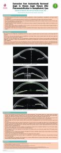
Bronchoscopy
Autopsy conclusion: Active granulomatous inflammation present involving a large cavitary lesion within the left lung, and vessels in this region were also thickened and inflamed consistent with GPA
• Major depressive disorder
Discussion
Medication: Mycophenolate mofetil 1gm po bid, Prednisone 10 mg po daily, Lexapro, Nexium. Family Hx: Stage 4 breast cancer in mother, biliary cancer in father.
• Focal subglottic airway stenosis and parenchymal cavitary lesions are well-recognized manifestations of GPA • Rarely pseudopolyps, submucosal tunnels or luminal synechial bands can occur • However, contiguous involvement of lung parenchyma and large airways coalescing into a single cavitary lesion is an uncommon presentation • Our case demonstrates that GPA can present as a single cavitary lesion involving the lung parenchyma and central airways.
Social Hx: Never smoker. Denied use of alcohol or illicit drug use. She writes documentation for software in the computer industry. Work up: • CT scan showed bilateral reticulonodular infiltrate and large cystic bronchiectasis in the lingula as shown on the right. • ANCA, MPO, PR-3 antibodies were negative • PFT: FVC 0.81 L (22% predicted), FEV1 0.65 L (20% predicted), FEV1/FVC 0.80, TLC 1.5 L (29% predicted), DLCO 4.9 mL/mmHg/minute (20% predicted). • BAL: positive for HSV1 by PCR • Bronchoscopy: Severe architectural distortion on left upper lobe and left lower lobe as shown on the right. Hospitalization Course: • Treated with rituximab x 1 • Developed rapid and progressive respiratory failure and expired
Top left: Left lung with extensive fibrosis and large cavitary lesion. Top right: Pulmonary vasculitis with transmural thickening. Bottom left: Pulmonary vasculitis with destruction of elastic lamina. Bottom right: Left lung cavitary lesion with vasculitis and granulomatous inflammation.
Reference Top left: Lingula orifice, looking into cavity with no visible airway Top right: Left mainstem bronchus with connective tissue band. Bottom left: Lingula cavity with few remaining small airway orifice (blue arrow) Bottom right: left mainstem bronchus.
1. Daum, Timothy E., et al. "Tracheobronchial involvement in Wegener's granulomatosis." American Journal of Respiratory and Critical Care Medicine 151.2 (1995): 522-526. 2. Polychronopoulos, Vlassis S., et al. “Airway involvement in wegener’s granulomatosis.” Rheumatic Disease Clinics of North America 33 (2007):755-775 3. Lohrmann, Christian.,et al. “Pulmonary manifestations of wegener granulomatosis:CT findings in 57 patients and a review of the literature.” European Journal of Radiology 53 (2005): 471-477