Energy dispersive X-ray spectroscopy (EDS, EDX or EDXRF) is an analytical technique used for the ... Core shell ionisation: chemical microanalysis by. X-ray, Auger ... In addition their low energy makes them easily absorbed. SEM. TEM ->.
Electron Microscopy Advanced Techniques 1. High-Resolution TEM 2. Analytical EM 3. 3D Microscopy, Special Techniques, Trends
Spring 2010
Experimental Methods in Physics
Marco Cantoni
Introduction to EDX Energy Dispersive X-ray Microanalysis (EDS, Energy dispersive Spectroscopy)
Spring 2010
Experimental Methods in Physics
Marco Cantoni
summary • Energy dispersive X-ray spectroscopy (EDS, EDX or EDXRF) is an analytical technique used for the elemental analysis or chemical characterization of a sample. It is one of the variants of XRF. As a type of spectroscopy, it relies on the investigation of a sample through interactions between electromagnetic radiation and matter, analyzing x-rays emitted by the matter in response to being hit with charged particles. Its characterization capabilities are due in large part to the fundamental principle that each element has a unique atomic structure allowing x-rays that are characteristic of an element's atomic structure to be identified uniquely from each other. • To stimulate the emission of characteristic X-rays from a specimen, a high energy beam of charged particles such as electrons or a beam of X-rays, is focused into the sample being studied. At rest, an atom within the sample contains ground state (or unexcited) electrons in discrete energy levels or electron shells bound to the nucleus. The incident beam may excite an electron in an inner shell, ejecting it from the shell while creating an electron hole where the electron was. An electron from an outer, higher-energy shell then fills the hole, and the difference in energy between the higher-energy shell and the lower energy shell may be released in the form of an X-ray. The number and energy of the X-rays emitted from a specimen can be measured by an energy dispersive spectrometer. As the energy of the Xrays are characteristic of the difference in energy between the two shells, and of the atomic structure of the element from which they were emitted, this allows the elemental composition of the specimen to be measured.
Experimental Methods in Physics
Spring 2010
Marco Cantoni
3
Marco Cantoni
4
Basics of EDX •
a) Generation of X-rays
•
b) Detection
•
•
Spring 2010
Si(Li) Detector, EDS
c) Quantification
EDX in SEM, Interaction volume Monte-Carlo-Simulations EDX in TEM
d) Examples
Experimental Methods in Physics
X-ray generation: Inelastic scattering of electrons at atoms Eelectron_in > Eelectron_out
SE
inner shell ionization ••Characteristic Characteristic X -ray X-ray emission •• Continuum X Continuum XXray ray production production ((Bremsstrahlung Bremsstrahlung ,, Synchrotron) Synchrotron)
SE, BSE, EELS Experimental Methods in Physics
Spring 2010
Marco Cantoni
5
Core shell ionisation: chemical microanalysis by X-ray, Auger electron and Electron Energy Loss Spectrometries e-
Designation of x-ray emission lines L1
L2
K
L3
+ e-
M5 M4 M3 M2 M1
Ionisation
1ps
La1
eRX
La2
L1M1M2 L3 L2
KL2L3
Kα2
L1 K
L1
L2 L3
+
K
L1
L2 L3 Ka1
+
Ka2 Kb
KL1L2 KL1L3 KL2L3 K
Rayons X Emission X Spring 2010
Electrons Auger
Emission Auger Experimental Methods in Physics
Marco Cantoni
6
Emission of characteristic X-ray and Auger electron
Spring 2010
Experimental Methods in Physics
Marco Cantoni
7
Experimental Methods in Physics
Marco Cantoni
8
Forbidden transitions ! quantum mechanics: conservation of angular momentum
Spring 2010
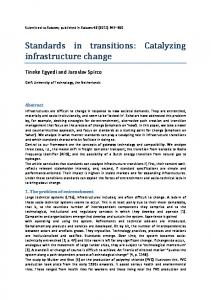
Efficiency of X-ray generation Relative efficiency of X-ray and Auger emission vs. atomic number for K lines
Ionization cross-section vs. overvoltage U=Eo/Eedge (electron in -> X-ray out)
Light elements Auger Spectroscopy
Heavy elements EDS
TEM -> Cu-K 8.1kV, HT 15kV U = 15/8.1 = 1.85
Light element atoms return to fundamental state mainly by Auger emission. For that reason, their K-lines are weak. In addition their low energy makes them easily absorbed.
Spring 2010
SEM
To ionized the incident electron MUST have an energy larger than the core shell level U>1. To be efficient, it should have about twice the edge energy U>2.
Experimental Methods in Physics
Marco Cantoni
9
Characteristic lines: Moseley's Law EDS range ~ 0.3-20 keV
! To assess an element all detectables lines MUST be present!!! known ambiguities: Al Kα = Br Ll S Kα = Mo Ll Spring 2010
Experimental Methods in Physics
Marco Cantoni
10
Moseley‘s law for K-series • Frequency ν of X-rays emitted from K-level vs. atomic number
ν = 2.481015(Z −1)2 E= hν et λ=c/ν with the Planck constant:h=6.626 068 76(52) × 10-34 J·s and 1eV = 1.6 10-19 J So, lets measure the Xrays emitted from my Energy of characteristic Xsample and determine the ray composition ! But how to detect it ? -> Element
Qualitative EDX-Analysis Spring 2010
Experimental Methods in Physics
Marco Cantoni
11
b) Detection of X-rays (EDX)
Spring 2010
Experimental Methods in Physics
Marco Cantoni
12
Right: Si(Li) detector Cooled down to liquid nitrogen temperature
modern silicon drift (SDD) detector: no LN cooling required Spring 2010
Experimental Methods in Physics
Marco Cantoni
13
Marco Cantoni
14
X-Ray energy conversion to electrical charges: 3.8eV / electron-hole pair in average electronic noise+ imperfect charge collection: 130 eV resolution / Mn Ka line
• • • •
Detector acts like a diode: at room temperature the leak current for 1000V would be too high ! The FET produces less noise if cooled ! Li migration at room temperature ! ->Detector cooling by L-N
Spring 2010
Experimental Methods in Physics
EDX spectrum of (K,Na)NbO3 Characteristic X-ray peaks
Continuum, Bremsstrahlung
Max Energy, 10keV
Electron beam: 10keV Experimental Methods in Physics
Spring 2010
Duane-Hunt limit Marco Cantoni
15
Detection limit EDS in SEM • Acquisition under best conditions – Flat surface without contamination (no Au coating, use C instead) – Sample must be homogenous at the place of analysis (interaction volume !!) – Horizontal orientation of the surface
0.5 %at Sn in Cu
– High count rate – Overvoltage U=Eo/Ec >1.5-2
• For acquisition times of 100sec. : detection of ~0.5at% for almost all elements Spring 2010
Experimental Methods in Physics
Marco Cantoni
16
(K,Na)NbO3 Continuum, Bremsstrahlung
l a e d i t o N !
Overvoltage, 10keV
Duane-Hunt limit Spring 2010
Experimental Methods in Physics
Marco Cantoni
17
(K,Na)NbO3
Spring 2010
Spectrum
Na
K
Nb
O
Total
Spectrum 1
8.19
Spectrum 2
9.59
10.1 8 8.66
Spectrum 3
7.82
9.54
Spectrum 4
9.79
9.37
Spectrum 5
8.86
9.35
Spectrum 6
9.46
9.07
Spectrum 7
8.89
Spectrum 8
8.60
10.2 5 9.40
20.7 0 20.7 5 21.1 3 20.3 6 20.7 7 20.6 3 20.3 7 20.8 6
60.9 3 61.0 0 61.5 1 60.4 8 61.0 2 60.8 4 60.4 9 61.1 4
100.0 0 100.0 0 100.0 0 100.0 0 100.0 0 100.0 0 100.0 0 100.0 0
Max.
9.79
Min.
7.82
21.1 3 20.3 6
61.5 1 60.4 8
Experimental Methods in Physics
10.2 5 8.66
Marco Cantoni
18
c) Quantification • First approach: compare X-ray intensity with a standard (sample with known concentration, same beam current of the electron beam) • ci: wt concentration of element i • Ii: X-ray intensity of char. Line • ki: concentration ratio
Spring 2010
Experimental Methods in Physics
Yes, but….
ci Ii = = ki cistd Iistd
Marco Cantoni
19
Intensity ~ Concentration…?
How many different samples…?
Spring 2010
Experimental Methods in Physics
Marco Cantoni
20
Spring 2010
Experimental Methods in Physics
Marco Cantoni
21
Electron Flight Simulator
Spring 2010
Experimental Methods in Physics
Marco Cantoni
22
Casino
Spring 2010
Experimental Methods in Physics
Marco Cantoni
23
X-rays generated X-rays detected
Spring 2010
Experimental Methods in Physics
Marco Cantoni
24
When the going gets tough…..
Quantification Correction matrix
ci Ii [Z × A × F ] std = std = ki ci Ii •
•
•
"Z" describe how the electron beam penetrates in the sample (Zdependant and density dependant) and loose energy "A" takes in account the absorption of the X-rays photons along the path to sample surface "F" adds some photons when (secondary) fluorescence occurs
Experimental Methods in Physics
Spring 2010
Marco Cantoni
25
Flow chart of quantification Measure the intensities and calculate the concentrations without ZAF corrections Calculate the ZAF corrections and the density of the sample Calculate the concentrations with the corrections
Is the difference between the new and the old concentrations smaller than the calculation error?
Yes !
no
Spring 2010
Experimental Methods in Physics
Marco Cantoni
stop
26
Correction methods: • • • •
ZAF (purely theoretical) PROZA Phi-Rho-Z PaP (Pouchou and Pichoir) XPP (extended Puchou/Pichoir)
• with standards (same HT, current, detector settings) • Standardless: theoretical calculation of Istd • Standardless optimized: « hidden » standards, user defined peak profiles
Spring 2010
Experimental Methods in Physics
Marco Cantoni
27
Quantitative EDX in SEM •Acquisition under best conditions –Flat surface without contamination, horizontal orientation of the surface (no Au coating, use C instead) –Sample must be homogenous at the place of analysis (interaction volume !!) –High count rate (but dead time below 30%) –Overvoltage U=Eo/Ec >1.5-2
•For acquisition times of 100sec. : detection of ~0.5at% possible for almost all elements
•Standardless quantification •possible with high accuracy (intensities of references under the given conditions can be calculated for a great range of elements), test with samples of known composition, light elements (like O) are critical… •Spatial resolution depends strongly on HT and the density of the sample
Spring 2010
Experimental Methods in Physics
Marco Cantoni
28
Modern EDX systems: • • •
User friendly interfaces New and more powerful electronics (stability of calibrations, higher count rate) Drift compensation for long acquisition times (element mapping on CM300 at high mag, “sitelock”)
Spring 2010
• • •
Synthesized spectra (spectrum overlay) easier identification Advanced element mapping: Spectral imaging (data cube), selection of elements and regions post-acquisition Powerfull reporting and Export tools (Word, Powerpoint, html, tif etc.)
Experimental Methods in Physics
Marco Cantoni
29
Spectrum imaging Data cube
Synthesized spectrum Extraction of element maps
Spring 2010
Experimental Methods in Physics
Marco Cantoni
30
EDS in TEM PZT bulk
20nm thick PZT
High spatial resolution !
Spring 2010
Experimental Methods in Physics
Marco Cantoni
31
Marco Cantoni
32
EDS in TEM • •
Thin samples -> correction factors weak (A and F can be neglected) Very weak beam broadening > high spatial resolution ~ beam diameter (~nm)
High energy: artifacts !
Spring 2010
Experimental Methods in Physics
STEM point analysis PbMg1/3Nb2/3O3 (bulk)
Processing option : Oxygen by stoichiometry (Normalised) Spectrum
Mg
Si
Spectrum 1 Spectrum 2 Spectrum 3 Spectrum 4 Spectrum 5 Spectrum 6 Spectrum 7 Spectrum 8 Spectrum 9
30.02 19.15 6.01 5.65 5.63 5.98 5.55 5.49 5.63
13.32 7.96
Max. Min.
30.02 5.49
13.32 7.96
Nb
Pb
O
Total
4.11 12.49 12.39 12.48 13.66 12.45 12.96 12.19
11.72 22.13 22.67 22.52 20.11 22.66 21.84 23.04
56.66 57.06 59.37 59.29 59.36 60.25 59.34 59.72 59.14
100.00 100.00 100.00 100.00 100.00 100.00 100.00 100.00 100.00
13.66 4.11
23.04 11.72
60.25 56.66
All results in Atomic Percent
Spring 2010
Experimental Methods in Physics
Marco Cantoni
33
STEM linescan
Pb(Zr,Ti)O3 (thick film), slight Pb excess
Spring 2010
Experimental Methods in Physics
Marco Cantoni
34
STEM Element Mapping PMN/PT 90/10 (bulk)
Spring 2010
Experimental Methods in Physics
Marco Cantoni
35
Marco Cantoni
36
Artifacts
how to recognize/minimize them
Spring 2010
Experimental Methods in Physics
Analytical TEM of multifilament Nb3Sn superconducting wires Superconducting Nb3Sn cables for high magnetic fields 10-20T: increase current density, lower cost Potential Applications: NMR, Tokamak fusion reactors Large Hadron Collider (LHC), CERN Typical cable: 1 x 1.5mm cross-section 121x121 filaments of Nb3Sn in a bronze (Cu/Sn) matrix
0.5 mm
Prof. R. Flükiger, V. Abächerli, D. Uglietti, B. Seeber
Dept. Condensed Matter Physics (DPMC), University of Geneva Experimental Methods in Physics
Spring 2010
Processing
“Nano”-engineering: controlled creation of “imperfections” of nm scale (coherence length)
„bronze route“
Cu and Ti are believed to play an important role at the grain boundaries: „dirty“ grain boundaries = pinning • •
Ti Ti
Nb3Sn
Is it possible to detect Cu and Ti at the grain boundaries ? What is the difference between the grain boundaries depending on where the additives are added to the unreacted material ?
Heat treatment
Nb Ta Cu,Sn bronze
Marco Cantoni
Nb
Cu,Sn SEM: reacted filament (1 out of 14‘000) Spring 2010
Experimental Methods in Physics
Marco Cantoni
38
Typical problems: thinning of heterogeneous specimens: selective thinning
bronze
Nb3Sn filament
Cross-section, polished mechanically to 30 um, ion milled until perforation Spring 2010
STEM, Dark field: core of filament too thick, preferential etching of bronze matrix
Experimental Methods in Physics
Marco Cantoni
39
Preparation by Focused Ion Beam defining and cutting of lamella
15um
“Lift-out” Spring 2010
Experimental Methods in Physics
TEM grid, 3mm diameter
Marco Cantoni
40
Preparation by Focused Ion Beam final thinning, “two windows”
20um
“two windows, 5x5 um” um”
Top view: final thickness of 4040-60 nm Experimental Methods in Physics
Spring 2010
Marco Cantoni
41
Marco Cantoni
42
Specimen preparation by focused Ion Beam (FIB): large areas with uniform thickness ideally for EDX Analysis in the TEM (STEM mode)
15um
thickness:40-50nm
EDS, element maps
SEM (FIB) Ion milling
STEMSTEM-DF
STEM, Bright field
FIB Sample #21
Spring 2010
Experimental Methods in Physics
Spot analysis Line profile
„Nb3Sn“ bronze Nb
Tc/Jc „useful“
Point
Ti %at
Nb %at
Sn %at
Ta %at
1
0.1
79.7
17.1
2.9
2
0.4
79.2
17.8
2.4
3
0.8
77.8
18.5
2.7
4
1.8
75.1
20.8
2.1
5
0.5
76.5
20.9
1.9
6
0.2
74.3
23.1
2.2
7
1.6
73.1
23.4
1.7
8
1.2
73.7
22.8
2.1
9
0.9
70.4
26.4
2.1
Sample #21
Sn Experimental Methods in Physics
Spring 2010
Marco Cantoni
43
grain boundaries ? Ti/Cu
EDX line-scan Cu
Nb
Ti
Ta
Cu and Ti at the grain boundaries: width ~ coherence lenght (4nm) possible pinning centers !!
Sn Sample #21
Spring 2010
Experimental Methods in Physics
Marco Cantoni
44
grain boundary without Ti
Nb
Cu
Ti
Ta Quantitative Line-scan
Sample #24 Sn
Spring 2010
Experimental Methods in Physics
Marco Cantoni
45