32nd Annual International Conference of the IEEE EMBS Buenos Aires, Argentina, August 31 - September 4, 2010
Electrical Circuit Models of the Human Respiratory System Reflect Small Airway Impairment Measured by Impulse Oscillation (IOS) Michael D. Goldman, Homer Nazeran, Senior Member, IEEE, Carlos Ramos, Emily Toon, Katrina Oates, Diana Bilton, Erika Meraz, Nazila Hafezi and Bill Diong, Senior Member, IEEE
Abstract—The use of the forced oscillatory input impedance parameter, frequency-dependence of Resistance (fdR), to assess small airway impairment (SAI) has not been widely accepted due to concern about the effects of “upper airway shunt” on oscillometric resistance and low frequency reactance. On the other hand, recent medical studies suggest that low frequency reactance is a very sensitive index of treatment intervention directed at small airways. The present study was undertaken to analyze and compare Impulse Oscillometry (IOS) resistance and reactance data with model-derived indices of small airway function from two models of the respiratory impedance, one with, and the other without an element for upper airway shunt capacitance. Fifty six patients with stable chronic obstructive lung disease of varying severity due to Cystic Fibrosis (CF) and 21 patients with asthma were evaluated by IOS testing. IOS data were input into the augmented RIC (aRIC) model with an upper airway shunt capacitance, and the extended RIC (eRIC) model, without a shunt capacitance element. Model-derived indices were compared between the two models for CF patients separately from asthma patients. We conclude that IOS indices of SAI are modeled equally well with or without upper airway shunt capacitance, and do not seem to be dependent on upper airway shunt capacitance.
I. INTRODUCTION
R
airflow measured by lung function tests of maximal forced expiration, spirometry, may provide useful analysis of airflow obstruction; but in children, spirometry is known to be within normal limits in the majority of asthmatic children not suffering from an acute exacerbation [1]. In adults with asthma or Chronic Obstructive Pulmonary Disease (COPD), peripheral airways are often inflamed and impaired; but no clinically useful index of small airway impairment (SAI) is yet established [2]. Forced oscillation (FO) has been reported to provide indices that reflect small airway impairment, SAI [3]; and Impulse Oscillometry, IOS, using aperiodic repetitive pulses of pressure at the mouth, shows changes in low frequency reactance in response to inahled corticosteroid (ics) aerosols delivered to small airways in medical clinical trials [4-6]. RESPIRATORY
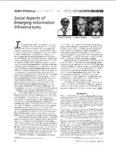
resistance (R) and reactance (X) spectra at oscillation frequencies, from 5 to 25 Hz. Questions of the relevance to SAI of the fall in R with increase in oscillation frequency, (frequency-dependence of R, fdR), and low frequency X have impeded diagnostic use of FO. Some reports attributed fdR to upper airway artifact, or shunt compliance and offered ameliorating approaches. Whereas upper airway shunt compliance may affect the magnitude of fdR, application of oscillations around the entire head and upper neck, or pressing firmly on the cheeks with both hands to support the cheeks failed to remove fdR in patients with airflow obstruction [7-9]. The unique contribution of airway compliance in parallel with airway resistance causes fdR in the presence of increased peripheral resistance [10], stimulating our research group to utilize the Mead respiratory system model to analyze data obtained by Impulse Oscillometry (IOS) testing in adults and children with airflow obstruction [11-14]. We reported that the Mead model yields spurious results for calculated lung and chest wall compliance and therefore used derivative models which exclude these parameters [14]. The augmented RIC, aRIC, model includes shunt capacitance for the upper (extrathoracic) airway. The extended RIC, eRIC, model excludes shunt capacitance [13]. Figure 1 shows the original Mead model and derivative models used. The present study was undertaken to compare the performance of eRIC and aRIC model parameters in reflecting IOS R and X data in patients with known chronic airflow obstruction to assess potential effects of upper airway shunt capacitance, Ce, on model-derived parameters of peripheral airway mechanics.
IOS pressure pulses at the mouth generate oscillations in flow, analyzed by fast Fourier transform, FFT, to calculate Manuscript received April 1, 2010. This work was supported in a small part by NIH/NIEHS #1 S11ES013339-01A1(Pilot 1) to HN. M.G., H.N., C.R., E.M., N. H. are with the University of Texas at El Paso, El Paso TX, 79968 USA. (tel 915-747-8937, email
[email protected]). E. T., K. O., D. B. are with Papworth Hospital, UK. B. D. is with the Engineering Department, Texas Christian University, Fort Worth, TX, USA ).
978-1-4244-4124-2/10/$25.00 ©2010 IEEE
Fig. 1. Mead model with solid-line circles around capacitative elements for the lung (Cl) and chest wall (Cw). Deleting these two elements results in the aRIC model. Further deletion of Ce, the capacitative element for extrathoracic “shunt” compliance results in the eRIC model.
2467
II. METHODS A. Patients. Clinically stable adolescent and young adult patients with Cystic Fibrosis (CF) were studied at annual review visit at Papworth Hospital, UK. In parallel studies in the US, adult asthmatic patients were studied before and after treatment with inhaled corticosteroids (ics) and bronchodilator. Forced oscillation utilized impulse oscillation (IOS, Jaeger Wuerzburg, Germany) to measure resistance and reactance between 5 and 25 Hz.
electrical circuit models, which provide both large (central) airway parametrers, (Rc and Inertance, I) and small (peripheral) airway R and C (Rp and Cp, respectively). Figure 3 shows the values of Rp from aRIC plotted as a function of Rp from eRIC. There is a close correspondence between the two models’ estimates of Rp (slope = 0.96, with r = 0.99).
B. Impulse Oscillometry. The IOS pneumotachometer was calibrated for volume daily using a 3-liter syringe. At weekly intervals, stability of pressure calibration was confirmed using a 0.2 kPa/L/s reference resistance following the volume calibration. Subjects underwent 3 to 6 replicate IOS tests of 60 seconds each. IOS tests were performed with cheeks supported by voluntary contraction of the circumoral muscles to decrease upper airway shunt dissipation of oscillations while avoiding uncomfortable mechanical loading of the chest wall by patients’ lifting their arms and pushing on the cheeks [8, 15]. C. Data analysis. IOS pressure, flow, and volume data were analyzed off-line to reject data adversely affected by swallowing, coughing, or airflow leak [16]. We omitted the first 20 seconds of each test to avoid previously observed variability at the onset of testing [17]; an average of 3 – 5 tests was calculated. We tabulated IOS resistance and reactance between 5-25 Hz. We selected the fall in resistance between 5 and 20 Hz (R5-R20, “frequencydependence of resistance,” fdR), and integrated low frequency reactance (AX) as the IOS indices most related to small airway function [4-6, 16, 18-20]. III. RESULTS Figure 2 shows the nature of the primary resistance and reactance spectra outputs from IOS tests. Note that, R falls significantly with increasing frequency from 5 to 20 Hz; and X magnitude decreases with increasing frequency from 5 Hz to resonant frequency, Fres, (14.6 Hz).
Fig. 2. Resistance (circles) and reactance (crosses) at all frequencies between 5 and 25 Hz in 56 patients with CF. Scales for R and X at left, in kPa/L/s. Vertical Standard Error (SE) bars shown.
The area under frequency axis, from 5 Hz to Fres, and above the reactance (X) curve is defined as integrated reactance, AX. Mean (range) of R5-R20 was 0.11 kPa/L/s (0.01-0.26). Mean (range) of AX was 0.72 kPa/L (0.1-3.1). The entire R and X spectral ranges can be analyzed using
Fig. 3. Model-derived Rp from aRIC model on Y axis as a function of Rp from eRIC model on X axis for CF patients.
Similarly close correspondence for models’ estimates of Cp was found, with slope = 0.95 and r = 0.99. Relationships between aRIC and eRIC estimates for large airway resistance (Rc) showed a slope of 0.95, r = 0.97, and for I, a slope of 1.03, r = 0.95. These results demonstrate a close similarity of parameter estimates for Rc, I, Rp, and Cp from the two models. More importantly, we may relate Rp to the IOS index R5-R20, and Cp to the IOS index AX. Figure 4 shows our ‘landmark’ IOS index Cp as a function of AX for both models and figure 5, Rp as a function of R5-R20 for both models.
Fig 4. Cp as a function of AX for eRIC and aRIC models in CF patients.
The regressions for Cp as a function of AX are nearly indistinguishable, with very tight correlations between Cp and AX, slightly better for eRIC (r = 0.93) than aRIC (r = 0.88). Figure 5, below, shows comparable data for Rp, as a function of R5-R20.
2468
aRIC and eRIC models, we also list the larger errors observed when using a model consisting only of inertance and capacitance in series with resistance, RIC. Table 1. Average total errors for fitting 3 models to IOS data acquired from 56 CF and 21 asthma patients.
Dataset Fig 5. Rp as a function of (R5-R20) for eRIC and aRIC models in CF patients.
CF Asthma Both Most severe asthma
Again, the regressions for both models are closely similar, but with lesser correlations between Rp and R5-R20.
Total Average Error RIC eRIC aRIC model model model 0.016 0.004 0.003 0.045 0.004 0.002 0.031 0.004 0.003 0.062 0.004 0.002
Because asthmatic patients may show substantially greater IOS abnormalities than the results observed in CF, we made the same comparisons in asthmatic patients. Rc, Rp, and Cp slopes were 1.07, 0.82, 0.96, and correlation correficients were 0.99, 0.99 and 0.90, respectively. Asthma patients were more severely affected, in general, than CF patients; and their IOS resistance and reactance spectra are shown in figure 6. The overall shapes of IOS resistance and reactance spectra were similar to those in CF patients; but Rrs is higher, and falls more with oscillation frequency; and low frequency Xrs magnitude is greater, with greater AX, and a higher resonant frequency. Mean (range) for R5-R20 is 0.20 kPa/L/s (0.12-0.46), and for AX is 2.4 kPa/L (0.8-7.5). These IOS indices are consistent with more severe SAI in asthmatics than in CF patients. In the 4 most severely affected asthmatic patients, the IOS resistance spectrum showed a positive frequency dependence above 15 Hz. In the presence of positive
Fig. 6. Resistance (circles) and reactance (crosses) at all frequencies between 5 and 25 Hz in 21 patients with Asthma. Scales for R and X at left, in kPa/L/s. Vertical Standard Error (SE) bars shown.
frequency-dependence of Resistance (fdR), aRIC fits IOS data with less error than eRIC. Table 1 and the chart below it show the average total errors for model fitting of IOS data in CF and asthma patients, and separately for the 4 most severe asthma patients who manifested clear positive frequencydependence of resistance above 15 Hz. In addition to the
As shown previously [13], the RIC model does not allow for change in resistance with oscillation frequency; and therefore is not appropriate for patients with airflow obstruction in whom resistance is frequency-dependent. Errors in the CF patients and the entire group of asthmatic patients for aRIC and eRIC were all within what is considered a good fit to the IOS data; but eRIC errors in asthmatic patients were 40% greater before bronchodilator (BD), and 20% greater after BD than errors for aRIC. In the most severely affected asthmatics aRIC error was twice as large as in group average asthmatic data, while eRIC error was 3 times as large as group average. Despite increased error in the severe asthmatic patients, the ‘landmark’ eRIC model-derived parameter, Cp, differed from aRIC Cp by only 10% with a correlation coefficient r = 0.99 between the two, while aRIC large airway resistance Rc, and Rp both differed by 28% from eRIC R and Rp, with r = 0.99 and 0.96, respectively. Importantly, with respect to the clinical significance of model-derived parameters, we found in the entire group of asthmatic patients a very close correspondence between both models’ estimates of changes in Rp, Cp, and Rc from pre- to post-bronchodilator. For Rp, the slope was 0.94, with r = 0.99, for Cp, slope was 1.12, with r = 0.97, and for Rc, slope was 0.88, with r = 0.84. Thus, both aRIC and eRIC modelderived parameters change in a closely similar manner in asthmatic patients during treatment with bronchodilators. IV. DISCUSSION The present study was undertaken in unselected patients with chronic airflow obstruction resulting from CF and adult
2469
asthmatic patients being screened for a medical clinical trial of inhaled corticosteroids (ics) plus bronchodilator therapy. Mean IOS data were not severely abnormal in CF; but were more severely abnormal in asthma. The ranges of IOS measures were very wide in both patient groups, owing to varying severity of illness. Over these widely ranging data from IOS tests, model-derived parameters for R, I, Rp, and Cp were remarkably similar, independent of presence of a model element corresponding to the upper airway shunt capacitance, with I (large airway inertance) differening significantly in only the most severely obstructed asthmatics. These data are consistent with only minimal contributions from upper airway shunt capacitance to the model-derived parameters Rp and Cp, most closely related to IOS indices reflecting SAI, namely fdR (R5-R20) and integrated low frequency reactance from 5 Hz to Fres (AX), despite absence of manual (hand) support of the cheeks during IOS testing (subjects were asked to support their cheeks by voluntary contraction of circumoral muscles). We analyzed the severe asthmatics specifically because they commonly manifest positive frequency dependence of IOS R above 15 Hz. In the aRIC model, the shunt capacitance, Ce (see figure 1) permits this positive frequency dependence of R; but does not require it. In contrast, the eRIC model demands a monotonic decrease in R with increase in oscillation frequency. In CF patients and less severe asthmatics, R decreased monotonically with increase in oscillation frequency; and model-derived Ce was uniformly small, of the order of 1-2x10⁻³. In the 4 most severe affected asthmatic patients Ce was 4-5x10⁻³. Nevertheless, in these patients, similarly close values of R, Rp, and Cp were observed with the eRIC model as compared to the aRIC model. Only I (large aiway inertance) was substantially different.
expiratory effort during resting breathing [8]. Despite the potential for small cheek movements to contribute to the upper airway shunt, model-derived parameters were remarkably similar whether a shunt capacitance was included or not. Experimentally, it was shown in [8] in patients with chronic airflow obstruction, that elevating the arms pressing on the cheeks manually caused an increase in the abnormalities in fdR and low frequency reactance, instead of “correcting” the presumed shunt capacitance effect. The controversy over significance of upper airway shunt capacitance reflects primarily the concern over early FO results using the “head generator” method, compared to patients firmly supporting their cheeks manually. In [9] constriction of the vocal aperature caused negative fdR, as was observed in patients with chronic constriction in the trachea after tracheostomy [23] with severe constrictions. In [23], an artifactual increase in X magnitude was associated with this severe tracheal constriction. In contrast, the commonly observed mild vocal cord constriction observed during IOS testing in normal subjects in our laboratory does not result in negative fdR in normal subjects. Use of the head generator in [9] increased overall values of R at all frequencies, and shifted the X-frequency curve to the left, but it did not eliminate negative fdR. In contrast, manual cheek support [8] did not decrease fdR in moderate airflow obstruction, while in severe obstruction it increased fdR and shifted the X-frequency curve to the right.
Our model analyses are restriced to the two derivatives of the Mead model (figure 1), based on experimental evidence showing by direct measures of oscillatory pressures, that 80% or more of the pressure applied to the trachea in living dogs was dissipated across large airway resistance when oscillatory pressures reached airways of 2-mm diameter [21]. At volumes corresponding to resting breathing in awake sitting humans, these experimental measurements of pressure show that little or no pressure is available to distend the lung or chest wall. Our research team has recently performed a circuit analysis of the Mead model with the same conclusions [22]. Thus, we suggest that the derivative models used are appropriate for the small-pressure-smallvolume output of Impulse Oscillometry (IOS).
We conclude that attempts to correct upper airway artifact are not uniform, do not invariably eliminate negative fdR in patients with airflow obstruction; and have variable effects on the X-frequency spectrum. Head generator FO creates unusual facial, ear and neck sensations. Neck pressure, pressure gradients across the tympanic membrane, involuntary vocal cord constriction and posture change may contribute to the observed positive fdR. Thus head plethysmographs may compress the neck, and may change posture, breathing pattern, functional residual capacity (FRC), and vocal cord aperture. Effects of head generator on R and X in patients with airflow obstruction could conceivably result from subtle vocal cord constriction. We suggest the head generator is not an optimal gold standard for normal resting quiet breathing. Finally, an extensive comparison between the head generator and standard input impedance methods concluded that the standard method of applying forced oscillation pressures at the mouth was acceptable for measuring respiratory impedance in epidemiological studies [24].
Patients in the present study breathed naturally, as those in the original study of Forced Oscillation [15] while firmly contracting their lips and circumoral musculature to support the cheeks. Whereas this method allows some small movements of the cheeks, we believe it avoids an important artifact associated with the adverse effects of upper arm muscles on the chest wall, making it more uncomfortable for patients to breathe and leading to “forced” instead of relaxed
Early interventional studies focusing on fdR reported increased negative fdR in circumstances of peripheral airway abnormality, resulting from pharmacologic intervention, gas density changes or recovery from acute heart attacks [2527]. Recent evidence demonstrates that AX (integrated low frequency reactance from 5 Hz to Fres) improves with pharmacologic treatment targeting peripheral airways [5, 6]. Our group has also reported very close correlations between
2470
fdR and AX [13]. In the present patients with both CF and asthma, we found that fdR was closely related to AX (r = 0.93, for CF, and r = 0.96 for asthma). Thus, we suggest that fdR and AX, low frequency R and X respectively, provide useful reflections of peripheral airway mechanical function. In the present study in patients with CF or asthma, we find that the model-derived indices Rp and Cp are closely correlated with these IOS indices. In relation to IOS indices reflecting SAI, we find good correlations between Rp and fdR and Cp and AX. That between Rp and fdR is clearly inferior to that between Cp and AX. We suggest this reflects the relatively limited sampling of R at only two frequencies, while AX is an integrative index over all frequencies below resonance. We find that model-derived Rp and Cp (as well as R and I) are closely comparable between aRIC and eRIC, reflecting only small model-derived values of aRIC Ce in CF and most asthmatic patients. In asthmatic patients with severe obstruction, the model error in fitting the R spectrum is increased when using the eRIC model, but optimized values of R, Rp and Cp do not differ substantially from those provided by aRIC; and changes in these parameters from pre- to post-BD are similar with both models. There is a significantly larger error in model fit for eRIC than aRIC, but overall errors are small in magnitude. Previous work from our research group included oscillation frequencies of 35 Hz, in which larger discrepancies in total error of eRIC relative to aRIC were found [11-14]. Recent information from the IOS manufacturer noted systematic discontinuities in the resistance spectrum, casting doubt on the validity of IOS spectra at frequencies > 25 Hz [28]. For this reason, we limited the analyses in the present studies to 25 Hz. Increasing the number of model parameters tends to decrease total error of model fit. However, it is desired to avoid ‘overparameterization’ in respiratory models. We suggest that, in addition to magnitude of error, an equally, if not more important, criterion for model appropriateness is the reliability of model-derived parameters relative to basic physiological data. In the present studies in patients with asthma or CF, model parameters from eRIC appear reliable, and slightly more closely correlated with IOS physiological indices, compared to aRIC; and eRIC is a more parsimonious model, making it inherently more attractive. The closely similar changes in model-derived parameter estimates during treatment interventions in asthmatic patients (CF patients were not evaluated pre- and posttreatment) provides further reassurance that the less complex eRIC model is suitable for medical evaluation of treatment. Finally, we have not included normal subjects in the present study. Early studies of FO have shown some positive frequency-dependence of R over the entire spectrum of R [29]. In this circumstance, eRIC will require constraints to allow optimization of model parameters, not done in the present study.
V. CONCLUSIONS The present studies analyzed two “derivative” models obtained from the well-known Mead model by deleting the capacitances of the lung and the chest wall. This is justified by experimental evidence of 80% or greater pressure dissipation from the trachea to 2-mm diameter airways, and further valided by circuit analysis of the Mead model using IOS data [22]. The aRIC model includes a parallel shunt airway capacitance element which in the present work, was generally small in magnitude and did not significantly affect the estimated values of large and small airway resistances, small airway capacitance and inertance in patients with CF and asthma. In more severely obstructed patients, with positive frequency dependence of R above 15 Hz, model fit with eRIC shows significantly greater error than does aRIC. Further work in a large number of severely obstructed patients is required before we can conclude that the eRIC will be suitable for use across a wide spectrum of airflow obstruction severity, and including normal subjects with positive frequency dependence of R. REFERENCES [1] J. D. Spahn, R. Cherniack, K. Paull, E. W. Gelfand. “Is forced expiratory volume in one second the best measure of severity in childhood asthma?” Am J Respir Crit Care Med 2004 169: 784-6. [2] M. Contoli, J. Bousquet, L. Fabbri, H. Magnussen, K. Rabe, N. Siafakas, Q. Hamid, M. Kraft. “The small airways and distal lung compartment in asthma and COPD: a time for reappraisal.” Allergy 2010; 65:141-51 [3] T. Takeda, T. Oga, A. Niimi, H. Matsumoto, I. Ito, M. Yamaguchi et al. “Relationship between Small Airway Function and Health Status, Dyspnea and Disease Control in Asthma.” Respiration. 2009 Sep 22. [Epub ahead of print] [4] G. Larsen, W. Morgan, G. Heldt. “Impulse oscillometry versus spirometry in a long-term study of controller therapy for pediatric asthma.” J Allergy Clin Immunol 2009; 123:861-7. [5] M. Yamaguchi, A. Niimi, T. Ueda, M. Takemura, H. Matsuoka, M. Jinnai et al. “Effect of inhaled corticosteroids on small airways in asthma: investigation using impulse oscillometry.” Pulm Pharmacol Ther. 2009;22:326-32. [6] M. Hoshino. “Comparison of Effectiveness in Ciclesonide and Fluticasone Propionate on Small Airway Function in Mild Asthma.” Allergol Int. 2010;59:59-66 [7] R. Peslin, C. Duvivier, J. Didelon, C. Gallina. “Respiratory impedance measured with a head generador to minimize upper airway shunt.” J Appl Physiol 1985; 59:1790-5 [8] M. Cauberghs, K. P. Van de Woestijne. “Effect of upper airway shunt and series properties on respiratory impedance measurements.” J Appl Physiol. 1989;66(5):2274-9. [9] M. Cauberghs, K. P. Van de Woestijne. “Changes of respiratory input impedance during breathing in humans.” J Appl Physiol. 1992;73(6):235562. [10] J. Mead. “Contribution of compliance of airways to frequency dependent behavior of lungs.” J. Appl. Physiol. 1969, 26, 670–3. [11] A. Rajagiri, B. Diong, M. Goldman, H. Nazeran. “Can Asthma in Children be Detected by the Estimated Parameter Values of the Augmented RIC Model?”, Proceedings of the IEEE-Engineering in Medicine
2471
and Biology Society (EMBS), 28th Annual International Conference, pp. 5595-5598, New York City, USA, Aug 30-Sep 3, 2006
[29] K. E. Finucane, S. V. Dawson, P. D. Phelan, J. Mead. “Resistance of intrathoracic airways of healthy subjects during periodic flow.” J Appl Physiol 1975; 38:517-30
[12] T. Nguyen, B. Diong, H. Nazeran, M. Goldman. “A Study of IOS Data Using Two Mead-related Models of Respiratory Impedance”, Proceedings of the IEEE-Engineering in Medicine and Biology Society (EMBS), 29th Annual International Conference, Lyon, France, pp. 10781081, Aug 23- 27, 2007 [13] B. Diong, H. Nazeran, P. Nava, M. Goldman. “Modeling Human Respiratory Impedance”, IEEE Engineering in Medicine and Biology, Special Issue on Respiratory Sound Analysis, Vol 26, No. 1, pp 48-55, 2007 [14] B. Diong, A. Rajagiri, M. Goldman, H. Nazeran. “The Augmented RIC Model of the Human Respiratory System”, J Med Biol Eng Comp, 47:395-404, 2009 [15] A. B. DuBois, A. Brody, D. Lewis, B. Burgess. “Oscillation mechanics of lung and chest in man.” J Appl Physiol 1956;8:587-94 [16] G. Skloot, M. Goldman, D. Fischler, C. Goldman, C. Schechter, S. Levin, et al. “Respiratory Symptoms and Physiologic Assessment of Ironworkers at the World Trade Center Disaster Site.” Chest, 2004; 125:1248-1255 [17] E. Toon, D. Bilton, M. D. Goldman, K. E. Oates. “Reproducibility of Impulse Oscillometry (IOS) within and between tests.” Proc. Am Thoracic Soc. 2005; 2:A34 (abstract) [18] M. Goldman. “Clinical Application of Forced Oscillation.” Pul Pharm Ther, 2001; 14: 341-50. [19] M. Goldman, C. Saadeh, D. Ross. “Clinical Applications of Forced Oscillation to Assess Perhiperal Airway Function.” Res Physiol Neuro 2005; 148:179-94 [20] H. Smith, P. Reinhold, M. Goldman. “Forced oscillation technique and impulse oscillometry.” Eur. Respir. Mon. 2005; 31:72-105 [21] P. Macklem, J. Mead. “Resistance of central and peripheral airways measured by a retrograde catheter.” J. Appl. Physiol. 1967;22:395-401 [22] C. Ramos, H. Nazeran, M.D. Goldman, B. Diong. “Circuit Analysis Justifies a Rduced Mead’s Model of the Human Respiratory Impedance for Impulse Oscillometry Data.” Proceedings of the IEEE EMBS 32nd Annual International Conference, Buenos Aires, Argentina, August 31- September 4, 2010. [23] T. Horan, S. Mateus, P. Beraldo, L. Araújo, J. Urschel, E. Urmenyi, F. Santiago. Forced oscillation technique to evaluate tracheostenosis in patients with neurologic injury. Chest. 2001;120:69-73. [24] Y. Iwatsubo, H. Lorino, C. Hubert, C. Duvivier, R. Peslin, Q. T. Pham et al. “Measurement of respiratory impedance by forced oscillation: comparison of the standard and head generator methods.” Eur Respir J. 1994;7(5):901-6. [25] P. V. Bhansali, C. G. Irvin, J. A. Dempsey , R. Bush, J. G. Webster. “Human pulmonary resistance: effect of frequency and gas physical properties.” J Appl Physiol. 1979;47(1):161-8. [26] L. Brochard, G. Pelle, J. de Palmas, P. Brochard, A. Carre, H. Lorino, A. Harf. “Density and frequency dependence of resistance in early airway obstruction.” Am Rev Respir Dis. 1987;135(3):579-84. [27] B. Interiano, R. W. Hyde, M. Hodges, P. N. Yu “Interrelation between alterations in pulmonary mechanics and hemodynamics in acute myocardial infarction.” J Clin Invest. 1973; 52:1994-2006 [28] P. Reinhold, H.-J. Smith, A. Langenberg, P. Lekeux. “Measurement of respiratory impedance in healthy calves using the Impulse Oscillation technique – Physiological and Methodological aspects.” Vet J 1998; 155: 27-38
2472