AUGUSTINE Y. KIM AND HANS P. BLASCHEK*. Department ofFood ..... The double-stranded. pDM6 DNA from C. acetobutylicum NCIB 6444 may bethe.
Vol. 173, No. 2
JOURNAL OF BACTERIOLOGY, Jan. 1991, p. 530-535
0021-9193/91/020530-06$02.00/0 Copyright C) 1991, American Society for Microbiology
Isolation and Characterization of a Filamentous Viruslike Particle from Clostridium acetobutylicum NCIB 6444 AUGUSTINE Y. KIM AND HANS P. BLASCHEK*
Department of Food Science, University of Illinois, 580 Bevier Hall, 905 South Goodwin Avenue, Urbana, Illinois 61801 Received 8 June 1990/Accepted 3 November 1990
A single-stranded 6.6-kb DNA molecule complexed with protein was recovered from the supernatant of Clostridium acetobutylicum NCIB 6444. Electron microscopic examination of the DNA-protein complex revealed the presence of a ifiamentous viruslike particle, which was designated CAK1. The possible double-stranded plasmidlike replicative form and the single-stranded prophage were also recovered from the cell culture following alkaline lysis. CAK1 was released from the C. acetobutylicum cell culture in the absence of cell lysis. Polyethylene glycol-NaCl coprecipitation of the DNA-protein complex revealed the presence of single-stranded DNA complexed with protein in a manner rendering the DNA resistant to Bal 31 exonuclease. Proteinase treatment of CsCl density gradient-purified CAK1 resulted in recovery of DNase-sensitive single-stranded DNA. Tricine-sodium dodecyl sulfate-polyacrylamide gel electrophoresis of CAK1 demonstrated the presence of a 5-kDa major coat protein. Hybridization data indicated that the single-stranded DNA from CAK1 has homology with the M13 phage of Escherichia coli. An examination of various physical properties of CAK1 suggests that it is similar to the filamentous phage recovered from gram-negative microorganisms. Although infectivity or inducibility of CAK1 could not be demonstrated, to our knowledge this represents the first report of a nonlytic filamentous viruslike particle containing single-stranded DNA being recovered from a gram-positive bacterium.. more recently this plasmid (pDM6) was partially characterized (8, 12a). C. acetobutylicum NCIB 6444 was initially reported to contain more than one plasmid (22). Also, this strain continuously extruded single-stranded DNA (ssDNA) during cell growth (8), an event normally seen only in filamentous phage-infected gram-negative microorganisms (12, 16, 19). Furthermore, the identification of phage infecting clostridial species has received only limited attention (14, 17). C. acetobutylicum phages have been isolated from cells involved in abnormal fermentations. The phages appear to be lytic and to cause cell death during the fermentation (14, 17). However, characterization of C. acetobutylicum phage DNA has been hampered by the lack of a suitable genetic expression system (14). The identification and characterization of C. acetobutylicum phages ultimately may allow the construction of stable vectors. Vector constructs based on filamentous phage, such as Escherichia coli M13, have been developed for E. coli (2, 13, 16) and Bacillus subtilis (4). Infection by filamentous phage does not cause host cell lysis (12, 13, 16, 19, 23), and the replication systems resembled that of multicopy plasmids (2, 12, 13, 16) and provide for good expression of inserted DNA (2, 13, 16). Also, the observation that vectors based on filamentous phage DNA can be stably maintained without selection pressure (13) is of great importance for the genetic manipulation of industrially significant microorganisms. The objective of this study was to isolate and characterize a filamentous viruslike particle from C. acetobutylicum NCIB 6444 in preparation for its use in the development of a stable vector for this microorganism.
There continues to be growing interest in the metabolically diverse anaerobic clostridia used for producing chemicals from various feedstocks (18). The production of solvents by using C. acetobutylicum has been reviewed (1, 18, 28). Although the acetone-butanol-ethanol fermentation from C. acetobutylicum has a long history in commercial butanol production, our understanding of the microorganism is limited (28). One approach for understanding C. acetobutylicum butanol production and for increasing butanol productivity involves the application of molecular genetics. The development of suitable molecular genetic systems, specifically transformation and shuttle vector systems, is a prerequisite for expression of cloned genes in this microorganism and the ultimate construction of tailor-made C. acetobutylicum strains (1, 28). Transformation techniques based on electroporation (15) or protoplast formation (10, 17, 21) and shuttle vector construction, primarily with streptococcal plasmids, have been developed in the last few years (28). However, the strain-specific nature of the transformation systems developed, together with the instability of streptococcal plasmidbased vectors, limits the usefulness of these systems (28). Furthermore, the use of replication origins derived from other clostridial species is problematic because of their instability and limited function in C. acetobutylicum (21, 28). The search for indigenous extrachromosomal DNA, plasmids, and phages from C. acetobutylicum is important in order to understand the mechanism of gene transfer in this microorganism. Clostridial vector constructs based on native DNA are expected to result in stable genetic systems (7). The only indigenous C. acetobutylicum plasmid recovered to date was that from strain NCIB 6444 (18, 28). The initial discovery of plasmid DNA in C. acetobutylicum NCIB 6444 was reported by Truffaut and Sebald (22), and
MATERIALS AND METHODS
Bacterial strains, media, and culture conditions. C. aceto*
butylicum strains NCIB 6444 and ATCC 824 and E. coli strains DH5aF' and EM383 carrying M13mp9 were used in
Corresponding author. 530
VOL. 173, 1991
this study. The latter strain was kindly provided by S. Maloy, University of Illinois. The conditions used for C. acetobutylicum cell growth and maintenance were described previously (8). For the isolation of CAK1 and plasmid DNA, C. acetobutylicum was grown overnight in trypticase-glucose-yeast extract medium (TGY [8]) at 37°C under anaerobic conditions (85% N2, 10% C02, 5% H2; Coy Laboratory Products Inc., Ann Arbor, Mich.). Luria broth (L-broth [11]) was used for growth of E. coli. Isolation of a viruslike particle from C. acetobutylicum NCIB 6444. An overnight cell culture of C. acetobutylicum NCIB 6444 in TGY was centrifuged at 10,000 x g for 15 min in a Sorvall RC-2 centrifuge (Sorvall, Norwalk, Conn.). The supernatant was recovered and subjected to polyethylene glycol (PEG)-NaCl precipitation. The PEG-NaCl solution was prepared as 20% PEG 8000 (Sigma Chemical Co., St. Louis, Mo.) and 2 M NaCl (Sigma) and added to the C. acetobutylicum cell culture supernatant in a 1:1 (vol/vol) ratio (11, 27). The mixture was incubated at room temperature for 1 h. The precipitate was recovered following centrifugation at 10,000 x g for 30 min at 4°C and suspended in deionized water. For further purification of PEG-NaCl precipitate, CsCl was added at a concentration of 0.4 g/ml, and the mixture was applied to a discontinuous CsCl density gradient as described by Yamamoto et al. (27). The density of the CsCl density gradient was adjusted in order to compensate for the difference in density between E. coli filamentous phage and CAK1 (high density, 1.4 g/ml; middle density, 1.3 g/ml; and low density, 1.2 g/ml). The sample was centrifuged at 40,000 rpm for 20 h at 4°C with a 7OTi rotor (Beckman Instruments Inc., Fullerton, Calif.). Phage-containing bands were collected and dialyzed against TrisEDTA (TE) (8, 11) buffer for 24 h at 4°C. Purified CAK1 was further concentrated against 100% dry PEG and stored at 40C. DNA isolation from C. acetobutylicum and E. coli. Largescale isolation of extrachromosomal DNA from C. acetobutylicum and E. coli was carried out by the modified alkaline lysis methods described previously (7, 8), except that overnight cultures were used. RNase was added at a concentration of 50 ,ug/ml during the cell wall digestion step. In the case of C. acetobutylicum, the incubation times for lysozyme treatment of TE-glucose-washed cells and potassium acetate precipitation were twice as long as that used for E. coli. To test infectivity, randomly selected C. acetobutylicum or E. coli colonies were grown overnight in TGY or L-broth, respectively. Rapid screening of extrachromosomal DNA was carried out by a miniscale procedure described previously (7). Extracellular ssDNA was prepared by proteinase K (Bethesda Research Laboratories [BRL], Gaithersburg, Md.) digestion of purified CAK1. The CAKI suspension was adjusted to 0.001 M Tris and 0.025 M EDTA and incubated at 37°C overnight. Following phenol-chloroform extraction (7), the DNA was recovered by ethanol precipitation (11). DNA was subsequently suspended in deionized water and stored at 4°C. Physical characterization of purified CAK1. The density of CAK1 following discontinuous density gradient centrifugation was measured as described by Yamamoto et al. (27) with a Bausch & Lomb ABBE-3L refractometer (Bausch & Lomb Instruments Inc., Rochester, N.Y.). The purity of the DNA was examined by determining the OD280/OD260 ratio (11) with a DU-40 spectrophotometer (Beckman). Electron microscopy. Electron microscopic examination of CAK1 was performed by the phosphotungstic acid negative staining technique (5). Formvar carbon-coated 200-mesh
VIRUSLIKE PARTICLE IN C. ACETOBUTYLICUM
531
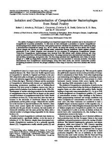
grids were set on one drop of diluted, purified CAK1 suspension or diluted C. acetobutylicum cell culture for 1 to 3 min. Excess fluid was removed by absorption onto filter paper. The dried grids were floated on a drop of 2% phosphotungstic acid staining solution (pH 6.5) for 1 to 3 min, and excess phosphotungstic acid was removed as above. The dried grid was examined with an electron microscope (JEOL model 100C). Restriction enzymes and exonuclease Bal 31 digestion. All restriction enzymes including exonuclease Bal 31 were purchased from BRL and the reactions were carried out as described by the manufacturer or by Maniatis et al. (11). Gel electrophoresis. DNA was electrophoresed in 0.7 or 1.0% GTG SeaKem agarose (FMC Corp., Marine Colloids Div., Rockland, Maine) with Tris-acetate running buffer (pH 8.1) (11) at 100 V constant voltage. The gel was photographed as described previously (7). Protein associated with discontinuous density gradient-purified CAK1 was subjected to 16.5% Tricine-sodium dodecyl sulfate (SDS)-polyacrylamide gel electrophoresis (PAGE) as described by Schugger and Von Jagow (20). The gel was composed of a 16.5% acrylamide running gel (7.5% [vol/vol] glycerol) overlaid with a 10% spacer and a 4% stacking gel. The protein sample was diluted with sample buffer (8% SDS, 24% [vol/vol] glycerol, 100 mM Tris, 4% [vol/vol] mercaptoethanol, 0.001% Coomassie brilliant blue R) and boiled for 90 s prior to loading to dissociate viral proteins. Electrophoresis was carried out at 85 V (constant voltage) with Tricine-Tris running buffer (20) as the cathode buffer. Gels were stained by silver staining (Sigma) as described by the manufacturer and photographed with type 57 Polaroid high-speed (3,000 ASA) film. The molecular weight of isolated protein was determined by using protein size markers (Sigma) and known M13 coat proteins as markers (16). Southern hybridization. DNA fractionated by 1.0% agarose gel electrophoresis was transferred to nitrocellulose as described previously (8). Purified ssDNA from CAKI was biotin labeled by using the random primer labeling kit (Bio-Rad Laboratories, Richmond, Calif.) with biotin-7dATP (BRL). The denaturation step of template DNA was omitted prior to the DNA-primer reannealing reaction, and the DNA primer and biotinylated probe were recovered by ethanol precipitation. The filter was hybridized with probe, and the DNA was detected by using the BlueGENE nonradioactive nucleic acid detection system (BRL). RESULTS Isolation of a viruslike particle. ssDNA was continuously released from C. acetobutylicum NCIB 6444 during cell growth (8). Cytoplasmic leakage during growth was initially believed to be responsible for the presence of extracellular ssDNA, since alkaline lysis of C. acetobutylicum NCIB 6444 also allowed recovery of ssDNA. Although the existence of ssDNA generating plasmids in gram-positive microorganisms is not unusual (3), the extrusion of ssDNA is a characteristic apparently limited to C. acetobutylicum. The PEG-NaCl precipitate of the C. acetobutylicurf NCIB 6444 supernatant contained DNA and protein (A2601A280 = 1.4) but no RNA. The DNA-protein complex was unable to enter the agarose gel during electrophoresis (Fig. 1, lane a) and was resistant to DNase (data not shown). These results suggest that the DNA-protein complex is released from C. acetobutylicum NCIB 6444 and is not present in the supernatant simply because of cellular leakage. Agarose gel electrophoresis of the CsCl density gradient-purified PEG-
532
KIM AND BLASCHEK
FIG. 1. Agarose gel electrophoretic analysis of CAK1. Lanes: a, CsCl density gradient-purified CAK1; b, CAK1 following proteinase K treatment; c, plasmid pDM6 recovered from C. acetobutylicum NCIB 6444. Arrow indicates the position of ssDNA.
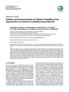
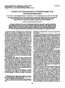
NaCl precipitate following proteinase K treatment and phenol-chloroform extraction resulted in migration of the DNA to a position which corresponds to that previously reported for pDM6 ssDNA (8) (Fig. 1, lanes b and c). CsCl discontinuous density gradient centrifugation of the PEG-NaCl precipitate demonstrated that CAK1 had a density of 1.35 g/ml, compared with a density of 1.75 g/ml for plasmid DNA from C. acetobutylicum. In order to examine the structure and release of the DNA-protein complex, PEG-NaCl-precipitated and CsCl density gradient-purified CAKi from C. acetobutylicum cell culture was subjected to electron microscopic analysis. Examination of electron photomicrographs of C. acetobutylicum NCIB 6444 cell cultures revealed the existence of a threadlike structure which was clearly distinguishable from C. acetobutylicum flagella (Fig. 2A). Electron photomicrographs of CsCl-purified CAKi showed the presence of homogeneous threadlike particles with a small amount of contaminating flagellar fragments (Fig. 2B). The threadlike particles were ca. 1,000 nm in length and 50 to 100 nm in diameter (Fig. 2). The electron photomicrograph shows that CAKi is structurally similar to filamentous phage present in gram-negative microorganisms. DNA in CAK1. The DNA from CAK1 (Fig. 1, lane b) was identical to the extracellular ssDNA which was recovered from the supernatant of C. acetobutylicum NCIB 6444 by using a DNA purification procedure which included proteinase K treatment and denaturation with SDS (8). The molecular size of ssDNA was estimated to be ca. 6.6 kb, as determined by plasmid pDM6 restriction enzyme digestion analysis and comparison with ssDNA derived from coliphage M13mp9 (data not shown). CAKl-associated proteins. The identification of proteins associated with CAKi ssDNA was carried out by subjecting CsCl density gradient-purified CAK1 to 16.5% Tricine-SDSPAGE. Initial attempts to identify a CAKi coat protein by the gel technique described by Laemmli (9) were not successful because of the poor resolving ability of this system for low-molecular-weight proteins. Figure 3 (lane b) shows the presence of 5.0- and 32-kDa proteins associated with CAK1. The 5.0-kDa protein was similar in size to the 5.2-kDa major coat protein reported for filamentous viruses recovered from gram-negative microorganisms (12, 16, 24, 25). However, it appeared that CAKi did not contain the minor coat proteins responsible for infectivity which are
J. BACTERIOL.
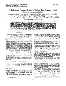
present in M13 (Fig. 3, lane a) (16). CAK1-associated proteins were sensitive to detergent and solvent treatment and protected the DNA from Bal 31 nuclease (data not shown). DNA homology with filamentous phage M13mp9 from E. coli. The physical characteristics of CAKi showed great similarity to those of the filamentous phages isolated from gram-negative microorganisms (Table 1). The overall similarity suggested that the 6.6-kb ssDNA in CAKi may have homology with the DNA from other filamentous phage (12, 25). Extrachromosomal DNA was subsequently isolated from an M13mp9-infected E. coli EM383 cell culture. The extrachromosomal DNA from E. coli and C. acetobutylicum NCIB 6444 was subjected to agarose gel electrophoresis and blotted onto nitrocellulose. A probe was prepared from the ssDNA of CAKi by the random primer labeling method without denaturation of DNA. The ssDNA from CAKi showed homology with the replicative form (RF) of M13mp9 phage (Fig. 4B, lane b) as well as with the RF of the pDM6 plasmid (Fig. 4B, lane a). However, restriction enzyme digestion demonstrated differences between plasmid pDM6 and the M13 RF plasmid (data not shown). Possible homology with the E. coli-derived lacZ gene in M13mp9 was ruled out (data not shown). The hybridization results between CAKi and M13 suggest that the origin of the two DNAs may be the same, as predicted from other filamentous phage sequencing data (25). Absence of biological activity of CAK1. CAKi recovered from the supernatant of C. acetobutylicum NCIB 6444 and CsCl density gradient-purified CAKi failed to demonstrate plaque formation during infectivity experiments with E. coli DH5aoF' and C. acetobutylicum ATCC 824 despite the similarity that CAKi has with filamentous phage from gramnegative bacteria (Table 1). It is not clear whether CAKi is a defective phage or whether the host system does not have the necessary infection factors (i.e., receptor in E. coli DH5aF' or sex pili in C. acetobutylicum ATCC 824). Also, the possibility of unsuitable experimental conditions, which were mainly an adaptation from the E. coli infection protocol (2, 11, 13, 19, 23), is not ruled out. At the same time, the transformation of plasmid pDM6 into C. acetobutylicum ATCC 824 and E. coli DH5aF' by electroporation (7) was attempted in order to examine the replication functions and to modify CAKi by passage through host systems which may allow infectivity of CAK1. However, the lack of a suitable selective marker during transformation experiments made recovery of transformants difficult. Incorporation of a suitable antibiotic marker into CAKi may solve this problem. In any case, plaque formation was not observed. In order to prove that CAKi is a nonlytic defective phage, we attempted to induce the lytic cycle by exposing the cells to UV light (14, 19, 25, 26). When a lawn of C. acetobutylicum NCIB 6444 cells was exposed to 256-nm UV light, induction was not observed. Although extensive UV exposure resulted in cell death, there was no significant increase in recovery of CAKi from C. acetobutylicum NCIB 6444 (data not shown). Interestingly, the extrusion of the viruslike CAKi structure from C. acetobutylicum NCIB 6444 did not result in a significant alteration in growth rate compared with that of the ATCC 824 strain. This observation differs from that of phage-infected E. coli, which demonstrated delayed cell growth compared with the uninfected strain (19, 23). C. acetobutylicum NCIB 6444 demonstrated a faster growth rate and equivalent butanol production ability compared with the ATCC 824 strain (data not shown). Therefore, extrusion of CAK1 appears to have little effect on C.
VOL. 173, 1991
VIRUSLIKE PARTICLE IN C. ACETOBUTYLICUM
533
A
FIG. 2. Electron photomicrograph of negatively stained C. acetobutylicum NCIB 6444. (A) C. acetobutylicum NCIB 6444 cell culture; (B) CsCl density gradient-purified CAK1. Symbols: a, CAK1; b, flagella; f, fragments of flagella. Bars, 1,000 and 200 nm.
acetobutylicum NCIB 6444, especially as it relates to butanol production. In order to examine the replication mechanism of CAK1, which may be similar to that of other filamentous phages (i.e., ssDNA into double-stranded DNA, followed by ssDNA phage reproduction), a curing study with acridine orange was carried out as described by Hirota (6). Although acridine orange was a good agent for curing episomal DNA (6), the curing of plasmids from C. acetobutylicum NCIB 6444 was not observed and the production of CAKi was not
affected even at high doses (100 ,ug of acridine orange per ml). DISCUSSION
The results of this study suggested that CAKi is a defective, filamentous viruslike particle. The double-stranded pDM6 DNA from C. acetobutylicum NCIB 6444 may be the replicative form of ssDNA contained in CAK1. The presence of viruslike particles in other microorganisms is not
534
KIM AND BLASCHEK
a
J. BACTERIOL.
b
c
A
4
5~~~~4
2
9~~~~2
I.
20 W
14
FIG. 3. Tricine-SDS-PAGE of CsCl density gradient-purified CAK1. Lanes: a, M13mp19; b, CAK1; c, molecular size markers (shown in kilodaltons). Arrow indicates position of major coat
protein.
uncommon (14, 26). A surprising result from the experiments performed here was that the ssDNA derived from CAKi has homology with coliphage M13 ssDNA. Filamentous phage from gram-negative microorganisms are divided into two classes, 1 and 2, which appear to have derived from a single ancestor in that they have DNA homology and specific infectivity related to sex pili. However, to our knowledge, the existence of filamentous phage has not been reported previously for gram-positive, anaerobic bacteria. The recovery of a filamentous viruslike particle from C. acetobutylicum NCIB 6444 suggests that ssDNA
may be derived from the same ancestors as those of filamentous phage associated with gram-negative microorganisms. That they may have followed different evolutionary pathways is suggested by the restriction enzyme analysis com-
parison data between plasmid pDM6 and the replicative forms of filamentous phage (24). C. acetobutylicum NCIB 6444 may have been infected by a filamentous phage or acquired ssDNA during species differentiation. DNA comparison data between CAKi and other filamentous phage may help to answer the question of evolutionary relationships between C. acetobutylicum and gram-negative microorganisms. The mechanism for phage acquisition by C. acetobutyliis not clear. Possible explanations are that ancestral filamentous phage had a broad host specificity which was restricted to sex pili-related specificity and that C. acetobutylicum NCIB 6444 was accidentally infected by filamentous phage via an unknown mechanism. This is consistent with the observation that C. acetobutylicum NCIB 6444 is the only plasmid-containing strain of this microorganism described to date (8, 15, 18, 22, 28). The failure of CAKi to be infective in other C. acetobucum
TABLE 1. Comparison of various physical characteristics of CAKi with filamentous phages isolated from
gram-negative microorganisms
Density
Particle Diam
Length ingCsCI (gm) (nm)
Phage Phage in CsCl
CAKi
1.35 FilamentouSa 1.29L-1.32
1,000
870
~~~Nucleotides (kb)
protein (kDa)
6.6 6.6
5.0 5.234
Major coat
(nm)
5-8 5.5
B
a b
a Various filamentous phages recovered from gram-negative bacteria (12, 24). The major coat protein size was estimated from the amino acid sequences of gene VIII (24).
FIG. 4. (A) Agarose (1%) gel electrophoresis of extrachromosomal DNA recovered by the modified alkaline lysis method. Lanes: a, extrachromosomal pDM6 DNA isolated from C. acetobutylicum NCIB 6444; b, extrachromosomal DNA isolated from E. coli EM383. (B) Southern hybridization of same extrachromosomal DNAs as in panel A probed with biotin-labeled CAK1 ssDNA. Bottom arrow indicates monomer form of RF and top arrow indicates dimer form of RF.
tylicum strains may be explained in the same way. However, it is possible that CAKi is a defective filamentous phage. This is suggested from the failure to recover characteristic minor virion proteins related to infectivity during TricineSDS-PAGE (Fig. 3). On the other hand, the host system used during the infectivity test may not have suitable sex pili receptors. If the hypotheses that plasmids originated from phages and phages are tools for inter- and intraspecies genetic information transfer are valid (25, 26), the existence of viruslike particle in C. acetobutylicum may be significant evidence of the transition between phages and plasmids. It is possible that an unknown biological processing of phage during the long evolutionary process led tQ plasmid formation. The presence of ssDNA in a number of gram-positive microorganisms (3) may represent an intermediate transition state between phage and plasmid DNA. An understanding of the relationship between phage and plasmid DNA in terms of evolutionary processing may be facilitated once DNA sequence data for ssDNA-generating plasmids, including CAK1, become available. However, this hypothesis requires more examination. The similarity of CAKi to filamentous phages may increase the usefulness of a C. acetobutylicum vector containing a replication origin derived from CAK1. Infection with filamentous phages does not cause host cell lysis but does produce high copy numbers of the replicative form of the phages (23, 25). The RF of filamentous phage is replicated in a fashion similar to that of other multicopy plasmids and maintained stably over many generations. The E. coli M13 coliphage vectors are well characterized, and the use of these vectors has been explored in molecular genetics (2, 4, 13). If CAKi has nonessential regions where a foreign gene can be inserted, the recombinant RF form of CAK1 in a new host system can provide a good expression vector which can be stably maintained without selection pressure. The stability of plasmid DNA is an important priority in the generation of tailor-made industrial microorganisms. Recently developed vector systems for C. acetobutylicum require antibiotic pressure in order to maintain stability during fermentation (21, 28). In addition to the development of a stable vector expression system for C. acetobutylicum, the ability of CAK1 to generate ssDNA can be applied to inserted gene sequencing and mutation analysis experiments with base substitution for regulatory functional analysis.
VOL. 173, 1991
ACKNOWLEDGMENTS This work was supported in part by grant ICMB 89-0044-03 from the Illinois Corn Marketing Board, State of Illinois Competitive Value-Added grant 1-1-11963, University of Illinois Agricultural Experiment Station Hatch grant 50-313, the University of Illinois Research Board grant 1-2-69157, and a UIUC University Scholar grant to H.P.B. REFERENCES 1. Blaschek, H. P. 1989. Genetic manipulation of the clostridia. Dev. Ind. Microbiol. 30:35-42. 2. Gider, K. 1986. DNA cloning vectors utilizing replication function of the filamentous phages of Escherichia coli. J. Gen. Virol. 67:2287-2303. 3. Gruss, A., and S. D. Ehrlich. 1986. The family of highly interrelated single-stranded deoxyribonucleic acid plasmids. Microbiol. Rev. 53:231-241. 4. Guzman, P., and P. Youngman. 1988. Novel integrational vectors for Bacillus subtilis based on coliphage M13 and their use for the analysis of regulated promoters. Genet. Biotechnol. 2:299-303. 5. Haschemeyer, R. H., and R. J. Myers. 1972. Negative staining, p. 101-147. In M. A. Hayat (ed.), Principles and techniques of electron microscopy: biological applications. Van Nostrand Reinhold Co., New York. 6. Hirota, Y. 1960. The effect of acridine dyes on mating type factors in Escherichia coli. Proc. Natl. Acad. Sci. USA 46:5764. 7. Kim, A. Y., and H. P. Blaschek. 1989. Construction of an Escherichia coli-Clostridium perfringens shuttle vector and plasmid transformation of Clostridium perfringens. Appl. Environ. Microbiol. 55:360-365. 8. Kim, A. Y., A. A. Vertes, and H. P. Blaschek. 1990. Isolation of a single-stranded plasmid from Clostridium acetobutylicum NCIB 6444. Appl. Environ. Microbiol. 56:1725-1728. 9. Laemmli, U. K. 1970. Cleavage of structural proteins during the assembly of the head of bacteriophage T4. Nature (London) 227:680-685. 10. Lin, Y., and H. P. Blaschek. 1984. Transformation of heattreated Clostridium acetobutylicum with pUB110 plasmid DNA. Appl. Environ. Microbiol. 48:737-742. 11. Maniatis, T., E. F. Fritsch, and J. Sambrook. 1982. Molecular cloning: a laboratory manual. Cold Spring Harbor Laboratory, Cold Spring Harbor, N.Y. 12. Marvin, D. A., and B. Hohn. 1969. Filamentous bacterial viruses. Bacteriol. Rev. 33:172-209. 12a.Mattsson, D. Personal communication. 13. Messing, J., B. Gronenborn, B. Muller-Hill, and P. H. Hofschneider. 1977. Filamentous coliphage M13 as a cloning vehi-
VIRUSLIKE PARTICLE IN C. ACETOBUTYLICUM
14.
15.
16.
17. 18. 19. 20.
21. 22. 23. 24.
25.
26.
27.
28.
535
cle: insertion of a HindlIl fragment of the lac regulatory region in M13 replicative form in vitro. Proc. Natl. Acad. Sci. USA 74:3642-3646. Ogata, S., and M. Hongo. 1979. Bacteriophages of the genus Clostridium. Adv. Appl. Microbiol. 25:241-273. Oultram, J. D., M. Loughlin, J. K. Brehm, D. E. Thompson, and N. P. Minton. 1988. Introduction of plasmids into whole cells of Clostridium acetobutylicum by electroporation. FEMS Microbiol. Lett. 56:83-88. Rasched, I., and E. Oberer. 1986. Ff coliphages; structural and functional relationships. Microbiol. Rev. 50:401-427. Reid, S. J., E. R. Allcock, D. T. Jones, and D. R. Wood. 1983. Transformation of Clostridium acetobutylicum protoplasts with bacteriophage DNA. Appl. Environ. Microbiol. 45:305-307. Rogers, P. 1986. Genetics and biochemistry of clostridia relevant to development of fermentation processes. Adv. Appl. Microbiol. 31:1-60. Salivar, W. O., H. Tzagoloff, and D. Pratt. 1964. Some physicalchemical and biological properties of the rod-shaped coliphage M13. Virology 24:359-371. Schugger, H., and G. Von Jagow. 1987. Tricine-sodium dodecyl sulfate-polyacrylamide gel electrophoresis for the separation of proteins in the range from 1 to 100 kDa. Anal. Biochem. 166:368-379. Truffaut, N., J. Hubert, and G. Reysset. 1989. Construction of shuttle vectors useful for transforming Clostridium acetobutylicum. FEMS Microbiol. Lett. 58:15-20. Truffaut, N., and M. Sebald. 1983. Plasmid detection and isolation in strains of Clostridium acetobutylicum and related species. Mol. Gen. Genet. 189:178-180. Tzagoloff, H., and D. Pratt. 1964. The initial steps in infection with coliphage M13. Virology 24:372-380. von Wezenbeek, P. M. G. F., T. J. M. Hulsebos, and J. G. G. Schoenmakers. 1980. Nucleotide sequence of the filamentous bacteriophage M13 DNA genome: comparison with phage fd. Gene 11:129-148. Webster, R. E., and J. Lopez. 1985. Structure and assembly of the class I filamentous bacteriophage, p. 235-267. In S. Casjens (ed.), Virus structure and assembly. Jones and Bartlett Publishers, Inc., Portola Valley, Calif. Wood, A. G., W. B. Whitman, and J. Konisky. 1989. Isolation and characterization of an archaebacterial viruslike particle from Methanococcus voltae A3. J. Bacteriol. 171:93-98. Yamamoto, K. R., B. M. Alberts, R. Benzinger, L. Lawhorne, and G. Treiber. 1970. Rapid bacteriophage sedimentation in the presence of polyethylene glycol and its application to large-scale virus purification. Virology 40:734-744. Young, M., N. P. Minton, and L. Staudenbauer. 1990. Recent advances in the genetics of the clostridia. FEMS Microbiol. Rev. 63:301-326.