Vol. 263, No. 35, Issue of December 15, pp. 18911-18919, 1988 Printed in U.S.A.
THEJOURNAL OF BIOLOGICAL CHEMISTRY
0 1988 by The American Society for Biochemistry and Molecular Biology, Inc
Isolation and Characterizationof Human Lysosomal Membrane Glycoproteins, h-lamp-1 and h-lamp-2 MAJOR SIALOGLYCOPROTEINS CARRYING POLYLACTOSAMINOGLYCAN* (Received for publication, February 18, 1988, and in revised form, June 7, 1988)
Sven R. Carlsson$$, Jurgen Rothll, Friedrich Pillerz, andMinoru FukudaSII From the $ L a Jolla Cancer Research Foundation, Cancer Research Center, La Jolla, California 92037, the Department of Physiological Chemistry, §University of Umei, S-90187 Umei, Sweden, and the (Interdepartmental Electron Microscopy, Bwcenter, University of Basel, CH-4056, Basel,Switzerland
Two major lysosomal membrane glycoproteins with apparent M, 120,000 were purified from chronic myelogenous leukemia cells. These glycoproteins are major sialoglycoproteins containing polylactosaminoglycan and represent approximately 0.1-0.2% of total cell proteins. A monoclonal antibody specific to one of the glycoproteins and polyclonal antibodies specific to the otherglycoprotein were obtained. Immunoelectron microscopic examination of HeLa cells revealed that these twoglycoproteins mainly reside inlysosomes and multivesicular bodies. Immunoprecipitation experiments showed that a number of different cell lines express these glycoproteins. However, the apparent molecular weightsdiffered between cell lines; this probably representsdifferences in the amount of polylactosaminoglycan expressed by each cell line. As shown in thefollowing paper (Fukuda, M., Viitala, J., Matteson, J., and Carlsson, S . R. (1988)J.Biol. Chern. 263, 18920-18928) one of the glycoproteins is very homologous to thatof a mouse counterpart, m-lamp- 1. Thehumanform of this glycoprotein istherefore named human lamp-1 (h-lamp-1), while the other glycoprotein, towhich the monoclonal antibody wasmade, is called human lamp-2(h-lamp-2). Pulse-chase labeling experiments detected that hlamp-1 and h-lamp-2 are produced first as precursor forms of 87.5 and 84 kDa, and treatment withendo-8N-acetylglucosaminidase H (endo-H)or endo-/3-N-acetylglucosaminidase F (endo-F) reduced their molecular masses to39.5and 41.5 kDa, respectively. It was estimated that h-lamp-1 has 18 N-linked saccharides and h-lamp-2 16, based on the resultsof partial digestions with endo-F. These results indicate that the two lysosomal membrane glycoproteins are extensively modified by N-glycans, and some of these were found to have polylactosaminyl repeats and sialic acid. Human lamp-1andlamp-2,therefore,serve as good models for understanding polylactosaminoglycan formation and thebiosynthesis and processing of polylactosaminoglycan-containingglycoprotein.
Polylactosaminoglycans are heterogenous saccharides often having high molecular weights. They are distinguished from complex-type N-linked saccharides by having long side chains of Gal(31+4GlcNAc(31--*3repeats, which are susceptible to endo-(3-galactosidase(for review see Ref. 1). Polylactosaminoglycans carry various antigenic structures such as AB0 blood group antigens (2, 3), developmental antigens such as mouse F9 antigens (4) and human fetal(i) erythrocyte antigen ( 5 ) ,and tumor-associated antigens such as sialyl Le” (6). More recently, it has been shown that the lack of polylactosaminoglycan on the human erythrocyte anion transporter (Band 3) causes the glycoprotein to aggregate, resulting in abnormal membrane structures in a congenital dyserythropoietic anemia-type I1 (7, 8). In human erythrocytes, polylactosaminoglycans are attached to Band3and Band 4.5 (which includes glucose transporter) (2, 3, 5 ) , but not to glycophorins (9, 10). On the otherhand,little is known about the proteincarriers for polylactosaminoglycan in nucleated cells. Since only a limited number of glycoproteins contain polylactosaminoglycan, as shown on PA-1 human teratocarcinoma cells (ll), itis likely that the nature of a proteindetermines whether it is modified by polylactosaminoglycan. We have shown previously that glycoproteins with M, 120,000 are major carriers for polylactosaminoglycan in granulocytic cells (12). In order to characterize the molecules which carry polylactosaminoglycan in nucleated cells, we isolated those glycoproteins. The data presented in this report indicate that these glycoproteins are lysosomal membrane glycoproteins, a group of proteins extensively glycosylated by N-linked saccharides.
part by the payment of page charges. This article must therefore be hereby marked “aduertisement” in accordance with 18U.S.C. Section 1734 solelyto indicate this fact. I( To whom correspondence should be addressed La Jolla Cancer Research Foundation, 10901N. Torrey PinesRd., La Jolla, CA 92037.
Portions of this paper (including “Experimental Procedures,” Tables I and 11, and Figs. 2, 4, 7, and 12) are presented in miniprint at the end of this paper. Miniprint is easily read with the aid of a standard magnifying glass. Full size photocopies are included in the microfilm edition of the Journal that is available from Waverly Press.
-
-
EXPERIMENTAL PROCEDURES’ RESULTS
Purification of Lysosomal Membrane Glycoproteins and Production of SpecificAntibodies-Wehave previously shown that sialoglycoproteins with M, 120,000in granulocytic cells contain significant amounts of polylactosaminoglycan (12). In order to isolate the glycoprotein(s), total cell lysates were * This work was supported by Grant CA28896 from the National applied to a column of wheat germ agglutinin-agarose, and Cancer Institute (toM. F.), Grant B87-03X-07886-01from the Swedish Medical Research Council (to S. R. C.), and Grant3.396-086 from the bound glycoproteins were eluted with 100 mM N-acetylthe Swiss National Science Foundation and the Kanton Basel-Studt glucosamine. The eluted glycoproteins were subjected to pre(to J. R.). The costs of publication of this article were defrayed in
-
18911
18912
Polylactosaminoglycan-containingLysosomal Membrane Glycoproteins
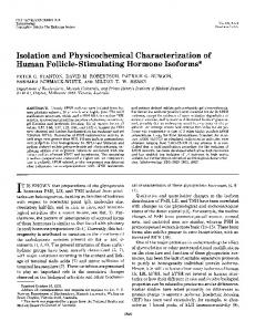
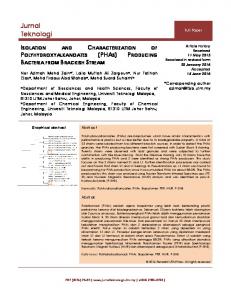
before and afterendo-&galactosidasetreatment. As shown in Fig. 3, all of the cell lines tested contain the lysosomal kDa membrane glycoproteins and two additional points were 200noted 1) the molecular weights of the glycoproteins differ 116- m v significantly among different cell lines, and 2) the suscepti92.5bility to endo-@-galactosidasediffers among cell lines tested. 66The latter characteristic appears to be directly correlated to 45the level of polylactosamine addition expressed by the different cell lines (see below). The glycoproteins were detected in 30all cells tested except erythrocytes. 20Detection of Precursors of Lysosomal Membrane Glycoproteins in HL-60 Cells-In order to obtain information on the FIG.1. Lysosomal membrane glycoproteins (lanes A and B ) polypeptide and carbohydrate moieties of the lysosomal memand leukosialin (lanes C and D ) before and after endo-@- brane glycoproteins, pulse-chase experiments were carried galactosidase treatment. The glycoprotein fraction containing hu- out. Fig. 4A shows that a precursor form of-85 kDa was man lamp-I and lamp-2 and leukosialin were labeled with '*'I. The detected immediately after the pulse labeling, and this prelabeled glycoproteins were incubated with (lunes B and D ) and without (lanes A and C)endo-B-galactosidase and subjected to SDS- cursor form wasgradually converted to themature form with 125,000. polyacrylamide gel electrophoresis (7.5-12.5% aceylamide gradient). a smeared, heterogenously migrating band of M, The conversion of the precursor to mature forms was found Gels were subjected to autoradiography. to have tM 45 min. An apparent heterogeneity in the time parative SDS2 gel electrophoresis. The material migrating required for processing of the precursor form to the mature between M, -100,000 and -130,000 was eluted from gels and form was noticed, and a small amount of the precursor form subjected to affinity chromatography with anti-leukosialin was still detected after 120 min of chase. It was also noted antibodies coupled to Sepharose. The glycoprotein fraction, that the precursor form appeared to consist of two closely which was not bound to thecolumn, appeared to be homoge- spaced bands (Fig. 4A). In order to elucidate the manner in nous when analyzed by SDS gel electrophoresis (Fig. 1, lane which the precursor and mature forms are glycosylated, the A ) . This purified glycoprotein fraction was initially used for same pulse-labeled glycoproteins were digested with endo-H immunization of a rabbit. The glycoproteins were isolated to remove N-linked carbohydrates. Fig. 4B shows that the from the total lysate, but further analysis showed them to be precursor form was converted to two bands with apparent molecular weights of -41,500 and -39,500. As shown by its lysosome-associated membrane proteins (lamp) with M, 120,000. As shown below, this purified glycoprotein fraction susceptibility to endo-H (compare a t 120 min with or without contained two lysosomal membrane glycoproteins with simi- endo-H), the mature form obtained after 120 min chase still lar molecular weights,and theantibodies produced werefound contained a small number of high-mannose saccharides. In order to estimate how many N-linked saccharides are to be specific to two lysosomal membrane glycoproteins, hpresent in the glycoproteins,the precursor form was digested lamp-1 and h-lamp-2 (see also Ref. 29). Lysosomal MembraneGlycoproteinsContain Saccharides with endo-F for increasing periods of time. From the results Susceptible to Endo-P-galactosidase-The lysosomalmemshown inFig. 5,19 bands were counted from the upper bands brane glycoprotein fraction was susceptible to endo-p-galac- of the precursor to the lowest band after digestion, whereas tosidase treatment (Fig. 1,lanes A and B),indicating that the 17 bands were counted from the lower band of the precursor glycoprotein contains polylactosaminoglycan (1, 8). In con- to the secondlowest band after digestion. These results, trast, leukosialin isolated from the same CML cells was barely obtained on both HL-60 and K562 cells, suggest that the affected by endo-&galactosidase treatment (Fig. 1, lanes C glycoproteins contain 16 to 18 N-linked saccharides, as furand D). The mobility of the lysosomal membrane glycoproHL-60 K662 HCT-16 PA-l HEp-02 IMR-90 teins in SDS-polyacrylamide gels was not significantly altered after sialidase treatment (Fig. 2, lanes C and G),while leuko' 1 2 3 ' 1 2 3'1 2 3'1 2 3 ' 1 2 3'1 2 3' kDa sialin was found as a more slowly migrating band after sialidase treatment (Fig. 2, lanes D and H). This increase of 200apparent molecular weight is characteristic of glycoproteins 116containing a high amount of 0-linked saccharides, as shown 92.5previously (14).These results suggest that lysosomal mem66brane glycoproteins and leukosialin differ significantly in glycosylation. Neither the lysosomal membrane glycoproteins nor leukosialin were affectedgreatly in mobility by reduction 45of disulfide bonds (Fig. 2, lanes E-H), indicating monomeric structures of the glycoproteins. Detection of LysosomalMembrane Glycoproteins in Various Human Cell Lines-A number of human cell lines were metabolically labeledwith [3H]glucosamineand subjected to imFIG. 3. Lysosomal membrane glycoproteins from different munoprecipitation with the lamp-specific antibodies. Immu- cell lines after treatmentwith (lanes 3)or without (lanes 2 ) noprecipitates were then analyzed by SDS gel electrophoresis
[email protected] glycoproteins were immunoprecipitated A 0 C D
-
-
-
The abbreviations used are: SDS, sodium dodecyl sulfate; CML, chronic myelogenous leukemia; h-lamp-1 and h-lamp-2, human lysosome-associated membrane protein-1 and protein-2; PMSF, phenylmethanesulfonyl fluoride; PBS, phosphate-buffered saline; endo-H, endo-0-N-acetylglucosaminidaseH; endo-F, endo-0-N-acetylglucosaminidase F.
by anti-(h-lamp-1 + h-lamp-2) serum from the indicated cell lines which were metabolically labeled with [3H]glucosamine. The immunoprecipitates were either untreated (lunes 2) or treated (lanes 3) with endo-0-galactosidase. The samples were analyzed by SDS-polyacrylamide gel electrophoresis (9%acrylamide gel) followed by fluorography. As controls, cell lysates were immunoprecipitated with normal rabbit serum (lanes I).
Polylactosaminoglycan-containingLysosomal Membrane Glycoproteins HL-60
18913
H digestion, however, h-lamp-2 migrated slower (greater Mr) than h-lamp-1 (Fig. 6C). Based on the difference in molecular weights before and after endo-H digestion, it was deduced kDa kDa that human lamp-1 and lamp-2 contain 18 and 16 N-linked oligosaccharide chains, respectively. Isolation of h-lamp-1 and h-lamp-2 by Affinity Chromutog92.5raphy Employing a Monoclonal Antibody-The anti-(h-lamp662) monoclonal antibody was coupled to Sepharose, and the 3 purified glycoprotein fraction, which contained human lamp451 and lamp-2, was applied to the column. The glycoprotein, -39.5 h-lamp-1, whichwas not bound, waspooled and purified further by Sephacryl S-300 gel filtration. The glycoprotein 30eluted from the column, h-lamp-2, was also subsequently purified by gel filtration. Each purified glycoprotein migrated as a single broad bandwhen examined by SDS gel electroFIG.5. Partial digestions with endo-F of lysosomal mem- phoresis. The glycoproteins were detected moreeasily by brane glycoprotein precursor forms from HL-60 and K562 periodate-Schiff reaction than by Coomassie Blue staining cells. Cells were labeled with [35S]methioninefor 10 min and chased (Fig. 7). Furthermore, purified lamp-2 showed a higher apparwith unlabeled methionine for 10 min. Lysosomal membrane glyco- ent molecular weight than lamp-1. Each of the purified glyprotein precursors were immunoprecipitated with anti-(h-lamp-l and coproteins was subjected to Edman degradation for determinh-lamp-2), and were untreated (lane I ), or treated with endo-F for 5 ing its NHp-terminal sequence, and both preparations showed min (lane 2 ) , 20 min (lane 3 ) , 45 min (lane 4 ) and 24 h (lane 5 ) . Similar resultswere obtained when treated with endo-H. Open arrow a single amino acid in each step of Edman degradation, confirming that each glycoprotein was highlypurified (Table shows unrelated coprecipitated radioactive material. I). A 6 C Amino Acid and Carbohydrate Composition of Human lump1 and lamp-2-Table I summarizes the characteristics of 1 2 3 1 2 3 1 2 3 human lamp-1 and lamp-2. Both glycoproteins contain large kDa kDa amounts of carbohydrate which contribute to about 60% of 200. each protein. The carbohydrate composition indicates that the glycoproteins contain mainly N-linked saccharides with -116-92.5 possibly a small amount of 0-linked saccharides, since N 92.5acetylgalactosaminewas detected. Both also contain a signif-66 66icant amount of sialic acid. Both lamp-1 and lamp-2 contain cysteine or cystine, while lamp-1 contains more methionine -45 45but less aspartic acid (+asparagine) than lamp-1 (Table 11). Webelieve that the glycoprotein fraction not bound by -30 anti-leukosialin antibody (the material shown in Fig. lA) consists of only lamp-1 and lamp-2 for the following reasons. First, the amino acid compositions of the startingmaterial is 0 clearly in ag-reement with the average of lamp-1 and lamp-2 FIG. 6. Immunoprecipitation of human lamp-1 and lamp-2 (Table 11). Second, the amino acid sequences obtained in the and their precursors. HL-60 cells were labeled with [35S]methionine for 10 min, and the label was chased with unlabeled methionine starting material gave 2 residues in each step,the same for 10 min (Band C) or for 120 min ( A ) . Cell lysates were immuno- residues which were found in purified lamp-1 and lamp-2. precipitated with anti-(h-lamp-2) antibody(lanes3 ) followed byantiImmunolocalization of Lysosomal Membrane Glycopro(h-lamp-1) antibodies (lanes 2 ) . H-lamp-1 and h-lamp-2 were coim- teins-Application of the two rabbit antisera, anti-(lamp-1 munoprecipitated with anti-(h-lamp-1 + h-lamp-2) antibodies (lanes lamp-2) and anti-(lamp-1)to ultrathinsections from HeLa or 1 ). The glycoproteins were subjected to SDS gel electrophoresis after MCF7 cells yielded the same pattern of staining. However, treatment with (C) or without ( A and E ) endo-H digestion. the anti-(lamp-1 lamp-2) serum gave consistently more intense labeling over all positive structures. Specific immuther discussed below. The results also suggestthat there is no difference in the number of N-glycans attached to lamps nolabeling was observed overlysosomes (as evidencedby cytochemically detectable acid phosphatase activity), greatly between HL-60 and K562 cells. varying in size and shape, and found either adjacent to the Isolation of Two Lysosomal Membrane Glycoproteins from the Purified GlycoproteinFraction-The results shown in trans side of the Golgi apparatus or in the remaining cytoFigs. 4 and 5suggested that thepurified glycoprotein fraction plasm (Figs. 8, A and C, and 9, A and B).Gold particles were actually consists of two different lysosomal membrane glyco- preferentially located at the luminal side of the lysosomal proteins. In fact, the monoclonal antibody, obtained by im- membrane as well as over amorphous material present in the munizing with the purified glycoprotein fraction, was found lysosome lumen. In parallel to these experiments, the Datura stramonium to react with only one of the precursors before endo-H digestion in an immunoprecipitation assay (Fig. 6B, lane 3). We lectin was employed in a cytochemical affinity technique for have designated the glycoprotein whichreacts with the mono- the detection of N-acetyllactosaminyl residues in lysosomes. clonal antibody h-lamp-2, and the one which does not react, It has been shown that D. stramonium lectin binds to Nh-lamp-1. This nomination isbased on the findings that acetyllactosaminyl residues and poly-N-acetyllactosamine human lamp-1 has an amino acid sequence homologous to (25, 26). As shown in Fig. 8, B and D, such residues were lamp-1 isolated from mouse(see the followingpaper, Ref. 30). detectable at the luminal side of the limiting membrane of Before endo-H digestion, the precursor of h-lamp-2 migrated variously sized and shaped lysosomes, as well as over the faster (lowerMI) than that of h-lamp-1 (Fig. 6B). After endo- amorphous content material. In similar types of lysosomes 1
2
3
~
K562
4
5
1
2
3
4
5
"
-
"
-
+
+
18914
Polylactosaminoglycan-containingLysosomal Membrane Glycoproteins
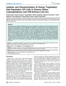
FIG.9. Immunolocalization of immunoreactivity for human lamp-1 and lamp-2 on Lowicryl K4M thin sections of HeLa cells with the protein A-gold technique. Gold particles are pre-
t
FIG.8. Comparison of the distributionof human lamp- 1 and lamp-2 immunoreactivity and N-acetyllactosaminyl residues in lysosomes of HeLa cells. In A , a group of lysosomes with immunolabel for lamp-1 and lamp-2 at theluminal side of the limiting membrane ( I ) or both the limiting membrane and content of the lumen (2) is shown. Structurally corresponding lysosomes (marked I and 2), but stained for N-acetyllactosaminyl residues with the D. stramonium lectin, are presented in B. Note the similar distribution of gold particles which is also evident from other two micrographs shown in C, lamp-1 and lamp-2 immunoreactivity, and D, N-acetyllactosaminyl residues. Anti-(h-lamp-1 h-lamp-2) serum was used in A and C. Magnification: A , X24,600; B, X28,200; C, X33,600; D, X27,600.
+
the pattern of immunolabeling for lysosomal membrane glycoproteins corresponded strikingly to the distribution of Nacetyllactosaminyl residues (compare Fig. 8, A with B, and C with D). In addition to lysosomes, other subcellular compartments showed positive labeling of gold particles. Multivesicular bodies exhibited specific labeling along their limiting membrane as well as at the level of the internal vesicles(Fig. 9C). Structures resembling peripheral endosomes were free of gold particles, although irregularly shaped vesicular structures closely located to them were labeled(Fig. 9, D andE ) . Plasma membrane labeling was only observed with anti-(lamp-1 + lamp-2) serum (Fig. 9B). This antiserum gave also weak immunolabeling over the trans side of the Golgi apparatus (Figs. 8C and 9F). No gold particles were detectable over the nucleus, mitochondria, or endoplasmic reticulum. When the antiserum was replaced by the preimmune serum, no gold particles were observed(not shown) confirming the specificity of the above described immunolabel. H-lamp-:! Derived from Different Cell Lines Also Shows Different Molecular Weights-As shown above, the apparent molecular weight of mature h-lamp-2 is larger than h-lamp1. The results shown in Fig. 3 could be, therefore, due to differences in the relative proportion of human lamp-1 and
sent at the limiting membrane and content of various lysosomal bodies ( A and B ) and multivescular bodies ( C ) . Vesicular structures with an electron-dense content (arrowheads in D and E ) are labeled, whereas other, lucent vesicles are free of gold particles (arrows in D and E ) . Plasma membrane labeling (arrow),preferentially associated with microvilli (arrowheads), and a positive lysosome are shown in B. The Golgi apparatus exhibits weak immunolabeling at the trans side and near smalllysosomal bodies ( F ) .Open arrow denotes plasma membrane. Anti-(h-lamp-1 h-lamp-2) serum was used in A , B, and F, whereas anti-(h-lamp-1)serum was used in C, D, and E. Magnifications: A , X21,300; B, X29,100; C, X29,lOO; D, X38,20O; E, X57,700; F, X44,200.
+
lamp-2 in different cells. In order to test this possibility, hlamp-2 produced in various cells was immunoprecipitated by the monoclonal antibody. As shown in Fig. 10, h-lamp-2 molecules from various cell lines exhibit significantly different molecular weights. These results indicate that both lamp-1 and lamp-2 give different molecular weights depending on cell types. Characterization of Carbohydrate Moiety of Human lamp-1 and lamp-2-In order to preliminarily characterize the carbohydrate moieties of human lamp-1 and lamp-2, glycoproteins were obtained by sequential immunoprecipitation of [3H]glucosamine-labeledK562 and HL-60 cells. After Pronase digestion and Sephadex G-50 gel filtration, glycopeptides obtained from lamp-1 of K562 cells showed essentially two peaks: the glycopeptides which eluted at the voidvolume (fractions 28-40) and those eluted at fractions 50-70 (Fig. 1I.A). The two glycopeptide fractions were separately subjected to mild alkaline degradation. The glycopeptides eluted at the void volume were converted to saccharides with low molecular weights after this treatment (Fig. 12A),indicating that these saccharides are 0-linked oligosaccharides. When the 0-linked oligosaccharides were applied to a Bio-Gel P-4 column,theyeluted atthe positionsconsistentwith NeuNAca2~3Gal/3l+3(NeuNAca2-~6)GalNAcOHand NeuNaca2+6(Galpl+3)GalNAcOH (datanot shown). In contrast, the glycopeptides which eluted in fractions 50-70 of
Polylactosamimglycan-containing Lysosomal Membrane Glycoproteins
18915
fromK562cells(Fig. 1lD) andthese glycopeptideswere susceptible to endo-&galactosidase treatment (Fig. 11F). In contrast, lamp-1 and lamp-2 glycopeptides ofK562 cells showed minimal susceptibility to endo-@-galactosidasetreatment (data not shown). When the 0-linked saccharides from HL-60 lamp-2 were subjected to Bio-Gel P-4 gel filtration, they eluted at the positions corresponding to NeuNAca2-
kDa 200-
3Ga1~1~4GlcNAc~1~6(NeuNAca2+3Gal~l+3)Gal116-
92.5-
66-
45-
FIG. 10. Immunoprecipitation of human lamp-2 from various cell lines. Human lamp-2 was immunoprecipitated by CD3 antibody from indicated cell lines which were metabolically labeled with [3H]glucosamine. Immunoprecipitation was isolated by using biotin-avidin interaction. The samples were analyzed by SDS-polyacrylamide gel electrophoresis (9% acrylamide gel) followed by fluorography. For all cell lines except U266 and Namalva, a volume of lysate containing 1 X lo6 cpm was immunoprecipitated. The lysates of U266 and Namalva contained one-third of the radioactivity.
NAcOH and Gal/31+4GlcNAc/31+6 (NeuNAccu2+3GalSl+ 3)GalNAcOH (Fig. 12B). Finally, the glycopeptides b eluted later than IgG glycopeptides, and they were susceptible to endo-H to yield high-mannose oligosaccharides which eluted between fractions 66 and 84 (Fig. 12C).These results indicate glycopeptides b are high-mannose saccharides. The glycopeptides obtained from HL-60 lamp-1 showed an intermediate elution profile between K562 lamp-2 and HL-60 lamp-2 glycopeptides. The N-glycans of HL-60 lamp-1 ( a of Fig. 1lC) are not as large as those of HL-60 lamp-2 and were only partially digested by endo-&galactosidase (Fig. 11E). These results suggested that both lamp-1 and lamp-2 of HL60 cells contain much more polylactosaminoglycan than those ofK562 cells, and lamp-2 contains more polylactosaminoglycan than lamp-1 in HL-60 cells. The latter data explain why mature lamp-2 is larger than mature lamp-1, even though the precursor of lamp-2 is smaller than thatof lamp-1. These results also indicate that human lamp-1 and lamp-2 contain 0-linked saccharides and high-mannose saccharides in addition to sialylated N-linked saccharides, some of which are polylactosaminoglycans. DISCUSSION
2-
30
40
50
60
70
90 80
30
40
50
60
70
80
90
Fraction Number
FIG.11. Gel filtration of glycopeptides obtained from human lamp-1 and lamp-2 of K562 and HL-60 cells. Pronase digests from h-lamp-1 ( A and C ) and h-lamp-2 ( B and D ) were subjected to Sephadex G-50 gel filtration. The glycopeptides, indicated by horizontal arrows, were subjected to endo-8-galactosidase digestion ( E and F) or endo-8-N-acetylglucosaminidaseH (see Fig. 12) and applied to the same column. A, K562 lamp-1; B, K562 lamp2; C,E,HL-60 lamp-1; D,F, HL-60 lamp-2. The elution positions of Band 3 polylactosaminoglycan ( M , -11,000), fetuin N-linked glycopeptides ( M , -2,900), and IgG glycopeptides (MI -1,700) are indicated by vertical arrows (Band 3, Fet, IgG). 3 denotes the elution positions of Gal@1+4GlcNAc~1+3Gal.
the Sephadex G-50 column were not changed after the same treatment (data not shown), and the elution positions of these glycopeptides are essentially the same as those eluted from fetuin. Similar results were obtained on the glycopeptides from lamp-2 of K562 cells (Fig. 11B). The glycopeptides obtained from lamp-2 of HL-60 cells, on the other hand, provided a different elution profile on Sephadex G-50 column chromatography. Three peaks wereobserved, and the second peak, which represents sialylyated Nglycans (indicated by a ) , eluted earlier than those obtained
The present report describes the isolation, characterization, and distribution of two human lysosomal membrane glycoproteins, h-lamp-l and h-lamp-2. These twoglycoproteins have polypeptide portions of about 40-kDa and are heavily glycosylated by N-glycans; h-lamp-1 and h-lamp-2 contain 18 and 16 N-glycans, respectively. Most strikingly, some of the N-glycans are polylactosaminoglycans. It is interesting to note that thepattern of distribution for lamps and D.stramonium lectin binding sites isstrikingly similar (Fig. 8).In both cases, a spot-like labeling is often seen along the luminal side of the lysosomal membrane. These results are consistent with the findings that D. stramonium lectin preferentially binds to poly-N-acetyllactosaminylstructures (25). Thus, our results obtained by cytochemistry and characterization of saccharides attached to h-lamp-1 and h-lamp-2 agree well and point toward the fact that human lamp-1 and lamp-2 are major sialoglycoproteins carrying polylactosaminoglycan. Similar glycoproteins have previously been detected by monoclonal antibodies in mouse (31, 32), rat (33, 34), and chicken (35, 36) cells. However, the previous studies did not characterize lysosomal membrane glycoproteins with M, 120,000 as carriers for polylactosaminoglycan. It is noteworthy that human lamp-1 and lamp-2 exhibit significantly different molecular weights depending on cell types (Figs. 3 and 10). For example, lamps from HL-60 cells showed muchhigher molecular weightsthan those from K562 cells. As shown in Fig. 5, however,the lamps contain the same number of N-glycans regardless of cell types. The results shown in Figs. 11 and 12 demonstrate that lamps from HL60 cells contain a significant amount of polylactosaminoglycan, whereas lamps from K562 cells essentially lack polylactosaminoglycan. These combined results indicate that the variation in molecular weightis most likely due to differential processing, particularly in the attachment of polylactosaminoglycan. The present results on the content of polylactos-
-
18916
Polylactosaminoglycan-containingLysosomal Membrane Glycoproteins
aminoglycan in lamp-1 and lamp-2 are also consistent with those reported previously on total cellular glycopeptides. It has been shown that K562 cells have minimal amounts of polylactosaminoglycan (37), whereas granulocytic cells (38), HL-60 cells (39), and PA-1 cells (11) express significant amounts of polylactosaminoglycan. Very recently, we have succeeded in isolating cDNAs for human lamp-1 (29, 30) and lamp-2 (30) and thesequences of those cDNAs are consistent with the conclusion that human lamp-1 and lamp-2 contain, respectively, 18 and 16 N-glycosylation sites. Furthermore, the attachment of polylactosamine at some of the glycosylation sites, make the carbohydrate moieties relatively bulky. Since the major portions of the molecules presumably reside in the luminal side of lysosomes, this large carbohydrate moiety probably serves to protect lysosomal membrane glycoproteins from degradation by lysosomal proteases. Experiments have also shown that h-lamp-1 and h-lamp-2 indeed are unusually resistant to a variety of different proteases (data not shown). The isolated glycoproteins contain a significant amount of polylactosaminoglycan and sialic acid, suggesting that the glycoprotein traverses the trans-Golgi cisternae (40). It is unlikely that the carbohydrate moiety is serving as a marker for targeting the molecules to lysosome, since tunicamycin treatment does not inhibit the transport of the glycoproteins to lysosomes (34). In this context,it is interesting to compare the present results with those obtained on mannose-6-phosphate receptors. As shown previously (41, 42), the receptorlysosomal enzyme complex exits from the Golgi apparatus, most likely from the trans-Golgi. Lysosomal enzymes have been shown to contain mannose 6-phosphate modification of carbohydrates, and aspecific receptor for this structureserves to route the enzymes toward the lysosome (43, 44). The receptor-enzyme complex is transported to a prelysosomal compartment where the enzymes are dissociated from the receptors (41, 42). The receptors then recycle to the Golgi apparatus and repeat the process, and thus, the mannose 6phosphate receptors do not reach lysosomes. On the other hand, lysosomal membrane glycoproteins are mainly present in lysosomes and much less are found in other compartments. These results suggest that a molecular signal for targeting of lysosomal membrane glycoproteins to lysosomes is different from that for mannose 6-phosphate receptor routing. In fact, no similarity exists in amino acid sequences between lysosomal membrane glycoproteins (see the following paper (30)) and mannose 6-phosphate receptors (45-48). Lysosomal membrane glycoproteins can also be detected in plasma membranes, multivesicular bodies, and possibly the trans-Golgi (Figs. 8 and 9; seealso Refs. 32, 36,and 49). It is possible that lysosomal membrane glycoproteins are somehow involved in the dynamics of lysosomes such as in the process of fusion of lysosomes with various other organelles. It is interesting to compare some of the properties of human lamp-1 andlamp-2 presented here to those reported by others. For example, Lewis et al. (33) showed the presence of 18 N glycosylation sites inrat lysosomal membrane glycoprotein(s) lgp 120 by endo-H digestion. They detected two protein bands on SDS-polyacrylamide gel electrophoresis after 20 h of digestion, and the ratio of these bands is approximately the same as seen here in Fig. 5, which represents lamp-1 and lamp-2. On -120-kDa glycoproteins of chicken and mouse cells, Lippincott-Schwartz and Fambrough (35) and Green et al. (40) showed a similar half-time for maturation of about 50 min and a similar doublet peptide after endo-H treatment. As shown in the following paper (30), the amino acid sequences of lamp-1 from human, mouse, and chicken are more than
50% identical. These results clearly indicate that lysosomal membrane glycoproteins in these different species are closely related to each other. Our studies show that human lamp-1 and lamp-2 are major glycoproteins of the total cell mass. Although we did not attempt to isolate glycoproteins specifically from lysosomes, the glycoproteins obtained were derived from lysosomes.Similarly, Hughes and August (32) isolated mouse lamp-1 from total membranes. Additionally, lamp-1 and lamp-2 are major polylactosaminoglycan-carrying glycoproteins in various cells, and we have found them each to contain asignificant amount of polylactosaminoglycan (Figs. 3, 8, and 11).These results led us to reevaluate some of the previous reports on polylactosaminoglycan-containing glycoproteins. For example, Dennis et al. (50) reported that metastic cells contain more GlcN A c P 1 4 branching on a-mannose thannonmetastatic cells. Since this branching is the most preferable site for polylactosamine extension (25, 51, 52), more polylactosamine is present in metastatic cells than nonmetastatic cells as aresult. These structures mainly reside on Gp 130 in mouse cells and it is likely that Gp 130 is the mouse counterpart of lamp-1 or lamp-2 since both Gp 130 and lamps represent major a carrier for polylactosaminoglycan. It has been reported from several laboratories that tumor cells invade surrounding tissue or penetrate the endothelial membrane by secreting lysosomal enzymes (53-55). It willbe intriguing to know how this increased secretion takes place in tumor cells and if any correlation exists between the secretion of lysosomal enzymes and surface expression of lysosomal membrane glycoproteins. Acknowledgments-We thank Bonnie Kirstein and V. Shaub for technical assistance, Dr. I. J. Goldstein for the gift of Datura stramonium lectin, Dr. Torgny Stigbrand for help with monoclonal antibody production, Dr. Per-Ingvar Ohlsson for the amino acid sequencing, Dr. Mark Williams for critical reading of the manuscript, D. Wey forpreparing the photographs, and Tami Clevengerfor secretarial assistance. REFERENCES 1. Fukuda, M. (1985) Biochim. Biophys. Acta 780,119-150 2. Krusius, T., Finne, J., and Rauvala, H. (1978) Eur. J. Biochem. 92,289-300 3. Jarnefelt, J., Rush, J., Li, Y. T., and Laine, R. A. (1978) J. Biol. Chem. 253,8006-8009 4. Muramatsu, T., Gachelin, G., Dammonville, M., Delarbe, C., and Jacob, F. (1979) Cell 1 8 , 183-191 5. Fukuda, M., Fukuda,M. N., and Hakamori, S. (1979) J. Biol. Chem. 254,3700-3703 6. Fukuda, M., Bothner, B., Ramsamooj, P., Dell, A., Tiller, P. D., Varki, A., and Klock, J. C. (1985) J. Biol. Chem. 2 6 0 , 1295712967 7. Fukuda, M. N., Klier, G., Yu, J., and Scartezzini, P. (1986) Blood 68,521-529 8. Fukuda, M. N., Dell, A., and Scartezzini, P. (1987) J. Biol. Chem. 262,7195-7206 9. Yoshima, H., Furthmayr, H., and Kobata, A. (1980) J.Biol. Chem. 255,9713-9718 10. Irimura, T., Tsuji, T., Tagami, S., Yamamoto, K., and Osawa, T. (1981) Biochemistry 20,560-566 11. Fukuda. M. N.. Dell, A., Oates, J. E., and Fukuda, M. (1985) J. Biof. Chem.260,6623-6631 12. Fukuda. M.. Koeffler. H. P.. and Minowada, J. (1981) Proc. Natl. Acad.’Sci.‘ U. S. A . 7 8 , 6299-6303 13. Roth, J. (1983) J.Histochem. Cytochem. 31,987-999 14. Carlsson. S. R., and Fukuda, M. (1986) J. Biol. Chem. 2 6 1 , 12779-.12786 15. Zacharius. R. H.. Zell. E. T.. Morrison. J. H.. and Woodluck, J. J. (1969) Anal.’Biockem. 30, 148-152 16. Greenwood, F. C., Hunter, W. M., and Glove, J. S. (1963) Biochem. J. 8 9 , 114-123 17. Kohler, G., and Milstein, C. (1975) Nature 2 5 6 , 495-497 18. Laemmli, U.K. (1970) Nature 227,680-685
Polylactosaminoglycan-containingLysosomal Membrane Glycoproteins 19. Updyke, T. U., and Nicolson, G.L. (1986) Methods Enzymol. -121,717-725 20. Fukuda, M., Carlsson, S. R., Klock, J . C., and Dell, A. (1986) J. Biol. Chem. 261.12796-12806 21. Fukuda, M., Lauffenburger, M., Sasaki, H., Rogers, M. E., and Dell, A. (1987) J. Biol. Chem. 262,11952-11957 22. Carlemalm, E., Garavito, R. M., and Villiger, W. (1982) J. Microsc. (Oxf.) 126, 123-143 23. Roth, J., Bendayan, M., Carlemalm, E., Villger, W., and Garavito, M. (1981) J. Histochem. Cytochem. 29, 663-671 24. Roth, J., Bendayan, M., and Orci, L. (1978) J. Histochem. Cytochern. 26,1074-1081 25. Cummings, R. D., and Kornfeld, S.(1984) J. Biol. Chem. 259, 6253-6260 26. Yamashita, K., Totani, K., Ohkura, T., Takasaki, S.,Goldstein, I. J., and Kobata, A. (1987) J. Biol. Chem. 262,1602-1607 27. Lucocq, J. M., Berger, E. G., and Roth, J. (1987) J. Histochern. Cytochem. 35,67-74 28. Roth, J., and Berger, E. G. (1982) J. Cell Bid. 93, 223-229 29. Viitala, J., Carlsson, S. R., Siebert, P. D., and Fukuda, M. (1988) Proc. Natl. Acad. Sci. U. S. A. 85, 3743-3749 30. Fukuda, M., Viitala, J., Matteson, J., and Carlsson, S. R. (1988) J. Bid. Chem. 2 6 3 , 18920-18928 31. Chen, J. W., Murphy, T., Willingham, M. C., Pastan, I., and August, J. T. (1985) J . Cell Bid. 101, 85-95 32. Hughes, E. N., and August, J. T. (1982) J. B i d . Chern. 2 5 7 , 3970-3977 33. Lewis, V., Green, S. A., Marsh, M., Vihko, P., Helenius, A,, and Mellman, I. (1985) J. Cell B i d . 1 0 0 , 1839-1847 34. Barriocanal, J. G., Bonifacino, J. S., Yuan, L., and Sandoval, I. V. (1986) J. Bid. Chem. 2 6 1 , 16755-16763 35. Lippincott-Schwartz, J., and Fambrough, D. M. (1986) J . Cell Biol. 1 0 2 , 1593-1605 36. Lippincott-Schwartz, J., and Fambrough, D. M. (1987) Cell 4 9 , 669-677 37. Fukuda, M. (1980) Nature 285,405-407 38. Spooncer, E., Fukuda, M., Klock, J. C., Oates, J. E., and Dell, A.
SUPPLEMENTALMATERIAL TO ISOLATION ANDCHARACTERIZATION OF HUMN L I S O S M l A L MEMBRANE GLYCOPROlClNS h - L a w 1 AND h-lamp-2: MAJOR SIALOGLYCOPROTEINSCARRYINGPOLYLACTOSMIHOGL~CAN by SYen R . CdrllPOn, Jurgen Roth. F r i e d r i c h P i l l e r
and Minoru FUkuda
EXPERINWAL PROCEDURES
18917
(1984) J. Biol. Chem. 259, 4792-4801 39. Mizoguchi, A,, Takasaki, S., Maeda, S., and Kobata, A. (1984) J. Bid. Chem. 259,11949-11957 40. Green, S., Zimmer, K.-P., Griffiths, G., and Mellman, I. (1987) J. Cell B i d . 1 0 5 , 1227-1240 41. Brown, W. J., Goodhouse, J., and Farauhar, M. G. (1986) J. Cell ~ i o i l 0 3 i2x-1247 , 42. Griffiths. G.. Hoflack. B.. Simons.. K... Mellman, I., and Kornfeld, S.(1988) Cell 52, 329-341 43. Kornfeld, S.(1986) J . Clin. Znuest. 77, 1-6 44. von Figura, S.,and Hasilik, A. (1986) Annu. Reu. Biochem. 55, 167-193 45. Dahms, N. M., Lobel, P., Breitmeyer, J., Chirgwin, J. M., and Kornfeld, S.(1987) Cell 5 0 , 181-192 46. Pohlmann, R., Nagel, G., Schmidt, B., Stein, M., Lorkowski, G., Krentler, C., Cully, J., Meyer, H. E., Grzeschick, K.-H., Mersmann, G., Hasilik, A., and von Figura, K. (1987) Proc. Natl. Acad. Sci. U. S. A. 84,5575-5579 47. Lobel, P., Dahms, N. M., and Kornfeld, S.(1988) J. Biol. Chern. 263,2563-2570 48. Oshima, A.. Nolan. C. M., Kyle. J. W.. Grubb, J. H., and Sly, W. s. (1988)'J. B i d Chem: 263; 2553-2562 49. Ho, M.-K., and Springer, T. A. (1983) J. Biol. Chem. 258, 636642 50. Dennis, J. W., Laferte, S., Waghorne, C., Breitman, M. L., and Kerbel, R. S.(1987) Science 236, 582-585 51. Sasaki, H., Bothner, B., Dell, A,, and Fukuda, M. (1987) J . Biol. Chem. 2 6 2 , 12059-12076 52. Van den Eijnden, D. H., Koenderman, A. H. L., and Schiphorst, W. E. C. M. (1988) J. Biol. Chem. 263, 12461-12471 53. Oosta, G. M., Farreau, L. V., Bealer, P. C., and Rosenburg, R. D. (1982) J . Bid. Chem. 2 5 7 , 11249-11255 54. Sloane, B. F., Rozhin, J., Johnson, K., Taylor, H., Crissman, J. D., and Honn, K. V. Proc. Natl. Acad. Sci. U.S. A. 83, 24832487 55. Niedbala, M. J., Madiyalakan, R., Matta, K., Crickard, K., Sharma, M., and Bernacki, R. J . (1987) Cancer Res. 47, 46344641
& v a t i o no fq l v c o c w o t e i n r bvmonoclonalantibody-Seoharose l f f l n i t v chromatoarauhl The membrane g l y c o p r o t e i n s . vas above mentlonedglycoprotein fraction. whlchcantalnedtwoIyroromal used f o r production o f m u s e monoclonal a n t l b o d i e r ( 1 7 ) . Among several clones. one clone, which p r o d u c e da n t i b o d i e sw i t ht h eh i g h e s ta f f i n i t ya g a l n r t one o ft h eg l y c a p r o t e i n r . was propagated antibody. c a l l e d (03 (IgGl. kappa), was and a n t i b o d i e s were produced I S a s c i t e s ~n mice.This p u r i f i e d on proteinA-Sepharore and coupled to Sepharore ( 4 w/ml g e l ) . The glycoproteins were parsedtwicethroughthe CUI-Sepharore m l u m w i t h 1 flow r a t e of 2 n l / h .A f t e r each c y c l et h e column M I S washed w i t h 50 m1 o f 0.5% sodium deoxycholate. 10 nll l r i r / H C l . pH 8.0 and bound m a t e r i a l was eluted w i t h 50 n i l diethylamine. pH 12. E l u t e df v a i t t o n s were ? m e d i a t e l yn e u t r a l i z e dw i t hr a l i dg l y c i n e . The Separatedglycoproteins were e r t e n r l v e l y d i a l y z e d a g a r n r t 6 1 s t ? l l c d water and lyophilized. The glycoprotein whichdvd not bind to the c o l u m i sc a l l e dh . l m p - I . h e r e a f t e r , whereas the bound and e l u t e d g l y c o p r o t e i n i s c a l l e d h - l a m p . 2 . ~
wyelogenOUS leukernla C e l l s (CHL) f o l l o w e de s ~ e n t i a l l yt h ep r o c e d u r ed e s c r i b e df o r 11oIation e; I e u k o r i a l i nf r o m HL-60 Cells ( 1 4 ) . Packed CML c e l l s sere h o m g e n i t e d In I Y O ] . of 2% NP-IO. 150 M NaCI, 1 M PIISF, 50 nll TriP/HCl. pH 7.4. A f t e ri n c u b a t i o n on i r e f o r 30 .in. the homogenate was c e n t r i f u g e df o I 20 mln. at 2OOOxg. TO the supernatant w a s added SOS and EDTA to f i n a l c o n c e n t r a t i o n s 0.4% and 5 rd r e s p e c t i v e l y i was r e n t r l f u g e d f o r 1 h. at io0,ooorg and i h e __I_...I__... ly
"..I.I-..
Amino a c i d and carbohvdrate a n a l v r i r - h i n o a c i d and c a r b o h y d r a t e a n a l y l ~ r were c a r r i e d out a$ described i n 1 P r e v i o u s r e p o r t I l k ) . The a n a l y r i r Of saccharidesbySephrder 6-50 and Bio-Gel f-k gel f l l t n t i o n 1 1 s p e r f o m e d as d e s c r i b e dp r e v i o u s l y (20 21). Standard0-linkedsaccharides were o b t a i n e df m m9 r a n u l o c y t l r Cells as descvibed 1 2 0 ) . S t i n d a r d high-mannosesaccharides were obtained frem acute myelogenaur leukemia c o l l r b y endo-H d i g o r t i a n o f g l y c o p e p t i d e r ( u n p u b l i s h e d results).
Polylactosaminoglycan-containing Lysosomal Membrane Glycoproteins
18918
A
1
2
1 3
B 2 3
kDa 200-
. The (25.26) in
Icctln was used for th. detection O f N-acetylIactoslmlnyl resl6ues I 110 step cytOChuIcal I f f l n l t y tcchnlquc (Egea and Roth. In prcparrtlon). B r e i f l y .u l t r a t h l n srctlonr of L w l c r y l K4M nbc6ded HCLl cells Incubated w l t h the puvcl l u t l n (75 #+I, 15 .In) f o l l a d by rlnser r l t h PBS and lncubatlon wlthovaucold-gold complex (0.0. 525 nm 0.25, 30 .In). S p e c l f l c l t y Of I e c t l n rtalnlnq was controlled by prelncubatlon of the l e c t l n r l t h 0vD.uc0I6. I I I ~ ~ w ~ o o ~ o . u c O I ~or N-aceytllact o r r l i n e . and by pletreatment o f the t h i n sectlons r l t h endo F ( 2 7 ) .
=re
-
11692.566-
A B C D
4 5-
E F G H
kDa 20011692.566-
45-
1
R
a
A
f
kDa 20011692.50645J 3 0 1 0 Y l M 7 0 8 ) S Q I W l
F~acIm
FIG. 12
p
n 1 m - 1 md I-.
A. Sephadex G-50 gel f l l t r a t i o n Of alkallneborohydrlde-treatedglyc0p.ptld.s r l t h hlgh mlecular welghts ( F n c t l o n r 28-40 I n Flg. 111). 51mIlw p r o f l l e s were obtained fm Other
glycopeptlder r l t h hlgh nolecular welghts
(Fractions 28-40 I n Flp. 118. C and 0 ) .
8. 010-Gel P-4 gel f l l t n t l o n Of fractions 64-80 I n Flg. I2A. The 6 and 5 denote the elution posltlon o f NevWlc~2-3G~ldl~GlcN4c~l~(NeuN4~2-3G~lpl-3)G~lN4cWl and Gt1414G1cWbl-6(NeuNAc02-3Galbl-3) GaIHAcal.
0
- . . .* * . . . -
C. Sephadex 6-50gel f i l t r a t l o n Of g l y c o w p t l d r k In Flg. 110 after mdo-b-l-ac8tylglucosulnldase H treatment. Man8 and MJn5 denote theelutlon porltims O f Ilan8GlcN4cWl and nln5GlCN4CW.
0' 0' 10'20'30'45'60'90'120'120'
kDa
kDa 200'
11692.5-
-125 -05
6641.5 -38.5
45-
30-
20FIG. 1
h-
HL-60
cU.
Cells were labeled r l t h 3SS.methl~nin~for IO .In. and chased the radioactivity r l t h vnlabeled r t h l o n l n ef a r lndlcated prlodr o f tlr. The l y s o r o u l &ram glycoprolelnr e r e l u n o p r e c l p l t a t e d by mtl.(h-Iamp-l + h - l u p - 2 ) ~ n t k d l e sand malyzed by SOS-polyxrylulde gel elrctrophoreris ( 9 % Icrylamlde). As a control.the s u p l e taken after 120 .In of chase was ~ m n o p r c c i p l t a t c d r l t h noma1 r a b b l t scrv. (120' + NRS). 8. The Same s u p l e s I S p l n r l A were treated wlth endo-H ( + ) before analysis. The cootroll were untreated ( - ) . A.
18919
Polylactosaminoglycan-containingLysosomal Membrane Glycoproteins TMLE I
UURIICTERISTICS OF Hlwl I M P - I NCJLAW-2
1"l
lW.2
-ItOK
-1251:
Tup-1
llr Of nature glycoprotein
+ law-2' 10.6
of precursor glycoprotein
87.5N
84K
Ir of endo-ll dlgerted precursor
39.5u
41.5K
M I
N-terminal sequence N m r o f N-glycans
hNFllVKN61618
LELXLTDSEX.
16
8.6 10.4 6.8 4.9
6.8 8.1 2.1
7.8 2.1 4.2
Carhahydrate Cowosition.mler/nole FUCOIe
36.0
12.5
llrnnare
73.5
52.0
G111ctase
73.5
70.7
GlcNAc
98.9
95.1
GalM
15.4
8.2
Sialic acid
36.4
23.6
Percent carbohydrate
5%
8.8 2.6 4.4 2.1 4.4 5.2 d
1.9-2
l"l
10.7 (Il.l)b 1.7 ( 8.7) 10.3 (10.3) 8.2 ( 6.7) 5.0 ( 4.9) 7.8 ( 6.7) 8.6 ( 8.2) 1.7 ( 2 . 1 ) 7.2 ( 7.7) 2.7 ( 2.1) 3.4 ( 4.1) 8.8 ( 2.6 ( 4.5 ( 2.5 ( 4.1 ( 4.1 ( (
-
13.8 (14.S)b 10.6 (10.6) 9.0 ( 8.2) 8.2 ( 7.1) 3.7 ( 1.9) 7.0 ( 5.3) 1.0 ( 6.8)
1.7 7.2
9.0)
7.8
( 1.1) ( 7.6) ( 1.3) ( 5.0) ( 8.4)
2.6) 4.6) 2.1) 4.6) 4.4)
4.1
(4.1)
0.3)
1.4
5.1