6IATOI OCT CTC. ATC CCA. GAC ... OCT OTC AGC CTC GTC. CTC CTC ...... Wedlund, P. J., Aslanian, M. D., McAllister, C. B., Wilkinson, G. R. &. Branch, R. A. ...
Proc. NatI. Acad. Sci. USA
Vol. 83, pp. 8064-8068, November 1986 Biochemistry
Isolation and sequence determination of a cDNA clone related to human cytochrome P-450 nifedipine oxidase (gene cloning/polymorphism/dihydropyridines/calcium blockers)
PHILIPPE H. BEAUNE, DIANE R. UMBENHAUER, RICHARD W. BORK, R. STEPHEN LLOYD, AND F. PETER GUENGERICH* Department of Biochemistry and Center in Molecular Toxicology, Vanderbilt University School of Medicine, Nashville, TN 37232
Communicated by Elizabeth C. Miller, July 18, 1986
Enzymes and Antibodies. Human livers were obtained through the Nashville Regional Organ Procurement Agency,
and protocols were approved by the Vanderbilt Committee for the Protection of Human Subjects. Livers were perfused immediately after circulatory arrest, chilled on ice, and brought to the laboratory. The livers were cut in small pieces, frozen in liquid nitrogen, and stored at -70°C (8). P-45ONF was purified as described, and polyclonal and monoclonal antibodies were produced (7, 9). These antibodies recognized a single band migrating with purified P-45ONF when human liver microsomes were electrophoresed and immunoblotted (7, 9). Before use in screening the Xgtll library, antibodies were adsorbed twice overnight at 4°C with Escherichia coli (BNN97) (10) lysate bound to CNBr-activated Sepharose 4B (Pharmacia, Piscataway, NJ) in order to eliminate reaction with E. coli proteins; after immunoadsorption, the antibody reactivity toward P-45ONF and toward E. coli was assayed. Library Screening. The human liver cDNA phage Xgt11 expression library was a gift of G. A. Ricca and W. Drohan, Meloy Laboratories (Springfield, VA). It was screened with both polyclonal and monoclonal antibodies basically as described by Young and Davis (10); the nitrocellulose filters were developed with an immunochemical technique (11) using 4-chloro-1-naphthol in place of 3,3'-diaminobenzidine (12). Antisera or ascites fluids were used at dilutions of about 1:50. Positive plaques were checked and plaque-purified by at least two additional rounds of dilution and screening. High-titer phage stocks were purified through CsCl step and equilibrium gradients (13). Xgtll DNAs were prepared for restriction mapping and subcloning in phage M13 (14). Lysogens were prepared by infection of Y1089 E. coli with Xgtll phage clones, and colonies were selected for growth at 30°C and not 42°C. Fusion proteins were produced by initially growing cells in exponential phase at 30°C, heat-shocking them at 42°C for 15 min, and then growing them at 37°C in the presence of 10 mM isopropyl-,3-D-thiogalactose until the A600 stabilized (between 0.5 and 2 hr). Cells were then collected by centrifugation at 104 x g for 10 min, and the pellet was solubilized for electrophoresis, with subsequent visualization of bands by silver staining or immunoblotting. Subcloning and Sequencing. Xgtll DNA containing inserts was digested with EcoRI, HindIII, Sac I, or HindIII/Sac I and was subcloned in M13 phage as described (15), except that the UT481 strain was used instead of JM101 or JM103. E. coli UT481 was constructed by and obtained from C. Lark (Salt Lake City, UT). M13 plaques containing inserts were plaque-purified, and single- and double-stranded DNAs were prepared as described (15); the orientation was checked by asymmetric restriction digestion (i.e., Pst I or BamHI). Single-stranded M13 DNA was used as a template for DNA sequencing by the dideoxy termination method (16). DNA sequencing kits were obtained from New England Nuclear
The publication costs of this article were defrayed in part by page charge payment. This article must therefore be hereby marked "advertisement" in accordance with 18 U.S.C. §1734 solely to indicate this fact.
Abbreviations: P-450, liver microsomal cytochrome P-450; PCN, pregnenolone-16a-carbonitrile; kb, kilobase(s). *To whom reprint requests should be addressed.
Human liver cytochrome P-450NF is the form ABSTRACT of cytochrome P-450 responsible for the oxidation of the calcium-channel blocker nifedipine, which has been reported to show polymorphism in clinical studies. By screening a bacteriophage Xgtll expression cDNA library, we isolated two clones: NF95 with an insert length of 0.8 kilobases which gave a stable fusion protein and NF25 with an insert length of 2.2 kilobases. The two clones were both sequenced and shown to be identical in their overlapping section. The sequence of NF25 is 77% similar to that reported for a rat cytochrome '"P-45OPCN" cDNA (PCN = pregnenolone-16a-carbonitrile). The similarity decreases to 45-53% when the sequence is compared to human cytochromes P-450 belonging to other families [i.e., "pH P-450(1)," "P1-450," "P3-450," and "P-45OMP." The deduced amino acid sequence is 73% similar to that of rat cytochrome P-45OPCN, and the first 21 amino acids are identical to those reported for human liver cytochrome "P-450p." Sections of these clones were nick-translated and used as probes for analyses of human mRNA and genomic DNA. The number and size of bands indicate that P-45ONF belongs to a multigene family, the so-called pregnenolone-16a-carbonitrile-inducible family.
Cytochrome P-450 (P-450) plays an important role in the oxidation of drugs and carcinogens as well as endogenous substrates. Interindividual variation in oxidative metabolism can be attributed, at least in part, to the composition of individual P-450 forms, and these differences are partly due to genetic factors. Since 1977, several genetic polymorphisms of drug oxidation have been demonstrated (1-3), and in some cases the involved form of P-450 has been identified in humans (4, 5). Recently Kleinbloesem et al. (6) reported that oxidation of nifedipine, a vasodilator and calcium-channel blocker, was distributed in a polymorphic manner-17% of the Dutch population studied were phenotypically poor metabolizers. Recently we identified and purified the human liver P-450 form that is responsible for this oxidation of nifedipine (7). This protein, P-45ONF, was shown to be related or identical to P-450s previously isolated from human liver in this laboratory (8). To better understand the mechanism underlying this polymorphism, we used polyclonal and monoclonal antibodies to screen a human liver bacteriophage Xgtll cDNA expression library. The selected clones were analyzed, sequenced, and used to prepare nick-translated probes for the analysis of mRNA and genomic DNA.
MATERIALS AND METHODS
8064
Biochemistry: Beaune et aL Hindm EcoRI
NF 25
I
Proc. Natl. Acad. Sci. USA 83 (1986)
HindZ
Eco RI
ToqI
sacI
EoR/
ToqI Bo HIf
II
I
II
8065
II
Eco RI
Sac I
Hindm
PitI 1
5'1 .. .. _, vI-I .0
I9
3'
probe 3
[probe 3
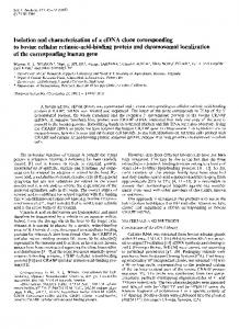
FIG. 1. Restriction map of clones NF95 and NF25 and sequencing strategy. The hatched region is the coding sequence. Arrows indicate the direction and extent of sequence determined for cDNA fragments subcloned in M13mp19 or M13mpl8. Recognition sites for restriction nucleases are indicated. Except for Taq I, these were all determined experimentally and confirmed by computer analysis of the sequence.
SacI
Sac I Eco RI/ Pst I
NF 95
I
probe 2
Hindm
of
\ Eco RI
I 9 IMiM //A3
M,
4z
A 1
3
200bp
probe
and used according to the supplier's instructions except that the incubation temperature was 370C and no NaCl was included in the buffer. Additional sequencing primers were 18-mers or 20-mers synthesized with a BioSearch Sam-One Series II DNA Synthesizer; oligonucleotides were purified as described by Lloyd et al. (17). Sequences were compared with the aid of computer access to the protein sequence data base of the National Biomedical Research Foundation.t Blotting Analysis of RNA and DNA. Genomic DNA and total RNA were isolated from the same preparation by using the CsCl cushion method described by Chirgwin et al. (18). The RNA was collected as a pellet on the bottom of the centrifuge tube, dissolved in water, and precipitated with 0.1 vol of sodium acetate and 2 vol of ethanol. The DNA was collected as a viscous solution at the interface between the CsCl and the homogenate. This solution was extracted once with phenol/CHCl3, 1:1 (vol/vol) and once with CHCl3 and then was precipitated with ethanol. The DNA was dissolved in 10 mM Tris HCl buffer, pH 8.0/1 mM EDTA, treated with proteinase K (50 pg/ml) in 0.1% sodium dodecyl sulfate, and reextracted with phenol/CHCl3 and CHCl3 as above. The purified DNA was then precipitated with ethanol. For Southern blots, 20 jug of genomic DNA was cut with various restriction enzymes and electrophoresed through a 0.8% agarose gel. The gel was processed by the Wahl et al. (19) modification of the Southern (20) procedure. The DNA was transferred to GeneScreenPlus (New England Nuclear) and processed as suggested by the supplier. For RNA blot analyses, 15 /ig of RNA was submitted to agarose/ formaldehyde electrophoresis as described (21). RNA was then transferred to GeneScreenPlus (New England Nuclear) and processed as suggested by the supplier. DNA and RNA blots were probed with nick-translated DNA inserts (=108 dpm/,tg). DNA fragments were purified from agarose gels basically as described (22). Hybridization and washings were performed as described by the supplier. Filters were then autoradiographed with Kodak XAR film (Kodak) with two screens from 3 to 7 days at -70'C. tNational Biomedical Research Foundation (1986) Protein Sequence Data Base of the Protein Identification Resource (Washington, DC), Release No. 8.0.
RESULTS AND DISCUSSION Isolation and Characterization of cDNA Clones. A human liver cDNA expression library in bacteriophage Xgtll was screened with polyclonal and monoclonal antibodies raised against P-45ONF. After the screening of a total of 3 x 10⁵ plaques, 15 clones were positively identified by both antibodies. These clones were characterized by insert length and by the presence of fusion proteins in lysogen preparations. Two clones were selected: NF25, the insert ofwhich was long A X
B
x
95
_+_+-
C 95
+
-
+
00-
1. 6
0.8 pmol 68
P-4()
_
43_ -
_
P-450
;
__
_
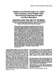
FIG. 2. (A and B) Electrophoresis and immunoblotting of Xgtll lysogen proteins: immunoblotting with anti-P-45ONF (A) and silverstaining of the gel (B). Lanes: X, E. coli lysogen infected with Xgtl1 bacteriophage without insert; 95, E. coli lysogen infected with Xgtll clone 95; -, cells grown at 30TC pelleted and electrophoresed; +, cells grown at 30TC, heat-shocked 15 min at 42TC, and treated with isopropyl-p-D-thiogalactose. Molecular mass is shown in KDa. P-450 and Gal indicate the migration positions of P-45ONF and 3-galactosidase, respectively. The arrowhead at the right indicates the migration of the fusion protein. Clone NF25 gave results similar to those in lanes X. Development of immunoblots with anti-(3galactosidase visualized 3-galactosidase in lane X+ and the fusion protein with a higher molecular mass in lane 95+. (C) Various amounts of purified P-45ONF (from sample HL 93) (7) were spotted on nitrocellulose with a "dot" apparatus, incubated with a 1:50 dilution of rabbit antibody raised against /-galactosidase/clone NF 95 fusion protein (see text), and developed as described (11, 12).
8066
Biochemistry: Beaune et al. O~~~~CACAGCAA----- AGAGCAACACAG
I 2
62
T A TTCGCATGC ~~~~~~~~~TT
N Ala 6IATOI OCT AC EATOG
136 151
m
Low CTA 00
Oly 36 226 21 266l 341 316
Proc. Nati. Acad. Sci. USA 83 (1986)
Lea TTG T Pie
Lym
AAA
Val GTG
Asp
Tyr
TAT T
Pll. Gly
GGA C
ti.
LOn
Pro CCA T Set
CTC ATC O CT Lea
Gly
GGA
Him
Tie ACC
CAT
G
Aim AAT C Tie
Val OTG A A lie
TOG
Lew CTA
Va1 GTG
Tip
CC Arg Lea
lie AT? GOG Val
TTG C
Asp
Lea
GAC CT Ala
Gly
Plie TTT C Leau
Lyn
Gin
AAA C
GAA
Ala
TTG CTC
SeW
GCC ACA Tie
Gly
Him
TCA A Tie
CGCA
CAT
CC
Met ATG CTG Lea
Lea
CTT
CGlu
(;AA Pit.
TTT C
-
CCC
CC.(TCAAA(;CAAC;ACTCAC;ACGAC.At;ACATAACTAAGC-AAAGTAG-TG AG C. CA A CA T TCT CCTGCACGC
A
Tip Leu Lea LeU TOO CTT CTC CTC. C
Tie
ACC
C.
Lys
Lea AG
Lys
AAG A
AAC
Val
CTT
Gly
C.CA
Set
Tyr
TCC A AT
Him
TAC
CAT T
Tyr Amp Gly GAT, GOT
Tyr
TAT T
Lys
Tyr
Cyn
Sec
TAT T
TCT
TTT A Lea
Gie CAA
Gin CAC AT Met
pro CCT
Pie
Tie
Val CTC
TTC
Cy$
Pie
CCC T
Pie TGT C
Gly
AAG T Met
414 42?
Lys 463 4696
Set AGT
Ala lie GCC ATC TO V.1
Gly
GGA
Lys
AAA G
Sar TCT Len CTC
lie ATA C Val
Ala GCT
Lys
Gin GAG
AAG
C.
Tip Va1
T
Lea
Gin GAG
Amp
GAT
Gin GAA
Gin GAA
CCC
C
C
A
Lyn
Tip
;i62 COO 35 A A Gin
651 634 m" 763
Tie ACT C
Ly"
AAG
Gin GAA 0 Set AGC
Ala GCA 0
Tie ACA
Len Lys AAG CTT A T C
Gin GAG
Tie ACA
Set TCA
Pie TTT
Len TTA
Avg
AGA
Lys Met ATG
V.1 CTC T
Pro CCT C
i. ATC
Gly
Lym
GGC
Pro CCT
AAG
Vat GTG
Gly
GGA
Pie TTT
Asn AAC
Amp
CAT
Val CTC
CCC
Pro CCA
Ilie
ATT A
Len
CTT TA
Tyr 636 641
Lys
AAA T C
Avg
AGO A
Met ATG
Tyr 66?
O16
Amp Ser GAC TCT A TO Aun Ain
Gin CAG T His
Gin Val GAA GTA G AG Met
Lyn
Len
Ann
TTA
AAT C
Tie ACC
Amp
GAC T
Pie TTT
Lea TTG T
CAT
lie ATC
Cys TGT
Lea TTG A Met
Aie OCT
lie ATC
C
Avg
ACC C
Pro CCT C.A
Lea
Amp Avg
O AT
Amp
lie lie SeW lie Plie 663 TCA AT? ATC TTT ATT 676 C T
TCA T
Pie
TTT
AAA
Amp
Ain
GCT
ACT AA
Lyn
Tyr Tyr
TAT
Tie
ACT
His CAC
pro TIe Tyr 1161 CCC ACC TAT
Amp
GAG A
Set
Lea CTC T
111?T
Pre
lie
Ala
Net 1176 CCA ATT OCT
1166
C
ATO AT
ACT
Avg AGA
Val
GTG
Lev CTT
Lev
CTA A C Met
Gin
CAC
Tip
Vat
1236 000 TOG CTC 1255 CA
Val GTC C
Tyr
TAT
Gin
Met
ATG ~~~~~~CAG C A Gin
Avg ACC
Va1 CTC
A
Met ATC
Ile
ATT
1324
Pie TTC T
Set 13? AG?
133
1446
Lev
CTC 0
564
im"
Gin GAA
Avg Pro Oly Avg GCA CCC ACA
Set
Pro
~~~~~CCATAGC CT
Pie Set Avg Lys AGAC TTC AGC AAC Aso
AAC
Cym
TCC
AnC Aim
LeW CTT
Gin
CAC
1462 C A
Iils 1S3i
pro CCT A
Len
CTT
GIN CAA
Aim
AAC
Pie
TTC
Set
lie ATT
Ann AAC
Ann AAT C
pro CCA C
Pie TTC
Pie
Pro CCA
Pie Lden TTT CTC C CT C
TTT C
TCC T
CAC T
Avg
Gin
AGA A
GAA T
Lys
Amp
Lys AAG
His CAC T
Lys
AAA C
pro
Tyr
TAT
AC
Lys
AAA
Pro CCA
Tle
Lys
AAG C.Cia
Pro
Val
Val
CTT CTT A
lie
Cys
TOT
C
Hib
Lys
Lym
CAT
AAA
AAG
PNW TTT
Amp
Met ATG
lie ATC
Tie
Tyr
Gly
TAT
GGA
Lys
TIe ACA
C
Len
CTA
46
66 so
Amp
GAC A
Pie
Cly
Pro CCA
Va1 GTC
TTT
CCT
The
C
AAA 0
Gln
Gly
GGA
Pie
TTT
met
ATG
G A
lie
Sec
Lea TTC Vat
CTG
Lea CTC Leau
TTG
Sec
TCT C
Val GTG A
pro CCA C
Tie
ACC C
Tyr
TAC
Set
AGC
Pie
TTC
Amn Lea AAT CTG ACGT C T
Met ATG
Tyr Amp
Gin CAA A C
AT
Anm Lym
AAA
its
Tie
136
ACC
Avg
AGO
Val
GTG
Amp
pro CCC T
Pie TTT
Vat GTG
Gin GAA 0
Aim AAC A
Tie ACC
Val GTC C
Pie TTT
pro CCA
Pie TTC
Len CTC
Len TTA C Pie
AGA A
Lym
Sar TCT T Pie
lie ATC C lie Val
Lys
Tie ACA CT
Val
Len
Val GTT TCA Set
Tie ACA T lie
Ann AAT G A Gin
Pie TTT
Avg Val Amp CGA CYG CAT
Pie
TTC
Len
CTT
Avg
AAA
LYS Gin CAG
Len
CTG
Met
ATG
GTA C
161
164
164 26?
26? 236 236 213 213
l~e ATT
216
Met
216
TC
Met
t4t
A
lie ATC
CAC T
lie ATA C Val
92 its
GOO Gly
Lys
OAT
92
136
Avg
AGA
Lys
Set TCA
le
26 ISO
2
Tie
Gin
Val
Lea
Tyr
Amp
Met
C
Val
TAT C
CTT C
C-AC ATC GTC
Cyt
Lyt
AAA
Lya AAA
Amp CAT
Ca Vat CTT CAC
Lea CCT (t.TT
His CAC
Avg CCT
Amp CAC
~~~~~~~~TGC T Ala
Ann
T
Cia
CAA
ACC
Ala
Val
Ann
GTC T
SWt
C
C
Ann
AAC
CTC
Tie
ACA
C-in CAC
Ilie
T Val
C
Pro
Le TTA C
Hle ATT
Pr CCC
CTC
Ilie Ann ATC AAT
Gly CCC
Met
Pie
T
Lys
y
i
AAG TAC TGG C
CATC
Ann
AAr pro CCC T
Met
ATC
Lys
AAA
Lea
CTT C
Lea
CTC
Lya
AAA
Len
TTA
TTC T
G
Cia
Tie
Val CTA
De
lie
36
AT
Ly AAA
344 36
~~~~ ~~~ ~y Mot 361 ~~~~~~~~~~~~~Val i i r i 1
ACA
GAG
CCT GAG AAG CCA Tyr
TAC
Tie
ACA
TCT
Len Ain Lea CCT CTA C
pro CCC
lie
Pie
Gin Gly
436
Av
Val
436
TTT GGA _____
AT AGA CT A ~~~~~~~ C
set Len GCA Cly ACC TTA C
Set
ACT
GCC
414
437
~T Lyn 446 ~~~~~~~~~~~~~~~~~~~~~~~~ Gly
GGA
CA Gin
ACC
Pi TTC
Tyr
His pro G~~~~~~ ~ ~~~ ~~~~~~~in Pro
Val
ATC
Avg AGA
ACGC
Amp lie Tyr CAT CCT TAC ATA TTC
Met ATC
Len
GAA
C
lie ATA
C.
Set Len
T
Tie
Lea
Pro CCA
322 4 345
Gin
AAT
Lys Set Min TCA Avg CAT Val CAG AmP Gly .AC CTT ACC GCC CA
lie
C
Avg
A
Amp
AAC
Lys
Him
Po Gin Gin lie Ain Amp Lea Pro Aim Lys Ain Val A CACTTT C PArAoCAAT ATAC C C
GAG A
AAA
LeU
TCC
GIn
T
A
pro CCT
23 23
GAA
Lys CAC AAC C. Cly Cly Met Avg Pie Ala CCC ATC ACC TTT CCT
Pihe
pro CCC
Leau
CTC
T
T
Lys Cyn TCC TTC AAA CCT TCT C C
Cin
CAA
pro CCT
Tyr TAT C
Ain Len Sec Amp Len Gin Len V.1 Ala Gln CCT CTG TCC OAT CTG GAG CTC GTG 0CC CAA C AAA AC0 Tie
C A~~~~~~~~~~~~Gi Cin
CCA
CGla
CAA
Pro CCT A A
Amp CAT
SW
1366
Tie
ACA A
CAT C
Cly
CCA
Am Gly
Cly
pro CCC
Val
Tie
TCA C C Ala
A
C C ~~~~A Tie Lys
OAT
Lea
CTC
Tie Gin Ln Tie Set Sec Val Len Set Pie lie Met Tyr CiG ACC ACT ACG ~~~~~~~~~ACC GTT CTCT TCC TTCT CATT ATG TAT GAA CTG ~~~~ ~~ ~~~~~~ C C C A CA C C C ~ ~ ~~~~~~A
Gly
CCC
Val Gin Amp Gin Lea Lys CAT GTC CAG CAG AAA CTG ~~ ~~~~ A CA A
Tie
Cly
GCCO
ACA C
CCA
Leau Val GTG
pro CCT
Amp
C
C
Pro
Ain 1632 GCC 1646
Met
ATC
Lys
TCT C
LeAM
CTC
Lys
C A Met
GAA C
TTA AT
A
A
Gin
- --
Pro CCA T
Val Ala Amp Pie Gly AGT CC T C
AA
Va1
GTC.
Vat
C AT lie
Gin Lyn
Set Len Gin Avg Amp Tie Cin AAA GAA ACT CCC CTC GAA CAT ACA CAA C cc C C T TC GT Tie Amp See Val Sec Gin Tie Amnt Gin Sec Lys His
AAT
CAC
AA
le ATC C Val
C
Gln
Ala
Ilie ATT
Pie 716 7?!
Avg ACA
AAC
TCC
Amp
C-AC T
T
Pie Avg
Asn AAC
ACA
Phe TTT C
Lys Lea CTC
GTG C
Avg
Avg
CT
L~ys
Met
ATG AA
TOT
Phe Sat ACT AA
lie ATT
C.
Ala Val Set Lea OCT OTC AGC CTC A
Gin
Ann
GGC C
T
Len
CTT
462
463 ~~~~~~~~~~~~~~~~~~~~~~~~~~~~~~~ Clv Ain 563 CCA CCC
ITGA
A1 S
ATT
T0
TTCCT-AACCACTT. -- -CT;CTTTCCT C C C
CTC.
AA
FIG. 3. Nucleotide and deduced amino acid sequences of NF25 (upper line) and comparison with rat P-45OPCN sequence (24) (lower line). Only the differences from NF25 are indicated in the rat P-450pcN sequence. Initiation and termination codons are boxed. The cysteine-containing peptide thought to bind heme (25) is underlined. Dashes indicate the three-nucleotide gap introduced to increase the base and amino acid match.
enough to code for the entire protein (Fig. 1), and NF95, which gave a very strong antibody response and produced a fusion protein recognized by both anti-p3-galactosidase and monoclonal and polyclonal anti-P-450NF antibodies (Figs. 1 and 2). The increase in molecular weight of the fusion protein (compare p-galactosidase) correlated well when compared to
the length of the insert (Fig. 2 A and B). Antibodies were raised against the fusion protein from NF95 by transferring the fusion protein to nitrocellulose paper, solubilizing the nitrocellulose in dimethylsulfoxide, and injecting this material into rabbits (23). These antibodies recognized P-45ONF (Fig. 2C). NF25 contained two internal EcoRI sites, which
Biochemistry: Beaune et al.
Proc. Natl. Acad. Sci. USA 83 (1986)
were used for subcloning into M13, while NF95 did not contain any internal EcoRI sites. Three probes were prepared by nick-translation of restriction fragments of insert DNA (probes 1, 2, and 3; see Fig. 1) obtained after subcloning in M13. These were used to confirm the alignment of the subinserts of NF25 by Southern blotting of Xgt11 DNA that had been cut with EcoRI and electrophoresed through a 1% agarose gel. Probe 1 recognized the same subinsert as probe 2 (data not shown). All other Xgtl1 clones selected with antibodies were recognized by probe 1. Sequence Analysis of NF95 and NF25. NF25 and NF95, in phage M13, were sequenced by the dideoxy termination method (16) using the strategy indicated in Fig. 1. Each region represented by an arrow was sequenced between three and eight times. The sequence is shown in Fig. 3 along with the deduced amino acid sequence. The NF25 insert is 2.2 kilobases (kb) long and the sequenced portion represents 1606 nucleotides, including 68 nucleotides in the 5' untranslated region, 29 nucleotides in the 3' noncoding region, and an open reading frame of 1509 nucleotides, which corresponds to a protein of 503 residues. The termination codon is TGA, the most common termination codon published in known human and mouse sequences (26). The position -3 upstream from the initiation codon is a purine, as for all known eukaryotic sequences (27). The deduced amino acid sequence was compared to those of two P-450s, rat P-45OPCN (24) (PCN = pregnenolone-16acarbonitrile) and human P-450p (28); the latter is known to be highly similar or identical to P-45ONF by comparison of the procedures of purification (8, 28) and by immunochemical cross-reactivity (7, 28). The first 21 amino acids for NF25 (deduced sequence) and P-450p (28) are identical (Fig. 4). P-45OPCN (24) and NF25 share 77% nucleotide similarity, and only a one-codon gap has to be introduced (P-45OPCN has 504 amino acids instead of 503 for NF25). The NF25 nucleotide sequence shares 53%, 45%, 45%, and 47% similarity, respectively, with human pH P-450(1) (31), P1-450 (26), P3-450 (32), and P-450MP cDNA coding sequences (D.R.U., R.S.L., and F.P.G., unpublished results) (5), respectively, as analyzed with these programs, indicating that P-45ONF belongs to a family different from the phenobarbital- and 3-methylcholanthrene-inducible families-the rat and rabbit PCN-inducible P-450 gene family (25). A more extensive comparison of the sequences of the N-terminal region and C-terminal cysteine-containing peptide, a highly conserved region, is shown in Fig. 4. All P-450s of the PCN-inducible family A
Human
I
P-450
Human P-450p
Rat P-450 PCN
M DL L
LI
SALL
M D
M
Human P1-450
M L F P
Human P-450 B
M D S L V V
Rat P-450 PCN
Human
P1-450
Human P3-450
Human pH
P-450(1)
Human P-450
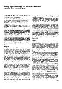
case for other hydrophobic P-450s, which may bind more sodium dodecyl sulfate than soluble proteins (34). A complete restriction map was obtained by computer analysis of the sequence and fitted perfectly with the experimental map (BamHI, Cla I, EcoRI, HindIII, Kpn I, Pst I, Pvu I, Sac I, and Sal I). NF95 was also sequenced and, in the overlapping region with NF25, no differences were seen, indicating that both clones were probably derived from the same mRNA. mRNA and Genomic DNA Analysis. Genomic DNA was cut with three different restriction enzymes, and the fragments were analyzed by Southern blotting (Fig. 5). Several bands were obtained with the three different cuts and the two nonoverlapping probes used (probes 2 and 3), even when relatively high-stringency hybridization conditions were used, indicating that P-45ONF very likely belongs to a multigene family. The size (50-60 kb with the two probes) and the numbers ofbands practically exclude the existence of a single gene. Liver samples HL 32 and HL 34 showed some small differences in Southern blotting patterns (Fig. SA); comparison with three other liver DNAs has not shown any specific pattern for poor or extensive metabolizers to date. The multiplicity of P-45ONF genes was confirmed by Northern blot analysis (Fig. SB), which shows that probe 1 recognizes three or four bands, indicating that there are several transcripts, the sizes of which (=2-4 kb) are all compatible with the size of the protein and the lengths of other P-450 mRNAs (25). Clearly the major size class is about 2.0 kb, although the number of distinct transcripts within this
20
A L I P D L A M E T W L L L A V S L V L
Rat P-450p
P-450
similar to human P1-450 and P-450MP. The heme ligand cysteine-containing peptide is more conserved within the gene families than within species: the nucleotide sequence similarity in this region can reach 96% within a family (P-45ONF versus P-45OPCN, Fig. 4). The cDNA-deduced amino acid composition was compared with the one obtained with purified P-45ONF (7); the difference index of 7.5 shows that they fit well (33). The amino acids that show the greatest deviation (glycine and alanine) are those that are known to generally yield discrepancies between amino acid analysis and cDNA-predicted sequences (34). Comparison of rat P-450PCN and P-45ONF compositions gave a difference index of 3.7, confirming the strong relationship between the two proteins. The deduced molecular weight of the P-45ONF protein is slightly higher than the one estimated by sodium dodecyl sulfate/polyacrylamide gel electrophoresis, as is the
I P D L A M E T W L L L A VI S L V L
Rabbit P-450 LM3c
Human
(human P-45ONF, rat P-450p, rat P-45OPCN, and rabbit P-450 LM3c) have similar N-terminal sequences, but they are not
10
M A L
D
G T W
TS
F M LN
I F
I
S L
ET
S MS A
L L A V V L V L SL L A A V
L A AS L V L
EFL
L A S V I F C
V L X L S XL L
L
L fffW K
F G S G P R N C
I
G M R F A L M N M K L A
F G N G P R N C
I
G M R F A L M N M K L A
F G M G KR KC V
G M G K
F S
I
R
R C
G N P N C
F S A G L
R
I
C
IG E T V A R W E VF LF IG E
8067
V L A K W E
IF L
F
F G E G L A K M E
LF L F
V G E
LF
A L A G M E
L
F
FIG. 4. Comparison of deduced amino acid sequences in two regions: N-terminal region (A) and the heme-binding cysteine-containing peptide (B) (25). Important similarities between the sequences are boxed. Amino acid sequences were obtained from the following sources in parentheses: rat P-45OPCN (24), rat P-450p (29), rabbit P450 LM3c (30), human P-450p (28), pH P450(1) (31), human P1-450 (26), human P3-450 (32), human P450Mp (D.R.U., R.S.L., and F.P.G.-unpublished results).
Biochemistry: Beaune et al.
8068
A a
Proc. Natl. Acad. Sci. USA 83 (1986) c
B -b
b
from the cDNA is identical to that of P-45ONF. However, the cloning and sequence determination should provide a basis for further studies on the basis of regulation of activities related to this P-450 in humans.
--
AAA
A AA a b a ba b
a
ba ba b
b
This research was supported in part by U.S. Public Health Service Grants CA 30907 and ES 00267. D.R.U. is the recipient of U.S. Public Health Service Individual Research Award ES 05340. F.P.G. is a Burroughs Wellcome Scholar in Toxicology (1983-1988).
a
i. !.
-
0
28S
* - 18S
*~~~~~~~~~~~~~~~~~~~~~~~~~....
FIG. 5. DNA and RNA analysis by blotting. Microsomes prepared from human liver samples HL 32 (lanes a) and HL 34 (lanes b) oxidized nifedipine at rates of 0.88 and 0.65 nmol of product formed per min/nmol of P-450, respectively. (A and B) Southern DNA blots. DNA was loaded (20 ,ug) and hybridized with probe 3 (A) and probe 2 (B). (C) RNA blots. RNA was loaded (15 ,ug) and hybridized with probe 1. Probes 1 and 2 gave similar results. Restriction enzymes used are indicated at the tops of the lanes. Ribosomal RNA markers are
indicated at 18S and 28S. Sizes
are
shown in kb.
zone is unknown, and we do not know conclusively which mRNA size-class codes for the protein. Our previous work suggested that catalytic activity is related to the amount of immunochemically detectable P-45ONF (7), and these results have been confirmed. Analysis of mRNA levels in nine liver samples (with probe 2) showed a 20-fold variation [after normalization of mRNA hybridization with an oligomer (50-mer) complementary to the 5' end of human serum albumin mRNA (35)]. However, the mRNA levels could not be correlated to either levels of nifedipine oxidase activity or immunochemically determined P-45ONF in microsomes prepared from this set of samples.
CONCLUSIONS While cDNA clones have been previously obtained for human P-450s (26, 31, 32), in no case has a sequence been obtained that is related to a P-450 protein that has been isolated and characterized in terms of physical properties and catalytic activity. We obtained a sequence for a cDNA related to a major human liver P-450 that appears to be involved in an oxidation polymorphism where a significant population is affected (6). The protein has been suggested to be inducible in humans (28), and other work in our laboratory indicates that P-45ONF is involved in a number of reactions, including 6,8-hydroxylation of testosterone and androstenedione, estradiol 2- and 4-hydroxylation, aldrin epoxidation (7), benzphetamine N-demethylation (7, 8), quinidine 3- and N-oxidation (36), and the oxidation of nifedipine and 17 other dihydropyridine analogs to pyridine compounds (37). Several related sequences are found in this gene family, and we cannot state with certainty that the coding sequence deduced
1. Maghoub, A., Dring, L. G., Idle, J. R., Lancaster, R. & Smith, R. L. (1977) Lancet R, 584-586. 2. Kupfer, A. & Preisig, R. (1983) Semin. Liver Dis. 3, 341-354. 3. Wedlund, P. J., Aslanian, M. D., McAllister, C. B., Wilkinson, G. R. & Branch, R. A. (1984) Clin. Pharmacol. Ther. 36, 773-780. 4. Distlerath, L. M., Reilly, P. E. B., Martin, M. V., Davis, G. G., Wilkinson, G. R. & Guengerich, F. P. (1985) J. Biol. Chem. 260, 9057-9067. 5. Shimada, T., Misono, K. S. & Guengerich, F. P. (1986) J. Biol. Chem. 261, 909-921. 6. Kleinbloesem, C. H., van Brummelen, P., Faber, H., Danhof, M., Vermeulen, N. P. E. & Breimer, D. D. (1984) Biochem. Pharmacol. 33, 3721-3724. 7. Guengerich, F. P., Martin, M. V., Beaune, P. H., Kremers, P., Wolff, T. & Waxman, D. J. (1986) J. Biol. Chem. 261, 5051-5060. 8. Wang, P. P., Beaune, P., Kaminsky, L. S., Dannan, G. A., Kadlubar, F. F., Larrey, D. & Guengerich, F. P. (1983) Biochemistry 22, 5375-5383. 9. Beaune, P., Kremers, P., Letawe-Goujon, F. & Gielen, J. E. (1985) Biochem. Pharmacol. 34, 3547-3552. 10. Young, R. A. & Davis, R. W. (1983) Proc. Natl. Acad. Sci. USA 80, 1194-1198. 11. Guengerich, F. P., Wang, P. & Davidson, N. K. (1982) Biochemistry 21, 1698-1706. 12. Guengerich, F. P., Dannan, G. A., Wright, S. T., Martin, M. V. & Kaminsky, L. S. (1982) Biochemistry 21, 6019-6030. 13. Yamamoto, K. R., Alberts, B. M., Benzinger, L., Lawhorne, L. & Treiber, G. (1970) Virology 40, 734-744. 14. Maniatis, T., Fritsch, E. F. & Sambrook, J. (1982) in Molecular Cloning: A Laboratory Manual (Cold Spring Harbor Laboratory, Cold Spring Harbor, NY). 15. Messing, J. (1983) Methods Enzymol. 101, 20-79. 16. Sanger, F. S., Nicklen, S. & Coulson, A. R. (1977) Proc. Natl. Acad. Sci. USA 74, 5463-5467. 17. Lloyd, R. S., Recinos, A., III & Wright, S. T. (1986) Biotechniques 4, 8-10. 18. Chirgwin, J. M., Przybyla, A. E., McDonald, R. J. & Rutter, W. J. (1979) Biochemistry 18, 5296-5300. 19. Wahl, G. M., Stern, M. & Stark, G. R. (1979) Proc. Natl. Acad. Sci. USA 76, 3683-3687. 20. Southern, E. M. (1975) J. Mol. Biol. 98, 503-517. 21. Alwine, J. C., Kemp, D. J. & Stark, G. R. (1977) Proc. Natl. Acad. Sci. USA 74, 5350-5354. 22. Benson, S. A. (1984) Biotechniques 2, 66-69. 23. Knudsen, K. A. (1985) Anal. Biochem. 147, 285-288. 24. Gonzalez, J., Nebert, D. W., Hardwick, J. P. & Kasper, C. B. (1985) J. Biol. Chem. 260, 7435-7441. 25. Adesnik, M. & Atchison, M. (1986) CRC Crit. Rev. Biochem. 19, 247-305. 26. Jaiswal, A. K., Gonzalez, F. J. & Nebert, D. W. (1985) Science 228, 80-83. 27. Kozak, M. (1984) Nature (London) 308, 241-246. 28. Watkins, P. B., Wrighton, S. A., Maurel, P., Schuetz, E. G., MendezPicon, G., Parker, G. A. & Guzelian, P. S. (1985) Proc. Natl. Acad. Sci. USA 82, 6310-6314. 29. Wrighton, S. A., Maurel, P., Schuetz, E. G., Watkins, P. B., Young, B. & Guzelian, P. S. (1985) Biochemistry 24, 2171-2178. 30. Koop, D. R., Morgan, E. T., Tarr, G. E. & Coon, M. J. (1982) J. Biol. Chem. 257, 8472-8480. 31. Phillips, I. R., Shepard, E. A., Ashworth, A. & Rabin, B. R. (1985) Proc. Natl. Acad. Sci. USA 82, 983-987. 32. Quattrochi, L. C., Okino, S. T., Pendurthi, U. R. & Tukey, R. H. (1985) DNA 4, 395-400. 33. Metzger, H., Shapiro, M. B., Mosimann, J. E. & Vintar, J. E. (1968) Nature (London) 219, 1166-1169. 34. Black, S. D. & Coon, M. J. (1986) in Cytochrome P-450: Structure, Mechanism, and Biochemistry, ed. Ortiz de Montellano, P. R. (Plenum, New York), pp. 161-216. 35. Dugaiczyk, A., Law, S. W. & Dennison, 0. E. (1982) Proc. Natl. Acad. Sci. USA 79, 71-75. 36. Guengerich, F. P., Muller-Enoch, D. & Blair, I. A. (1986) Mol. Pharmacol., in press. 37. Bocker, R. H. & Guengerich, F. P. (1986) J. Med. Chem., in press.