genes encoding four subunits of the spinach PSI ... sonication in 400 ul 50 mM HEPES-KOH pH 7.5, ... pellet was also sonicated into solution containing 50.
Plant Molecular Biology 10:435-445 (1988) © KluwerAcademic Publishers, Dordrecht - Printed in the Netherlands
435
Isolation and sequence of a tomato c D N A clone encoding subunit II of the photosystem I reaction center Neil E. Hoffman, 1, 2 Eran Pichersky, 3 Vedpal S. Malik, 4 Kenton Ko I and Anthony R. CashmorO
~Plant Science Institute, Department of Biology, University of Pennsylvania, Philadelphia, PA 19104, USA; 2permanent address: Department of Plant Biology, Carnegie Institute of Washington, 290 Panama Street, Stanford, CA 94305, USA; 3Department of Biology, University of Michigan, Ann Arbor, MI 48109, USA; 4philip Morris Research Center, PO. Box 26583, Richmond, VA 23261, USA Received 13 November 1987; accepted in revised form 20 Javuary 1988
Key words: electron transfer, light-harvesting complex I, membrane localization, photosynthesis, processing site, transit peptide
Abstract We report here the isolation and nucleotide sequence of a cDNA clone encoding a phtosystem I polypeptide that is recognized by a polyclonal antibody prepared against subunit II of the photosystem I reaction center. The transit peptide processing site was determined to occur after Mets0 by N terminal sequencing. The decuced sequence of this protein predicts that the polypeptide has a net positive charge (pI = 9.6) and no membrane spanning regions are evident from the hydropathy plot. Based on these considerations and the fact that subunit II is solubilized by alkali treatment of thylakoids, we concluded that subunit II is an extrinsic membrane protein. The absence of hydrophobic regions characteristic of thylakoid transfer domains furthermore implies that subunit II is localized on the stromal side of the membrane.
Introduction Photosystem I (Ps I) is the photosynthetic unit which mediates the light-activated transfer of electrons from plastocyanin to ferredoxin. The reaction center of P S I consists of six different polypeptides designated subunits I to VI in order of decreasing M r [4, 5, 25] and is associated with a light-harvesting antenna of chlorophyll a/b-binding polypeptides [23]. Subunit I, a homo or heterodimer of integral membrane proteins (M r - 65 000) encoded by chloroplast genes psaA and psaB [10], is bound to P700, the primary electron donor of P S I [20]. Recently it has been shown that the 65 kd polypeptide(s) also bears the 4Fe-4S cluster known as X [11, 15, 33]. Subunit VI is most likely the 8-9 kD polypeptide encoded by
chloroplast gene psaC [12] formerly designated ORF 81 in tobacco [35] and frxA in liverwort [27, 28]. This polypeptide has 9 cysteines [12, 27] and most likely contains the two 4Fe-4S clusters A and B [12, 14, 18, 22, 27] and mediates electron transfer to soluble ferredoxin [12, 20, 27]. It is somewhat hydrophobic but does not contain any amino acid stretches sufficiently long to span the membrane and is presumably located on the stromal side in the vicinity of ferredoxin. The genes encoding subunits I and V! have been mapped on the chloroplast genome and their nucleotide sequences determined in a variety of species [10, 16, 19, 28, 35]. The remaining four polypeptides, II, II, IV, and V, have relative molecular weights ranging from 10000 to 25 000 depending on the species [5, 24, 25]. Bengis and Nelson [5] have
436 proposed that subunit III is located at the lumenal side of the thylakoid membrane and is required for electron transport from plastocyanin to P700. The function of subunits II, IV, and V are unknown. Their membrane location is also uncertain; however they are all likely to be on the stromal side based on PSI topography studies that found several polypeptides in the size range of 14-19000 kD (possibly including subunits II, IV, and V) to be exposed on the stromal side [29]. Although the cloning of nuclear genes encoding four subunits of the spinach P S I reaction center has been reported [38, 40], sequences have yet to be presented. In the present study we report the DNA sequence of a tomato cDNA clone encoding subunit II of the P S I reaction center. We furthermore describe additional studies to determine the transit peptide processing site and polypeptide membrane orientation.
Materials and methods
cDNA cloning The tomato cDNA library was a gift from Dr Danny Alexander, ARCO plant Research Institute, Dublin, CA, USA. It was constructed according to the procedure of Alexander et al. [3] and was converted into an expression library in the lambda phage vector Charon 16 according to DellaPenna et al. [9].
Library screening, affinity purification of antibodies, western blots, electrophoresis, fractionation of P S I and PS 11, and nucleotide sequencing The procedures employed are described and referenced in Hoffman et al. [13]. Antibodies used include rabbit anti PSI-22 polyclonal prepared against Viciafaba 22 kD P S I polypeptides [13], rabbit anti subunit II polyclonal (gift of Dr Rachel Nechushtai) prepared against swiss chard subunit II of the P S I reaction center [24], mouse anti pea LHC II monoclonal M L H 1 and M L H 12 ([8], gift of Dr Sylvia Darr), and rabbit anti OEC-33 polyclonal (gift of Dr John Bennett), prepared against spinach
33 kD polypeptide of the oxygen-evolving complex [37].
In vitro transcription and translation, chloroplast import, and Southern blots. The procedures employed are described and referenced in Pichersky et al. [31].
Protein sequencing N-terminal Edman degradation using a gas phase sequenator was carried out at the Protein Sequencing Facility of the University of Pennsylvania on gel purified samples electroblotted to activated glass fiber filters [1] as modified [41] or on radiolabelled polypeptide recovered from thylakoids. Polypeptide radiolabelled with I_,[2, 3-3H]Ala (59 Ci/mmol) was produced using in vitro transcription and translation as described [31] with the modification that the translation reaction contained 20/~M aminooxyacetic acid to inhibit aminotransferase activity. Chloroplast import was performed as described [31]. Specifically 14 x 10 6 dpm precursor polypeptide was imported into 1 ml of intact chloroplasts containing 300 /~g chlorophyll. After import the chloroplasts (0.6 mg/ml chlorophyll) were incubated with thermolysin, 100 ug/ml, reisolated by centrifugation through 40°7o percoll, and thylakoids were prepared by successively washing the pellet in 0.1 M sorbitol5 mM EDTA and 0.1 M sorbitol-0.75 mM EDTA. Thylakoids were resuspended in water to 300 #1, delipidated by adding 1200/A cold HPLC grade acetone, and aliquoted into three tubes. After incubating on ice for 30 rain, the samples were spun in a microfuge for 5 rain. Supernatants contained no radioactivity and were discarded while the pellets were lyophilized and stored at - 2 0 °C. Immediately prior to sequencing, the pellets were dissolved in 50 /xl neat trifluoroacetic acid (Pierce Chemical Company, Sequana! grade).
Membrane localization Extraction of peripheral membrane proteins with
437 0.1N NaOH was essentially as described [30]. Destacked thylakoid membranes prepared from type B chloroplasts [23] were washed with a solution containing 5 mM HEPES-KOH (pH 7.5) and 10 mM EDTA. Membranes containing 500 ~g chlorophyll were centrifuged at 30000 RPM (50000 × g at rmax) in a TLA 100.3 rotor for 5 rain. The pellet was resuspended in 1 ml of 0.1N NaOH and kept on ice with periodic mixing for 30 rain at which time the sample was spun at 75000 RPM (300000 × g at rmax) for 30 min in a T L A 100.3 rotor. The clear supernatant was neutralized with HC1, protein was precipitated in 10% TCA for 30 rain on ice, the pellet was washed with 80% acetone and resuspended by sonication in 400 ul 50 mM HEPES-KOH pH 7.5, 1 mM CaC1 z. The dense green NaOH-insoluble pellet was also sonicated into solution containing 50 mM HEPES-KOH pH 7.5, 1 mM CaC12. Samples were prepared for electrophoresis by adding 100 ~1 5× SDS sample buffer (0.25 M Tris-HC1 pH 6.8, 0.25 M DTT, 50% glycerol, 10% SDS, 0.2% bromophenol blue) and heating for 2 min at 100 °C. Lanes were loaded with 15 t~l sample derived from an original sample containing 15 tzg of chlorophyll. For thermolysin treatment of right side out thylakoids, destacked thylakoids (125 ug chlorophyll) were resuspended in 100 ~1 of 50 mM HEPES-KOH pH 7.5, 1 mM CaCI 2 and incubated in either 100 or 500 ~g/ml thermolysin for 30 min at 0 °C. To protease-treat the lumen side of thylakoids, thylakoids were sonicated for 1 rain at a continuous output control setting #3 using a tapered microtip probe (Heat Systems W-385 sonicator), prior to treatment with thermolysin. After protease treatment samples were prepared for electrophoresis as described above.
Results
cDNA clone selection and identification In a previous report [13] we described the isolation of two cDNA clones by screening a cDNA expression library with an antiserum prepared against Vicia faba 22 kD-PS I polypeptides. One gene, cab6A, was identified as encoding a P S I CAB pro-
tein by virtue of its sequence homology to other CAB clones. The second clone, which is the subject of this report, has no sequence similarity to CAB6A, or to 12500 other sequences in the Genbank. Based on serological evidence (see below), we identify this clone as the gene encoding subunit II of the PS I reaction center and designate it psaD. Figure 1 shows an immunoblot analysis of P S I proteins from tomato and pea. The protein profiles of samples from tomato and pea used for this analysis are shown in Fig. 1, lanes 2 and 3. Lanes 4 and 5 of this figure show polypeptides which cross react with a monoclonal antibody prepared against pea PS II CAB polypeptides [8], but which also cross react with P S I CAB polypeptides. For pea samples (Fig. 1, lane 5), the lower four polypeptides which cross react are P S I CAB polypeptides while the upper bands are PS II CAB proteins contaminating the P S I preparation; the 4 P S I CAB bands recognized by the monoclonal antibody correspond to 4 bands stained for protein visible in lane 3. In tomato (lane 4), three P S I proteins strongly cross react with the monoclonal antibody while two weakly react with it. The three strongly reacting bands correspond to three bands stained for protein visible in lane 2 and appear to correspond to three bands in the pea sample. We note however that the strongest cross reacting polypeptide was the fastest migrating band in pea (lane 5) but the second fastest migrating band in tomato (lane 4). The antiserum employed for screening the library was made against P S I polypeptides of M r 22000 and we have designated it anti-PSI-22. This antibody cross reacted with two P S I polypeptides that were electrophoretically distinct in tomato but nearly comigrated in pea (Fig. 1, lanes 6, 7); this antiserum did not react with any PS II polypeptides in either organism. In both tomato and pea, the slower migrating band of the two is electrophoretically indistinguishable from the band that strongly reacts with the monoclonal antibody (Fig. 1, lanes 4 - 7 ) . The faster migrating band detected by anti-PSI-22 runs just ahead of the slower band in pea but is well separated from the slower band in tomato. This band is clearly not recognized by the anti-CAB monoclonal antibody suggesting that the antiserum consisted of two types of antibodies that recognize
438
Fig. 1. SDS polyacrylamide gel electrophoretic analysis of tomato and pea PSI fractions and of the protein encoded by psaD. Lane 1: molecular weight markers, bovine serum albumin 66.2 kD, ovalbumin 45 kD, carbonic anhydrase 31 kD, soybean trypsin inhibitor 21.5 kD, lysozyme 14.4 kD. Lanes 2-11: tomato PS 1 [2, 4, 6, 8, 10] and pea PSI [3, 5, 7, 9, 11]. Lanes 2 and 3: Coomassie blue stained gel. Lanes 4 and 5: Western blot with monoclonal antibody (MLH 12) made against a pea PS II CAB protein [8]. Lanes 6 and 7: Western blot with polyclonal antibodies made against 1I..faba PS I proteins M r 22000 [13]. Lanes 8 and 9: Western blot using PSI-22 antibodies affinity purified by Cab-6A phage lysates. Lanes 10 and 11:Western blot with PSI-22 polyclonal antibodies affinity purified by psaD phage lysates. Lanes 12 and 13: Fluorograph of processed and precursor translation products of tomato psaD.
unrelated proteins. This was confirmed by affinity purifying the a n t i b o d y using lysates o f the two isolated c D N A clones, phage clone cab-6A and phage clonepsaD, as ligands. A n t i b o d y PSI-22 affinity purified by clone cab-6A now only cross reacted with the slower migrating o f the two bands (Fig. 1, lanes 8 and 9). Likewise anti-PSI-22 affinity purified by clonepsaD only cross reacted with the faster migrating o f the two polypeptides (Fig. 1, lanes 10 and 11). Thus the antibodies could be resolved into two classes reacting with one or the other polypeptide. This allowed us to identify subunit II as the faster migrating b a n d in both t o m a t o and pea (Fig. 1, lanes 8-11). We further established that both C A B - 6 A and subunit II exhibit different electrophoretic m o bilities in pea and t o m a t o (Fig. 1, lanes 8 vs 9 and 10 vs 11). This was not so obvious for C A B - 6 A since in b o t h species a CAB-related polypeptide which is not e n c o d e d by cab-6A corresponds in mobility to
C A B - 6 A o f the other species (Fig. 1, lanes 4, 5; note that C A B - 6 A is the darker o f the two bands at 22 kD). We exploited the species variation in electrophoretic mobilities to determine if the protein encoded by phagepsaD corresponded to subunit II o f the P S I reaction center originally described by Bengis and Nelson [4, 5]. A monospecific polyclonal antiserum prepared against purified subunit II (provided by courtesy o f Dr Nechushtai) was found to react with the faster migrating o f the two bands recognized by o u r PSI-22 polyclonal antibodies (Fig. 2). As it is unlikely that the two antisera are reacting with distinct polypeptides which maintain precisely the same electrophoretic variation between pea and tomato, we conclude that phage psaD encodes subunit II o f the photosystem I reaction center. Tittgen et al. [38] have demonstrated that in spinach, subunit II is synthesized as a 26 kD precursor which is proteolytically processed to a 22 kD poly-
439 peptide during import into the chloroplast. We subcloned the phage psaD insert into pGem-4, in vitro transcribed the DNA, translated the RNA, and imported the resulting polypeptide into chloroplasts. The precursor polypeptide, M r 29000, was processed to a polypeptide, M r 20500, which was recovered from PSI fractions and was electrophoretically indistinguishable from tomato subunit II (Fig. 1, lanes 10 and 12) thus further confirming the identity of the clone. The nucleotide sequence of cDNA psaD is shown in Fig. 3. It contains 802 nucleotides followed by a poly A tail. The longest open reading frame, starting with methionine, encodes a protein containing 208 amino acids with a theroretical molecular weight of
Fig. 2. Proteins recognized by PSI-22 antibodies are also recognized by antibodies specific for subunit ll. All lanes were loaded with P S I proteins containing 3 p.g chlorophyll. Lanes 1 and 3: pea. Lanes 2 and 4: tomato. Lanes 1 and 2: Western blot with polyclonals against PSI-22 proteins [13]. Lanes 3 and 4: Western blot with polyclonal antibodies made against subunit lI [24].
i0
20
30
40
50
ACATACAATACATTGC TCAATT TTCCCCCAATTCCTCAATAATGGC M 80
90
i00
60
70
TATGGCAACTC~GCTTCCCTCTTCA A
ii0
M
A
T
Q
120
A
S
L
130
F
140
CCCC TCCTCTC TCCGTCCCCAAATCCACCACCGCTCCATGGAAACAATCCC
TCGTATCGTTC TCTACACC TA
T
L
P
P
L
S
150
V
P
K
160
S
T
T
A
170
P
W
K
180
Q
S
190
V
S
F
S
200
AGCAAC TCAAATCCACCGTC TCAGTAACCCGACCTATCCGTGCCATGGCTGAAGAAGCCCCAGC K
Q
L
K
220
S
T
V
S
230
V
T
R
240
P
I
R
250
A
M
260
A
&
E
~
E
E
K
P
90
A
P
A
300
G
F
T
310
P
P
Q
L
320
D
A
P
N
330
T
P
TGCCACCG
P
270
A
A
T
280
AAGAAAAAC CCGCTCCAGCGGGAT TTACCCCACC TCAATTGGACCCAAACACACCC E
T
210
P
2
T C C C C A A T C T T C G G TG S
340
P
I
F
G
350
360
GCAGCACCGGTGGGCTTCTTCGCAAAGCCCAAGTGGAA~TACGTCATCACATGGGAATCACCAAAGG G
S
T
G 370
G
L
L R 380
K
A Q 390
V
AACAGATC T T T GAGATGCCAACAGGTGGTGCTGC E
Q
I
F
E
440
M
P
T
G
450
G
A
E
E 400
F
Y
V 410
I
T
TATAATGAGACAAGGACCAAAT A
460
I
M
R
470
Q
G
P
N
480
W E 420
S
K
E
Q
C
L
A
L
G
T
R
L
R
S
K
Y
L
L
K
490 K
I
510 520 530 540 550 TT T T C C C C A A T G G C G A G G T G C A A T A T T T G C A T C C C A A A G A T G G T G T G T A C C C V
F
P
N
580
G
E
V
Q
590
Y
L
H
600
P
K
D
610
G
V
Y
P
620
E
G
V
50
G
Q
N
660
F 670
R
S
I 680
G
K
N 690
K
Y
Q
E
K
A
A
T TACAGGG F
V
630 S
L 500 Y
R
560 570 TGAAAAAGTGAACCCTGGTC
GTGAAGGAG TGGGT CAGAAC TTCAGATCAATTGGGAA~AACAAGAGTGCTAT R
N
K 430
T TGT TGAAAT TGGCAA
GGAAAGAACAG TGC T TAGCTC T TGGTACAAGG T TGAGATC TAAATACAAGATCAATTACCAGTT R
P
N
TGAAGTCAA I
700
P
G
640 E
V
K
6
TT T A C T G G G A F
T
G
710
720
AACAAGTGTATGATATTTAAGTTTGTGCACTTTTTTTTCTTTTTTTTTTTAAATTTATGATATTGTAATTGT K
Q
V
Y
D
730
I
740
750
760
770
780
790
TAATGGTTTTGTGATTATGTAGTGCATGTAACTTTATTTAGCACTACAAATACAGCATGGTCCATTAAATAT 800 T C A T C A T T G T (A n)
Fig. 3. Nucleotide sequence ofpsaD and the predicted amino acid sequence of presubunit II. The arrow indicates the chloroplast processing site of the transit peptide. Underlined a m i n o acids were determined in the third through fifth cycles of E d m a n degradation. Underlined nucleotides indicate the HindIIl site and EcoR1 site at 58 and 327 bp respectively.
440 23 kD (Fig. 3). As there are no stop codons upstream of this methionine, it is conceivable that cDNA psaD contains an incomplete reading frame. We believe that this is not the case for if the clone was missing a 5'-translated region, in vitro transcription and translation of the clone should result in a polypeptide with a truncated transit peptide which would not be expected to import very efficiently. In fact, this polypeptide is more efficiently imported into chloroplasts than PS II CAB precursors (data not shown), Therefore we conclude that it is likely that the clone does contain a complete reading frame and, in addition, 41 and 137 bp of 5' and 3'-non-translated regions, respectively. Southern blots of EcoRI-restricted tomato DNA, using as a probe a 0.27 kb HindlII-EcoRI fragment from the cDNA clone ofpsaD (nucleotides 58 - 331), reveals only one hybridizing fragment of 2.2 kb (data not shown). Restriction with other enzymes also gave a single band pattern. This suggests that subunit II is encoded by a single copy gene in tomato, as was previously found for the spinach genome [38].
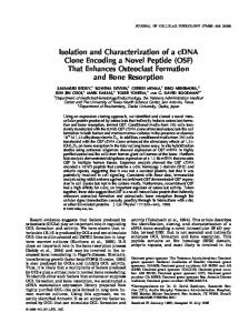
cleaved from the precursor, gel-purified subunit II was electroblotted to an activated glass fiber filter and subjected to five cycles of Edman degradation on a gas phase sequencer (Applied Biosystems). The results were ambiguous but suggested the sequence ?G,E,A,P. This sequence most closely corresponds to amino acids 51-55 of the amino acid sequence predicted from the cDNA (AEEAP) and predicts that processing occurs after Ms0. To further test this result, presubunit II was synthesized in a wheat germ extract in the presence of [3H]-AIa, processed in isolated chloroplasts, and subsequently sequenced. As only one radiolabelled polypeptide is detected upon electrophoresis of the thylakoid fraction following import and processing of presubunit II, a delipidated thylakoid fraction and not gel-purified protein was used for sequencing. When presubunit II polypeptide was labelled with [2, 3-3H]Ala, sequencing released label in fractions 1, 4, 6, 7, 13, and 15 (Fig. 4). Comparison with the amino acid sequence derived from the cDNA sequence confirms that the cleavage site occurs between Ms0 and Alas] both in the endogenous polypeptide and the polypeptide synthesized in vitro and processed in organello. Thus the mature polypeptide consists of 158 amino acids with a theoretical MW of 17.5 kD. Like most of the thylakoid membrane proteins, the theoretical
Processing site To determine the site where the transit peptide is
700 600 500 400
2O0 1 O0 0
*
0
I
*
I
I
I
2
4
6
8
I0
•
|
12
14
A E E A P A A T E E K P A P A
16
Fraction # Sequence
Fig. 4. Chloroplast processing site of presubunit II. Presubunit II labelled with [3H]-ala was processed in chloroplasts, subjected to automated Edman degradation, and radioactivity released W" cycle was counted. The amino acid sequence deduced from cDNA psaD beginning with A51 is indicated.
441 molecular weight of the mature polypeptide and the precursor is slightly smaller than the M r predicted from their migration in SDS-PAGE.
Structural aspects The transit peptide of presubunit II resembles other chloroplast transit peptides in having a somewhat hydrophobic region at the N-terminus (first third of this region) whereas the rest of the transit peptide is more polar and contains numerous positive charges. Mature subunit II contains high amounts of two helix-destabilizing amino acids glycine (10.1°70) and proline (9.4%), which presumably limit the extent of a helical secondary structure. Though subunit II is membrane-associated, the deduced sequence predicts that slightly over 50% of the amino acids are polar and the polypeptide has a net positive charge (pI = 9.6). There are three regions where the positive charges are clustered, i.e. amino acids 121-131,141-147, and 187-193 contain 3, 4, and 3 positive charges, respectively, and one region with 4 negative charges, i.e. amino acids 5 2 - 6 0 . The hydropathy plot, calculated by the method of Kyte and Doolittle [17] for a window size of 15, of this polypeptide predicts that the protein contains no membrane-spanning regions (Fig. 5). Thus, from the deduced protein sequence and the hydropathy plot, it appears that subunit II is an extrinsic membrane protein.
Membrane localization Extrinsic membrane proteins can be distinguished from integral membrane proteins by extractability of the former into 0.1 N NaOH. NaOH-soluble and NaOH-insoluble fractions were electrophoresed, electroblotted to nitrocellulose and probed with antibodies against subunit II, CAB3 (the most abundant PS II CAB polypeptide), and OEC-33 (Fig. 6). CAB3 and OEC-33 are integral and extrinsic membrane proteins, respectively [2, 34]. As expected both subunit II and the OEC-33 were soluble in NaOH, but not CAB3, supporting the notion that subunit II is also an extrinsic membrane protein. To assess
whether subunit II was on the lumen or stromal side of the thylakoids, we protease-treated either intact or sonicated thylakoids. We reasoned that since subunit II is an extrinsic protein, protease sensitivity in intact thylakoids would indicate a stromal location. If insensitive in this configuration but sensitive after sonication, which would expose the lumen side of the thylakoid to protease, the protein would most likely be on the lumen side. Unfortunately subunit II was relatively insensitive to protease in both intact and sonicated thylakoids though it was completely sensitive after NaOH extraction (Fig. 6, lanes 5 versus 6 and 8 versus 9). In contrast CAB3, which has a protease-sensitive site on the stromal side, was cleaved by thermolysin treatment of intact thylakoids. Truncated CAB3 is not detected in Fig. 6, lanes 6, 7, or 9 because the antibody employed, MLH1, recognizes an epitope on the N-terminal side of the membrane, was degraded by protease only after sonication (Fig. 6, lane 6 versus 9). Collectively these data imply that subunit II is protease-insensitive in its assembled conformation and hence it is unlikely that selective proteolysis will reveal the membrane localization.
Discussion
The PS I reaction center complex consists of polypeptides encoded by the chloroplast and nuclear genomes. A first step toward understanding the topogenesis of PSI and the functioning of individual subunits requires structural informations on each polypeptide. Structural information via sequence data was available only for both chloroplastencoded subunit I polypeptides (P-700 binding apoproteins) [10, 16, 19] and an 8 - 9 kD polypeptide [12, 27] presumably corresponding to subunit VI. In this report, we have extended the available structural information on the PSI reaction center to include the nuclear-encoded subunit II. The predicted protein structure suggests that subunit II is an extrinsic membrane protein and this was corroborated by NaOH extraction. Although not precisely identified as subunit II, a 19 kD spinach P S I polypeptide resembling subunit II in its SDS-PAGE pattern was shown to be exposed on the stromal side by labelling
442
2O
!
-20 i
I
i
i
I
1
I
i
i
i
I
40 i
i
i
I
I
I
80
I
I
I
I
I
120
I
I
i
i
160
I
i
200
4O
2O
0
-20
,
" ' "
I
1
30
I
i
I
i
I
60 i
i
i
i
i
90
I
I
I
I
I
120
I
I
I
150
40
C
20
-20 !
1
l
I
I
I
40
I
I
I
I
I
I
801
I
I
I
I
I
120
I
I
i
I
I
160
I
!
I
I
200
I
I
I
I
I
I
240
I
i
I
a
280
i
i
i
I
I
I
320
Fig. 5. Hydrophobicity plots of A) tomato presubunit II, B) spinach preplastocyanin [32], and C) arabidopsis 33 kD OE protein (K. Ko, unpublished results). The plots were calculated by the method of Kyte and Doolittle [17] with a window size of 15.
studies using an impermeant chemical modifier [29]. Additionally we speculate that the protein is localized on the stromal side since the amino acid sequence of the precursor does not contain regions characteristic of proteins which are imported across
the thylakoid membrane. Two such proteins are preplastocyanin [32, 36] and the precursor to OEC33([39], K. Ko, unpublished results), each of which contains a highly hydrophobic segment at the C terminal end of the transit peptide which is presumably
443
Fig. 6. Western blot analysis of thylakoid proteins extracted with alkali and/or protease treated using: A) Polyclonal antibody prepared against spinach 33 kD OE-protein [37]. B) monoclonal antibody (MLH1) prepared against pea LHC II and specific for CAB-3 [8]. C) Polyclonal antibody prepared against swisschard subunit II [24]. Lanes 1- 3: thylakoid proteins solublein 0.1 N NaOH. Lane 4: thylakoid proteins insoluble in 0.1 N NaOH. Lanes 5-7: intact thylakoids. Lanes 8-10: sonicated thylakoids. Lanes 1, 4, 5, and 8: no thermolysin treatment. Lanes 2, 6, and 9:100 ~g/ml thermolysin. Lanes 3 and 7:500 ~g/ml thermolysin. All lanes were loaded with protein derived from samples originally containing 15 gg chlorophyll.
needed for passage across the thylakoid membrane. The transit peptide of presubunit II does not contain such a domain (Fig. 5, compare A versus B and C). Based on these considerations it is likely that presubunit II is located on the stromal side of the thylakoid. This obviously excludes interactions of subunit II with lumen proteins such as plastocyanin. Early studies reported " b o u n d ferredoxin" in purified PS I reaction center preparations [5]. It has since been estimated that P S I contains 12 atoms of sulfur and iron per P-700 which is consistent with the presence of three 4 F e - 4S clusters, A, B, and X observed by epr experiments [20]. Based on a correlation between SDS solubilization of subunits IV, V, and VI with loss of epr signal, it was concluded that IV, V, and VI were the most likely candidates for F e - S apoproteins [5]. It now appears that the 4 F e - 4 S cluster, X, is associated with subunit I [11, 15, 33]. Recently, evidence based on 35S labelling [18], 59Fe labelling and amino acid analysis of isolated proteins have implicated an 8 kD polypeptide as the apoprotein of the A and B F e - S clusters [14, 22]. The polypeptide corresponds in size to that originally designated subunit VI [5] and in amino acid sequence to that predicted from a chloroplast gene in tobacco, psaC [12, 35], and liverwort, f r x A [27, 28]. In contrast, Bonnerjea et al. [6] suggested that F e - S centers A and B are bound to a spinach 19 kD polypeptide. This polypeptide is likely to correspond to subunit II as both are the second largest polypeptide in antenna-depleted spinach PSI preparations [6, 40]. We note that the predicted amino acid
sequence of tomato subunit II indicates that it is clearly unlikely for this role in that it contains only one cysteine, whereas a peptide containing two 4 F e - 4S clusters should contain a minimum of eight. Thus we favor the assignment o f F e - S centers A and B to the 8 kD polypeptide. The presence of only one cysteine in subunit II infers that this polypeptide does not bind Fe and does not participate in electron transfer reactions. Furthermore subunit II is also unlikely to bind chlorophyll as N a O H quantitatively extracts subunit II from membranes but removes no chlorophyll. What then is the function of subunit II, a protein conserved in antigenic determinants a m o n g the thermophilic cyanobacteria, green algae, and higher plants [24]? It has been speculated that subunit II serves as a template around which other PSI reaction center subunits assemble [25, 26]. The subunit stoichiometry has been determined to be two subunit I polypeptides for each subunit I I - V I [5]. Our data are not inconsistent with this notion of subunit II as a structural polypeptide provided we account for the peripheral location of the protein. In fact the association of subunit II with other proteins is a simple explanation to account for the insensitivity of subunit II to thermolysin digestion in the native state but not after N a O H extraction. One of the striking features of subunit I! is the presence of numerous positively charged residues which are clustered in three regions of the sequence. One negatively charged region exists within the protein which may interact with one of the positively charged
444 regions. The two remaining regions are most likely on the protein surface and may be important for binding other proteins. One possible candidate is ferredoxin, a stromally localized acidic protein that is reduced by P S I and that subsequently transfers electrons to ferredoxin NADP reductase for the reduction of NADP. Recently Zanetti and Merati [42] inferred from chemical cross linking studies that a 20 kD P S I polypeptide (that resembles subunit II in its SDS-PAGE pattern) indeed binds to ferredoxin. In keeping with this observation and in the absence of any known functional role for subunit II, it is tempting to speculate that subunit II is a structural protein that by virtue of its positive charge enables the negatively charged extrinsic protein, ferredoxin, to associate with the membrane-bound PS I reaction center. An anlogous situation may exist on the lumen side of the thylakoids between subunit III and the extrinsic protein, plastocyanin. In addition, subunit II may serve to correctly position functional subunits within the complex such as the 8 kD polypeptide that likely contains the A and B iron sulfur centers that presumably transfer electrons to ferredoxin [20]. At this time, however, we cannot exclude the possibility that subunit II has an unrecognized functional role and hence further studies are essential to establish the precise role of this polypeptide in the P S I reaction center.
Acknowledgements We thank Allan Place for helpful advice on protein sequencing and Sylvia Darr, Rachel Nechushtai, and John Bennett for their generous gifts of antibodies. K.K. gratefully acknowledges Neil Strauss (Department of Botany, University of Toronto) for support in the preparation of polyclonal antibodies. This work was supported by Department of Energy Grant DE-FG02-87ER13680 and National Institutes of Health Grant GM-38408 to A.R.C. In addition N.E.H. was supported by a National Science Foundation postdoctoral fellowship, E.P. by a National Institute of Health Postdoctoral Fellowship, and K.K. by a postdoctoral fellowship from the Natural Science and Engineering Council of Canada.
References 1. Aebersold RH, Teplow DB, Hood LE, Kent SB: Electroblotring onto activated glass. High efficiency preparation of proteins from analytical sodium dodecyl sulfate acrylamide gels for direct sequence analysis. J Biol Chem 261:4229-4238 (1986). 2. Akerlund HE, Jansson C: Localization of a 34000 and a 23 000 M r polypeptide to the lumenal side of the thylakoid membrane. FEBS Lett 124:229--232 (1981). 3. Alexander DC, McKnight TD, Williams BG: A simplified and efficient vector-primer cDNA cloning system. Gene 31: 79- 89 (1984). 4. Bengis C, Nelson N: Purification and properties of the photosystem I reaction center from chloroplasts. J Biol Chem 250:2783-2788 (1975). 5. Bengis C, Nelson N: Subunit structure of chloroplast photosystem I reaction center. J Biol Chem 252:4564-4569 (1977). 6. Bonnerjea J, Ortiz W, Malkin R: Identification of a 19 kDa polypeptide as an F e - S center apoprotein in the photosystem I primary electron acceptor complex. Arch Biochem Biophys 240:15-20 (1985). 7. Cashmore AR: The isolation of poly A+ messenger RNA from higher plants. In: Edelman M, Hallick RB, Chua NH (eds) Methods in Chloroplast Molecular Biology. Elsevier Biomedical Press, Amsterdam (1982) pp. 387-392. 8. Darr SC, Sommerville SC, Arntzen C J: Monoclonal antibodies to the light harvesting chlorophyll a/b protein complex of photosystem II. J Cell Biol 103:733-740 (1986). 9. DellaPenna D, Alexander DC, Bennett AB: Molecular cloning of tomato fruit polygalacturonase: analysis of polygalacturonase mRNA levels during ripening. Proc Natl Acad Sci 83:6420-6424 (1986). 10. Fish LE, Kuck U, Bogorad L: Two partially homologous adjacent light inducible maize chloroplast genes encoding polypeptides of the P70o chlorophyll a-protein complex of photosystem I. J Biol Chem 3:1413-1421 (1985). 11. Golbeck JH, Cornelius JM: Photosystem I charge separation in the absence of centers A and B. I. Optical characterization of center "A2" and evidence for its association with a 64 kDa peptide. Biochim Biophys Acta 849:16-24 (1986). 12. Hayashida N, Matsubayashi T, Shinozaki K, Sugiura M, Inoue K, Hiyama T: The gene for the 9 kD polypeptide, a possible apoprotein for the iron-sulfur centers A and B of the photosystem I complex, in tobacco chloroplast DNA. Curr Genet 12:247-250 (1987). 13. Hoffman NE, Pichersky E, Malik VS, Castresana C, Ko K, Darr SC, Cashmore AR: A cDNA clone encoding a photosystem I protein with homology to photosystem 1I chlorophyll a/b-binding polypeptide. Proc Natl Acad Sci (in press) 14. Hoj PB, Halkier BA, Moiler BL: Analysis of isolated PSI polypeptides for acid labile sulfide. In: Biggins J (ed.) Progress in Photosynthesis Research. Martinus Nijhoff, Dordrecht (1987) Vol 1I, pp. 53-56.
445 15. Hoj PB, Moiler BL: The 110 kDa reaction center protein of photosystem I, P700-chlorophyll a-protein 1, is an iron-sulfur protein. J Biol Chem 261: 14292-14300. 16. Kirsch W, Seyer P, Herrmann RG: Nucleotide sequence of the clustered genes for two P700 chlorophyll a apoproteins of the photosystem I reaction center and the ribosomal protein S14 of the spinach plastid chromosome. Curr Genet 10:843 -855 (1986). 17. Kyte J, Doolittle RF: A simple method for displaying the hydropathic character of a protein. J Mol Biol 157:105 -132 (1982). 18. Lagoutte B, Setif P, Duranton J: Tentative identification of the apoproteins of iron-sulfur centers of photosystem I, FEBS Lett 174:24-29 (1984). 19. Lehmbeck J, Rasmussen OF, Bookjans GB, Jepsen BR, Stummann BM, Henningsen KW: Sequence of two genes in pea chloroplast DNA coding for 84 and 82 kD polypeptides of the pbotosystem I complex. Plant Mol Biol 7:3 - 10 (1986). 20. Malkin R: Photosystem I. Ann Rev Plant Physiol 33: 455-479 (1982). 21. Maniatis T, Fritsch EF, Sambrook J: In: Molecular Cloning a Laboratory Manual. Cold Spring Harbor Laboratory, Cold Spring Harbor, N.Y., pp. 200-201. 22. Moller BL, Halkier BA, Hoj PB: The organization of the FeS acceptors of photosystem I. In: Biggins J (ed.) Progress in Photosynthesis Research. Martinus Nijhoff, Dordrecht (1987) Vol. II, pp. 49-52. 23. Mullet JE, Burke J J, Arntzen C J: Chlorophyll proteins of photosystem I. Plant Physiol 65:814-822 (1980). 24. Nechushtai R, Muster P, Binder A, Liveanu V, Nelson N: Photosystem I reaction center from the thermophilic cyanobacterium Mastigocladus laminosus. Proc Natl Acad Sci 80: 1179-1183 (1983). 25. Nechushtai R, Nelson N: Purification, properties, and biogenesis of Chlamydomonas reinhardii photosystem 1 reaction center. J Biol Chem 256:11624-11628 (1981). 26. Nechushtai R, Nelson N, Mattoo AK, Edelman M: Site of synthesis of subunits to photosystem I reaction center and the proton-ATPase in Spirodela. FEBS Lett 125:115 - 119 (1981). 27. Oh-oka H, Takahashi Y, Wada K, Matsubara H, Ohyama K, Ozeki H: The 8 kDa polypeptide in photosystem I is a probable candidate of an iron-sulfur center protein coded by the chloroplast gene frxA. FEBS Lett 218: 5 2 - 5 4 (1987). 28. Ohyama K, Fukuzawa H, Kohchi T, Shirai H, Sano T, Sano S, Umesono K, Shiki Y, Takeuchi M, Chang Z, Aota S, lnokuchi H, Ozeki H: Chloroplast organization deduced from complete sequence of liverwort Marchantia polymorpha chloroplast DNA. Nature 322: 572-574 (1986). 29. Ortiz W, Lam E, Chollar S, Munt D, Malkin R: Topography of the protein complexes of the chloroplast thylakoid membrane. Studies of photosystem I using a chemical probe and proteolytic digestion. Plant Physiol 77: 389-397 (1985). 30. Piccioni R, Bellemare G, Chua NH: Methods of polyacryla-
31.
32.
33.
34.
35.
-
36.
37.
38.
39.
40.
41. 42.
mide gel electrophoresis in the analysis and preparation of plant polypeptides. In: Edelman M, Hallick RB, Chua NH (eds) Methods in Chloroplast Biology. Elsevier Biomedical Press, Amsterdam (1982) pp. 985-1014. Pichersky E, Hoffman NE, Malik VS, Bernatzky R, Tanksley SD, Szabo L, Cashmore AR: The tomato Cab-4 and Cab-5 genes encode a second type of CAB polypeptides localized in photosystem II. Plant Mol Biol 9: 109-120 (1987). Rother C, Jansen T, Tyagi A, Tittgen J, Herrmann RG: Plastocyanin is encoded by an uninterrupted nuclear gene in spinach. Curr Genet 11: 171-176 (1986). Sakurai H, San Pietro A: Association of Fe-S center(s) with the large subunit(s) of photosystem 1 particles. J Biochem 98: 6 9 - 76 (1985). Schmidt GW, Bartlett SG, Grossman AR, Cashmore AR, Chua NH: Biosynthetic pathways of two polypeptide subunits of the light-harvesting chlorophyll a/b protein complex. J Cell Biol 91:468-478 (1981), Shinozaki K, Ohme M, Tanaka M, Wakasugi T, Hayashida N, Matsubayashi T, Zaita N, Chunwongse J, Obokata J, Yamaguchi-Shinozaki K, Ohto C, Torazawa K, Meng BY, Sugita M, Deno H, Kamogashira T, Yamada K, Kusuda J, Takaiwa F, Kato A, Tohdoh N, Shimada H, Sugiura M: The complete nucleotide sequence of the tobacco chloroplast genome: its gene organization and expression. EMBO J 5: 2043 - 2049 (1986). Smeekens S, de Groot M, van Binsbergen J, Weisbeek P: Sequence of the precursor of the chloroplast thylakoid lumen protein plastocyanin. Nature 317:456-458 (1985). Sutton A, Sieburth LE, Bennett J: Light-dependent accumulation and localization of photosystem II proteins in maize. Eur J Biochem 164:571-578 (1987). Tittgen J, Hermans J, Steppuhn J, Jansen T, Jansson C, Andersson B, Nechushtai R, Nelson N, Herrmann RG: Isolation of cDNA clones for fourteen nuclear encoded thylakoid membrane proteins. Mol Gen Genet 204:258-265 (1986). Tyagi A, Hermans J, Steppuhn J, Jansson C, Vater F, Herrmann RG: Nucleotide sequence of cDNA clones encoding the complete "33 kDa" precursor protein associated with photosynthetic oxygen-evolving complex from spinach. Mol Gen Genet 207:288-293 (1987). Westhoff P, Alt J, Nelson N, Bottomley W, Bunemann H, Herrmann RG: Genes and transcripts for the PT0o chlorophyll a apoprotein and subunit 2 of the photosystem I reaction center complex from spinach thylakoid membranes. Plant Mol Biol 2:95-107 (1983). Yuen S, Hunkapiller MW, Wilson K J, Yuan PM: SDS-PAGE Electroblotting. Applied Biosystems User Bulletin 25 (1986). Zanetti G, Merati G: Interaction between photosystem I and ferredoxin. Identification by chemical cross-linking of the polypeptide which binds ferredoxin. Eur J Biochem 169: 143-146 (1987).