(b) Dept. of Computer and Systems Science, Univ. of Rome âLa Sapienzaâ , Rome, ... in each subject by using the depth-weighted minimum norm algorithm (3).
Lecture NON INVASIVE BRAIN COMPUTER INTERFACE FOR COMMUNICATION AND CONTROL Fabio Babiloni (a,d), Donatella Mattia (a), Marco Mattiocco (a), Alessandro Timperi (a), Serenella Salinari (b), Maria G. Marciani (a,c), * and Febo Cincotti(a). (a) Fondazione Santa Lucia IRCCS, Roma, Italy. (b) Dept. of Computer and Systems Science, Univ. of Rome “La Sapienza” , Rome, Italy (c) Dept. of Neuroscience, Univ. of Rome “Tor Vergata”, Rome, Italy (d) Dept. of Human Physiology and Pharmacology, Univ. of Rome “La Sapienza”, Rome, Italy
Introduction Recently, it has been suggested that with the use of the modern high resolution EEG technologies (1) it could be possible to estimate the cortical activity associated to the mental imagery of the upper limbs movements in humans (2). However, a verification of such statement on a group of normal subjects has not been yet performed. The scientific question at the base of the present work is whether the estimated cortical activity related to the mental imagery of the upper limbs returns more useful features with respect to those obtained by using scalp EEG recordings. To address this issue, we performed high resolution EEG recordings during the imagination of upper limb movements in a group of five healthy subjects. Comparisons between the waveforms from scalp electrodes and those from the estimated cortical activity in particular Region of Interest (ROIs) were then performed. These comparisons returned information about the usefulness of the use of cortical activity for the recognition of mental states with respect to the use of the scalp recorded data.
Methods Five healthy subjects participated voluntarily in experiments, in which they were asked to perform the imagination of right finger movements when they perform the protrusion of their lips. EEG was recorded by using a high resolution EEG cap with 64 electrodes disposed accordingly to an extension of the 10-20 international system. Subjects were asked to imagine the movement of their right middle finger during the simultaneous protrusion of their lips. This provided the necessary EMG trigger to synchronize the average of the recorded movements. Eighty single EEG trials were recorded for each subject. Each single EEG trial was acquired from 2 second before the arrival of the visual trigger to 1 s after. For all subjects analyzed in this study, sequential MR images were acquired and realistic head models were generated. A cortical surface reconstruction was accomplished for each subject’s head with a tessellation of about 10,000 triangles on average. The estimation of cortical activity during the mental imagery task was performed in each subject by using the depth-weighted minimum norm algorithm (3). Such estimation returns a current density estimate for each of the five thousand dipoles constituting the modelled cortical source space. Each dipole returns a time-varying amplitude representing the brain activity of a restricted patch of cerebral cortex during the entire task time-
225
course. This rather large amount of data can be synthesized by computing the ensemble average of all the dipoles magnitudes belonging to the same cortical region of interest (ROI). Each ROI was defined on each subject’s cortical model adopted in accordance with its Brodmann areas (BAs). In the present study, the activity in the primary left and right motor area, related to the B.A. 4 for the lips as well as hand regions have been taken into account. Artifacts correction by visual and automatic inspection was performed on each single EEG trial recorded. Threshold criteria were used to discard EEG trials contaminated by electrooculogram (EOG) or EMG activity at the resting arms. Visual inspection has been used to discard trials with unusual subthreshold artifacts. On average, about the 10% of the acquired EEG trials were discarded in the recorded population. Each artifacts-free single trials was then subjected to the linear inverse procedure, and the time varying cortical distributions associated was estimated. The collapsing procedure explained above was then applied to retrieve the cortical waveforms related to each particular ROI analyzed.
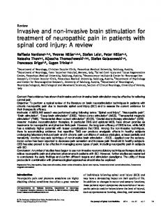
Results Figure 1 shows the event related potentials gathered from the scalp electrodes C3 and C4 (right bottom) and the event related mean current densities obtained from the bilateral MI-hand and MI-lips (right top and middle, respectively) areas for a representative subject (Subjet 5). Estimated current density underlying motor imagery is characterized by a negative slope peaking around 100ms before the EMG onset (0 Time) over the left M1 hand-ROI (namely contralateral to the imagined hand movements); no relevant activity was present over the right M1-hand ROI. As for the estimated current density underlying lip pursing, the negative slope involved bilaterally the M1-lips ROI. Scalp potential related to the task displayed a higher peak amplitude over C3 electrode lead with respect to C4. Analysis of the time varying current density distributions over the cortical mantle showed the presence of a bilateral negative (i.e. inward directed) activity on the supplementary motor area. The Table 1 reports the values of the measured current density in the different BAs examined at the peak of the motor potential for all the subjects employed in this study. Table I also reports the potential values obtained for the scalp potentials measured at the C3 and C4 leads, that are roughly placed on the central scalp areas overlying the primary motor areas. It is worth note as the potentials amplitudes at MP peak (gathered from C3 and C4 leads) are less unbalanced between left and right scalp areas when compared to the estimated current density activity in the primary motor areas related to the finger movements (BA4Rfinger, BA4Lfinger). Furthermore, the estimated cortical current density values in the primary motor areas related to the lips movements are rather symmetrical, i.e. the values are similar for the left and right ROI considered (BA4Llips and BA4Rlips, respectively). A statistical analysis of this unbalancing for the gathered scalp potentials as well as for the estimated cortical activity was then performed by using the t-paired Student test. Results obtained indicating; 1) a greater cortical activity estimated over the left primary motor cortical areas for finger movements (BA4Lfinger) with respect to the right one (BA4Rfinger), with a significance equal to p< 0.0011; 2) a statistical similar estimated cortical activity for the left (BA4Llips) and right (BA4Rlips) ROIs for the lips movement (p < 0.36); and a statistically significant differences between the MP peak for the scalp potentials gathered from the left scalp areas (C3) with respect to the right (C4) one, with a statistical significance of p