letter
© 2000 Nature America Inc. • http://genetics.nature.com
The glial cells missing-1 protein is essential for branching morphogenesis in the chorioallantoic placenta
© 2000 Nature America Inc. • http://genetics.nature.com
Lynn Anson-Cartwright1, Kerri Dawson1, Doug Holmyard1, Susan J. Fisher2, Robert A. Lazzarini3 & James C. Cross1,4,5,6
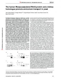
helix-loop-helix transcription factors Hand1 and Mash2 (refs 7–9), but the factors regulating syncytiotrophoblast differentiation are unknown. One candidate is Gcm1, which is expressed in a subset of chorionic trophoblast cells at E8.5 and later in the labyrinth10. In Drosophila melanogaster, gcm is necessary for differentiation of glial cells in the nervous system and scavenger cells in the immune system11–14. In mammals, the placenta is the major, if not exclusive, site of Gcm1 expression10,15–18. To investigate its function, we monitored Gcm1 expression during early chorioallantoic development. Chorionic trophoblast differentiation and morphogenesis begin only after chorioallantoic attachment1,19 (Fig. 1a), through interdigitation of the allantoic mesoderm and underlying fetal vessels with chorionic plate trophoblast cells to form simple branches1. These branches then elongate and bifurcate, and syncytiotrophoblast differentiation begins. We detected Gcm1 mRNA in clusters of 3–6 trophoblast cells at the flat chorionic plate stage (E8.5). These clusters were evenly spaced along the basal surface of the chorion and separated by 4–6 non-expressing cells (Fig. 1b,c). At later stages, Gcm1-expressing cells were present at sites where folding of the chorioallantoic surface was initiated (Fig. 1d,e). As the branches elongated, Gcm1 expression was limited to cells at the distal tip (Fig. 1f). The restriction of Gcm1 expression to sites of active morphogenesis suggested it as a regulator of chorioallantoic branching. Consistent with this hypothesis, Gcm1 expresTrophoblast stem cells reside in the chorion and differentiate into sion declines at E17, when growth of the labyrinth ceases10. either trophoblast giant cells or syncytiotrophoblast cells of the To test its function, we generated Gcm1-deficient mice by gene labyrinth5,6. Giant cell differentiation is regulated by the basic targeting (Fig. 2a,b). Heterozygous animals were viable, but
Trophoblast cells of the placenta are established at the blastocyst stage and differentiate into specialized subtypes after implantation1,2. In mice, the outer layer of the placenta consists of trophoblast giant cells that invade the uterus and promote maternal blood flow to the implantation site by producing cytokines with angiogenic3 and vasodilatory4 actions. The innermost layer, called the labyrinth, consists of branched villi that provide a large surface area for nutrient transport and are composed of trophoblast cells and underlying mesodermal cells derived from the allantois. The chorioallantoic villi develop after embryonic day (E) 8.5 through extensive folding and branching of an initially flat sheet of trophoblast cells, the chorionic plate, in response to contact with the allantois. We show here that Gcm1, encoding the transcription factor glial cells missing-1 (Gcm1), is expressed in small clusters of chorionic trophoblast cells at the flat chorionic plate stage and at sites of chorioallantoic folding and extension when morphogenesis begins. Mutation of Gcm1 in mice causes a complete block to branching of the chorioallantoic interface, resulting in embryonic mortality by E10 due to the absence of the placental labyrinth. In addition, chorionic trophoblast cells in Gcm1-deficient placentas do not fuse to form syncytiotrophoblast. Abnormal development of placental villi is frequently associated with fetal death and intrauterine growth restriction in humans, and our studies provide the earliest molecular insight into this aspect of placental development.
a Fig. 1 Morphogenesis and expression of Gcm1 in mouse placenta during early labryinth development. a, Stepwise progression of chorioallantoic placenta development. The allantois grows out from the posterior end of the embryo. Chorioallantoic attachment occurs at E8.5, and within a few hours the trophoblast cells of chorionic plate and mesodermal cells of the allantois begin to interdigitate to produce villi. These villi elongate and branch extensively during the succeeding days. Different stages are not drawn to scale. b, Gcm1 mRNA is expressed in a subset of trophoblast cells in the chorionic plate at E8.5. Mouse conceptuses were dissected at E8.5 and subjected to whole-mount in situ hybridization. Note the punctate pattern of purple staining in the chorionic plate. c,d,e,f, Expression of Gcm1 mRNA in the chorionic trophoblast cells during early chorioallantoic morphogenesis, from the ‘flat plate’ stage (c) to progressively more advanced stages. Shown are histological sections of chorioallantoic interface in specimens subjected to whole-mount in situ hybridization for Gcm1. al, allantois; ch, chorionic trophoblast cells; epc, ectoplacental cone.
b
c
d
e
f
1Program in Development and Fetal Health, Samuel Lunenfeld Research Institute, Mount Sinai Hospital, Toronto, Ontario, Canada. 2Departments of Anatomy, Stomatology, Pharmaceutical Chemistry, and Obstetrics & Gynecology, University of California, San Francisco, California, USA. 3Brookdale Center for Developmental & Molecular Biology, Mount Sinai School of Medicine, New York, New York, USA. Departments of 4Obstetrics & Gynaecology, and 5Molecular & Medical Genetics, University of Toronto, Toronto, Ontario, Canada. 6Present address: Department of Biochemistry and Molecular Biology,
Genes and Development Research Group, University of Calgary, Calgary, Alberta, Canada. Correspondence should be addressed to J.C.C. (e-mail:
[email protected]). nature genetics • volume 25 • july 2000
311
letter
© 2000 Nature America Inc. • http://genetics.nature.com
a
c
e
g
d
f
h
b
© 2000 Nature America Inc. • http://genetics.nature.com
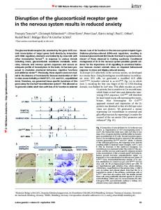
i Fig. 2 Function of Gcm1 in the mouse placenta. a, Gcm1 gene-targeting strategy. b, Southern-blot verification of correctly targeted ES cell clones. Mouse genomic DNA was cut with EcoRI and hybridized with a probe from a region flanking the 3´ arm of the targeting vector (probe B). Gross morphology of E9.5 (c,d) and E10.5 (e,f) embryos and chorioallantoic attachment (g,h) in Gcm1 mutants (–/–) and heterozygous littermates (+/–) are shown. Note that chorioallantoic attachment has occurred and umbilical cord vessels are evident in the Gcm1-deficient conceptuses. Bar, 1 mm. i–k, Histological sections of chorioallantoic interface at E8.5 (i,j) and E9.0 (k) in Gcm1 mutants (–/–) and heterozygous littermates (+/–). Note the extensive development of vessels lined by endothelium within the allantois (arrowheads). al, allantois; ch, chorioninc trophoblast cells; uc, umbilical cord.
Gcm1–/– embryos died in utero. Gcm1–/– embryos developed normally until E9.5 (Fig. 2c,d), but by E10.5 they were smaller than their littermates (Fig. 2e,f). No viable Gcm1–/– embryos were recovered after E10.5 (Table 1). The embryo proper showed no obvious abnormalities, consistent with the observation that Gcm1 expression is extremely low in the embryo10,15–17. Embryonic mortality appeared to be due to placental failure. The placentas of Gcm1–/– embryos developed normally until E9, but, thereafter, the labyrinth layer failed to form (Fig. 3). Attachment of the allantois to the chorionic plate and formation of a basement membrane between them were normal in mutant conceptuses, by both light (Fig. 3a) and electron (Fig. 4a) microscopy. Umbilical cord vessels were present (Fig. 2h), and the number, organization and density of allantoic blood vessels beneath the chorionic plate appeared to be normal in Gcm1–/– embryos (Fig. 2j,k). Morphogenesis of the chorioallantoic interface and intrusion of underlying fetal blood vessels was apparent by E9.5 in wild-type (data not shown) and Gcm1+/– (Fig. 3a) embryos, but the chorionic plate in Gcm1–/– mice remained flat and failed to interdigitate with the underlying allantoic mesoderm and blood vessels (Fig. 3a). The chorioallantoic interface remained flat up to three days after allantoic attachment (Fig. 3b, and data not shown), indicating that morphogenesis was blocked and not simply delayed. The trophoblast giant cell and spongiotrophoblast layers of the placenta appeared normal. In addition to failure of chorioallantoic morphogenesis, we did not see fusion of chorionic trophoblast cells to form syncytiotrophoblast in Gcm1 mutants, and the cells retained distinct cell membranes (Fig. 4c). The number of mitochondria, lipid droplets and electron-dense granules in chorionic trophoblast cells increase shortly after contact with allantoic mesoderm and before cell fusion19. These changes in ultrastructural features were not seen in Gcm1 mutants (Fig. 4a, and data not shown). In addition, expression of genes specific for chorionic trophoblast cells, including Esrrb, Tead5 (Fig. 4e), Cdx2, Dlx3 and Nr6a1 312
k
j
(data not shown) was normal. The chorionic trophoblast cells did not express genes characteristic of other trophoblast subtypes such as Pl1 and Tpbp, which are specific for trophoblast giant cells and spongiotrophoblast, respectively (Fig. 4e). These data suggested that the chorionic trophoblast cells had not altered their fate in the absence of Gcm1. In addition, the chorionic plate was of normal size for the E8.5–9.5 period, suggesting that Gcm1 is not required for proliferation of trophoblast stem cells. We conclude that Gcm1 is essential for early steps in chorioallantoic placental development. Several other gene mutations affect development of the placental labyrinth, but all of these other phenotypes are explainable by distinct mechanisms such as loss of chorionic trophoblast cells, failure of chorioallantoic attachment, delay (but not block) in chorioallantoic branching or decrease in the vascularity of the labyrinth1. The Gcm1 mutants provide the first molecular insights into two critical aspects of placental development. First, Gcm1 is the first gene shown to be critical for the formation of syncytiotrophoblast, one of the most abundant cell types in the mature placenta. Second, Gcm1 mutants are the first to show a complete block to chorioallantoic branching. Fusion of chorionic trophoblast cells to form syncytiotrophoblast begins a few hours after morphogenesis begins1,19, and thus the mechanistic connection of the two events was unexpected. Table 1 • Genotype of offspring derived from intercrosses of Gcm1+/– mice Age
No. of litters
Genotype Gcm1+/+
Gcm1+/–
Gcm1–/– 13 16 6b 7b
E8.5 E9.5 E10.5 E11.5
4 11 3 3
4 27 9 4
18 47 20 17
subtotal
44
102
41
E12.5 E14.5 weanling
1 4 23
4 6 81
5 16 158
subtotal
91
177
0
0 0 0
? 3a
4a 13a
aUnable to obtain DNA sample, resorptions. bSmaller than littermates, resorbing.
nature genetics • volume 25 • july 2000
© 2000 Nature America Inc. • http://genetics.nature.com
a
Fig. 3 Failure of chorioallantoic placental morphogenesis in Gcm1-deficient embryos. Histological sections (8 µm) from representative placentas at E9.2 (a) and E11.5 (b) were stained with haemotoxylin and eosin. Note that in heterozygous and wild-type (data not shown) conceptuses, the allantoic mesenchyme and chorionic trophoblast cells interdigitate to create villi (arrows). In contrast, the chorionic plate in Gcm1–/– embryos remains flat. The allantois is tightly associated with the chorion, although occasionally after E10.5 it separates due to haemorrhage (b). Arrowheads indicate nucleated fetal red blood cells. The dashed line indicates the outer surface of placenta lined by trophoblast giant cells. al, allantois; ch, chorionic trophoblast cells; lz, labyrinthine zone; m, maternal blood cells; sp, spongiotrophoblast. Bar, 100 µm.
© 2000 Nature America Inc. • http://genetics.nature.com
b
The elucidation of Gcm1 function provides an important insight into the steps of chorioallantoic morphogenesis. The precise mechanisms that underlie morphogenesis are unknown, but are thought to involve chorionic and allantoic cell differentiation and migration1,19. The predominant view of chorioallantoic morphogenesis is that allantoic mesoderm and blood vessels invade the chorionic plate and result in trophoblast differentiation19. The early expression of Gcm1 is restricted to focal sites in the chorionic plate that subsequently coincide with points of branch initiation, indicating that trophoblast factors specify the sites within the chorionic plate that undergo morphogenesis. Gcm1 mRNA expression is initiated before the allantois contacts the chorion10, but it is clear that interactions between the allantois and chorion are essential for subse-
Fig. 4 Failure of syncytiotrophoblast differentiation and tissue morphogenesis, but normal chorionic trophoblast differentiation, in Gcm1-deficient embryos. a–d, Transmission electron microscopy of chorionic trophoblast cells at the chorioallantoic interface at E8.5 (a; original magnification, ×3,200) and E9.5 (b,c; original magnification, ×7,400), and within the labyrinth at E10.5 (d; original magnification. ×3,200) in normal and Gcm1–/– conceptuses (original magnification, ×4,200). Note that in heterozygous (+/–) and wild-type (data not shown) littermates these cells fuse into a syncytium beginning around E9.2 (c), ultimately forming two layers of syncytiotrophoblast (ST-I and ST-II in e). In Gcm1deficient conceptuses, by contrast, distinct plasma membranes separate chorionic trophoblast nuclei (arrowheads in d). Asterisks indicate mitochondria. Arrows demarcate the basement membrane separating chorionic trophoblast from allantoic mesoderm. e, In situ hybridization analysis for Esrrb, Pl1, Tead5 and Tpbp mRNA expression in E9.5 placentas. al, allantois; ch, chorionic trophoblast cells; ec, endothelial cell; epc, ectoplacental cone; fRBC, fetal red blood cell; mRBC, maternal red blood cell. Bar, 100 µm.
nature genetics • volume 25 • july 2000
letter
e
a
b
quent development. For example, ultrastructural changes in chorionic trophoblast cells correlate with their contact with allantoic mesoderm19, and the chorionic plate does not undergo morphogenesis if the allantois does not attach1. Indeed, maintenance of Gcm1 expression may depend on contact with the allantois20. Our data indicate that Gcm1 has a central role mediating interactions between chorionic trophoblast and allantoic mesoderm cells. Our findings outline mechanisms that may explain and distinguish enigmatic pathologies of the human placenta. Pre-eclampsia and intrauterine growth restriction (IUGR) complicate 5–10% of human pregnancies, resulting in fetal or neonatal mortality and morbidity21. Paradoxically, two distinct pathologies have been described. A block in differentiation of extravillous cytotrophoblast cells, which normally invade and transform uterine arteries, is associated with pre-eclampsia and IUGR detected at term22. Homologues of the basic helix-loop-helix (bHLH) factors that regulate differentiation of invasive trophoblast cells in mice7–9 (giant cells) are expressed during extravillous cytotrophoblast differentiation in humans18. Thus, changes in the function of these factors may account for the cellular defect in pre-eclampsia. By contrast, reduced branching of chorionic villi is associated with a severe and early onset form of IUGR (ref. 23). In this case, normal bHLH, but defective Gcm1-mediated, functions are more likely to account for the pathology. GCM1 mRNA has been detected in human cytotrophoblast cells18 and we have localized expression to chorionic villi (unpublished data), suggesting that gene function is conserved between mice and humans. The prospect of describing all aspects of placental development in molecular terms is a significant advance for understanding abnormal human development.
c
d
313
letter
© 2000 Nature America Inc. • http://genetics.nature.com
© 2000 Nature America Inc. • http://genetics.nature.com
Methods Gene targeting. The 2.6-kb 5´ arm of the targeting vector (from the promoter region to exon 2) was generated by PCR using the primers 5´–CCGCTCGAG CGGTGCTGTTTAGCTCAGGCTA–3´ and 5´–TCCCCGCGGGGAGCTG AAGGGCTTGTTTTTC–3´ and the 1.9-kb 3´ arm was an SspI-BamHI fragment. These fragments were positioned to flank the neomycin resistance cassette (neor) in the vector pPNTloxP. Correct targeting resulted in deletion of gene sequence from exon 2 immediately upstream of the ATG codon to the middle of intron 5, and replacement with a lacZ reporter and the PGK promoter-neomycin resistance cassette. The construct was linearized by digestion with NotI. R1 ES cells were electroporated, subjected to positive-negative selection and screened by Southern-blot analysis24. We screened ES-cell clones for homologous recombination events with 5´ and 3´ flanking probes (Fig. 1). Of 146 clones analysed, 3 were confirmed to be heterozygous for the targeted allele (Gcm1+/–). Chimaeras were generated with heterozygous ES cell lines. Male chimaeras from two of these (4A9 and 11A10) were capable of germline transmission of the mutant allele. We generated heterozygous progeny by backcrossing to wild-type 129Sv females and outcrossing to CD1 females. We genotyped progeny by Southern blot and a PCR assay that identifies both mutant and wild-type alleles. A common antisense primer from distal intron 5 (primer B, 5´–GAGCTAGTAGCGCCCTGAAG–3´) was used in conjunction with sense primers specific for the wild-type allele from exon five (primer A, 5´–GATGAAGGGGAGACCAGCAG–3´) and to the mutant allele from the neor cassette (primer C, 5´–AAGGGCCAGCTCATTCCTCC–3´).
1. 2. 3.
4.
5. 6. 7. 8.
9.
10. 11.
12. 13. 14. 15.
16.
Cross, J.C. Genetic insights into trophoblast differentiation and placental morphogenesis. Semin. Cell Dev. Biol. (in press). Cross, J.C., Werb, Z. & Fisher, S.J. Implantation and the placenta: key pieces of the development puzzle. Science 266, 1508–1518 (1994). Jackson, D., Volpert, O.V., Bouck, N. & Linzer, D.I. Stimulation and inhibition of angiogenesis by placental proliferin and proliferin-related protein. Science 266, 1581–1584 (1994). Yotsumoto, S. et al. Expression of adrenomedullin, a hypotensive peptide, in the trophoblast giant cells at the embryo implantation site in mouse. Dev. Biol. 203, 264–275 (1998). Tanaka, S., Kunath, T., Hadjantonakis, A.K., Nagy, A. & Rossant, J. Promotion of trophoblast stem cell proliferation by FGF4. Science 282, 2072–2075 (1998). Rossant, J. & Ofer, L. Properties of extra-embryonic ectoderm isolated from postimplantation mouse embryos. J. Embryol. Exp. Morphol. 39, 183–194 (1977). Guillemot, F., Nagy, A., Auerbach, A., Rossant, J. & Joyner, A.L. Essential role of Mash-2 in extraembryonic development. Nature 371, 333–336 (1994). Riley, P., Anson-Cartwright, L. & Cross, J.C. The Hand1 bHLH transcription factor is essential for placentation and cardiac morphogenesis. Nature Genet. 18, 271–275 (1998). Scott, I.C., Anson-Cartwright, L., Riley, P., Reda, D. & Cross, J.C. The Hand1 basic helix-loop-helix transcription factor regulates trophoblast giant cell differentitation via multiple mechanisms. Mol. Cell. Biol. 20, 530–541 (2000). Basyuk, E. et al. The murine Gcm1 gene is expressed in a subset of placental trophoblast cells. Dev. Dyn. 214, 303–311 (1999). Hosoya, T., Takizawa, K., Nitta, K. & Hotta, Y. Glial cells missing: a binary switch between neuronal and glial determination in Drosophila. Cell 82, 1025–1036 (1995). Jones, B.W., Fetter, R.D., Tear, G. & Goodman, C.S. Glial cells missing: a genetic switch that controls glial versus neuronal fate. Cell 82, 1013–1023 (1995). Bernardoni, R., Vivancos, B. & Giangrande, A. Glide/gcm is expressed and required in the scavenger cell lineage. Dev. Biol. 191, 118–130 (1997). Bernardoni, R., Miller, A.A. & Giangrande, A. Glial differentiation does not require a neural ground state. Development 125, 3189–3200 (1998). Altshuller, Y., Copeland, N.G., Gilbert, D.J., Jenkins, N.A. & Frohman, M.A. Gcm1, a mammalian homolog of Drosophila Glial Cells Missing. FEBS Lett. 393, 201–204 (1996). Akiyama, Y., Hosoya, T., Poole, A.M. & Hotta, Y. The Gcm-motif: a novel DNAbinding motif conserved in Drosophila and mammals. Proc. Natl Acad. Sci. USA 93, 14912–14916 (1996).
314
Histology and in situ hybridization. We dissected mouse conceptuses at different gestational ages (day 0.5 defined as noon of the day in which a vaginal plug was detected). For routine histology and light microscopy, tissues were fixed overnight in 4% paraformaldehyde, embedded in paraffin and sectioned (7 µm). For electron microscopy, tissues were fixed overnight in 2% glutaraldehyde. For mRNA in situ hybridization experiments, conceptuses were collected in PBS and fixed overnight in 4% paraformaldehyde. We embedded tissues in paraffin and sectioned them. Adjacent sections (10 µm) were hybridized with digoxigenin-labelled riboprobes for Pl1 (ref. 25), Tpbp (ref. 26), Esrrb (ref. 27), Cdx2 (ref. 28), Dlx3 (ref. 29), Nr6a1 (ref. 29) and Tead5 (ref. 30). We detected signals using alkaline phosphatase immunohistochemistry and counterstained sections briefly with nuclear fast red. Whole-mount in situ hybridization was conducted using standard procedures. Acknowledgements
We thank Z. Basyuk for providing genomic clones; Y. Lu for histological sections; and A. Bernstein, J. Kingdom and J. Rossant for critical comments on the manuscript. The work was supported by grants from the MRC of Canada (to J.C.C.) and the NIH (to S.J.F. and to R.A.L.).
Received 15 February; accepted 13 April 2000.
17. Kim, J. et al. Isolation and characterization of mammalian homologs of the Drosophila gene glial cells missing. Proc. Natl Acad. Sci. USA 95, 12364–12369 (1998). 18. Janatpour, M.J. et al. A repertoire of differentially expressed transcription factors that offers insight into mechanisms of human cytotrophoblast differentiation. Dev. Genet. 25, 146–157 (1999). 19. Hernandez-Verdun, D. Morphogenesis of the syncytium in the mouse placenta. Ultrastructural study. Cell Tissue Res. 148, 381–396 (1974). 20. Hunter, P.J., Swanson, B.J., Haendel, M.A., Lyons, G.E. & Cross, J.C. Mrj encodes a DnaJ-related co-chaperone that is essential for murine placental development. Development 126, 1247–1258 (1999). 21. Cross, J.C. Trophoblast function in normal and preeclamptic pregnancy. Fetal Maternal Med. Rev. 8, 57–66 (1996). 22. Khong, T.Y., De Wolf, F., Robertson, W.B. & Brosens, I. Inadequate maternal vascular response to placentation in pregnancies complicated by pre-eclampsia and by small-for-gestational age infants. Br. J. Obstet. Gynaecol. 93, 1049–1059 (1986). 23. Krebs, C. et al. Intrauterine growth restriction with absent end-diastolic flow velocity in the umbilical artery is associated with maldevelopment of the placental terminal villous tree. Am. J. Obstet. Gynecol. 175, 1534–1542 (1996). 24. Wurst, W. & Joyner, A. Production of targeted embryonic stem cell clones. in Gene Targeting: A Practical Approach (ed. Joyner, A.) 33–61 (Oxford University Press, Oxford, 1993). 25. Colosi, P., Talamantes, F. & Linzer, D.I. Molecular cloning and expression of mouse placental lactogen I complementary deoxyribonucleic acid. Mol. Endocrinol. 1, 767–776 (1987). 26. Lescisin, K.R., Varmuza, S. & Rossant, J. Isolation and characterization of a novel trophoblast-specific cDNA in the mouse. Genes Dev. 2, 1639–1646 (1988). 27. Luo, J. et al. Placental abnormalities in mouse embryos lacking the orphan nuclear receptor ERR-β. Nature 388, 778–782 (1997). 28. Beck, F., Erler, T., Russell, A. & James, R. Expression of Cdx-2 in the mouse embryo and placenta: possible role in patterning of the extra-embryonic membranes. Dev. Dyn. 204, 219–227 (1995). 29. Morasso, M.I., Grinberg, A., Robinson, G., Sargent, T.D. & Mahon, K.A. Placental failure in mice lacking the homeobox gene Dlx3. Proc. Natl Acad. Sci. USA 96, 162–167 (1999). 30. Jacquemin, P. et al. Differential expression of the TEF family of transcription factors in the murine placenta and during differentiation of primary human trophoblasts in vitro. Dev. Dyn. 212, 423–436 (1998).
nature genetics • volume 25 • july 2000