Journal of Biomolecular NMR, 19: 287–288, 2001. KLUWER/ESCOM © 2001 Kluwer Academic Publishers. Printed in the Netherlands.
287
Letter to the Editor: Sequential assignment and secondary structure of the 14 kDa chemotactic protein CheY2 from Sinorhizobium meliloti Hubert Riepla,b , Birgit Scharfb , Rüdiger Schmittb , Hans Robert Kalbitzera & Till Maurera,∗ a Institut
für Biophysik und Physikalische Biochemie; and b Institut für Genetik, Universität Regensburg, Postfach, D-93040 Regensburg, Germany Received 15 December 2000; Accepted 2 January 2001
Key words: CheY2, protein structure, Sinorhizobium meliloti Biological context Motile bacteria are able to direct their swimming movement towards the most favourable chemical environment. This ability, known as chemotaxis, is mediated by a signal transduction pathway involving a set of cytoplasmic proteins and extracellular rotating helical flagella. CheA, an autokinase, activates a response regulator, CheY, by phosphorylation. CheY propagates the signal, which is sensed by the transmembrane chemoreceptors, to the flagellar motor. In response, Escherichia coli flagella reverse the direction of rotation, resulting in a tumbling and thus a change in the direction of movement. Dephosphorylation of CheY-P resets the signal and is accelerated by a phosphatase, CheZ. This is different in Sinorhizobium meliloti, where two response regulators, CheY1 and CheY2, are phosphorylated, with CheY2 being the chief regulator (Sourjik and Schmitt, 1998). The phosphorylated states of CheY1 and CheY2 are short-lived due to autophosphatase activity and in case of CheY2-P through a retrophosphorylation mechanism involving CheA and CheY1. Up to now, this retrophosphorylation as a new mechanism of adaptation is not fully understood. The striking differences in the two dephosphorylation reactions of the two response regulators, CheY (E. coli) and CheY2 (S. meliloti), respectively, and the fact that X-ray and NMR structures of the former have been determined (Stock et al., 1989; Santoro et al., 1995), instigated the present study of the molecular structure of CheY2 ultimately aimed at an understanding of interactions with other protein components of the system. We expect ∗ To whom correspondence should be addressed.
[email protected]
E-mail:
that a comparison of the 3D structures will provide clues toward an explanation of the different behaviours of the aspartyl-phosphates in the two different CheY molecules. Using chemical shift data from the sequential assignment of the Mg2+ -bound form of the unphosphorylated CheY2, the Chemical Shift Index (CSI) analysis suggests an (a/b)5 globular structure. This is confirmed by contacts found in the 2D NOESY spectrum.
Methods and experiments CheY2 was overexpressed from the plasmid pRU2313 (derivative of pTYB1, NEB) in M9 medium supplemented with glucose at 30 ◦ C. At mid-exponential phase, the medium was exchanged by fresh M9 medium supplemented with 1 g/l 13 C-glucose, followed by induction with 0.3 mM IPTG for 4 h. CheY2 was purified from the lysate after French press passage by affinity chromatography on Chitin-Agarose and gel filtration on Superdex HR75. NMR samples contained a 1.2 mM solution of CheY2 in 95% 1 H2 O/5% 2 H2 O in 20 mM NaPi , 5 mM MgCl2 , pH 6.9. All NMR measurements were conducted on a Bruker DXR600 spectrometer operating at 600 MHz proton frequency. The 2D spectra were recorded with 2048 data points in the proton and 512 data points in the 15 N dimension. All 3D data were acquired with 1024 data points in the proton dimension, 128 data points in the 13 C dimension using constant time evolution and StatesTPPI acquisition (Marion and Wüthrich, 1983) and 64 data points in the 15 N dimension with echo-antiecho type selection (Schleucher et al., 1993). Linear prediction (Barkhuijsen et al., 1985) in the indirect dimensions resulted in a spectral resolution of 5 Hz/data
288
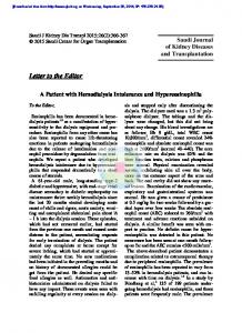
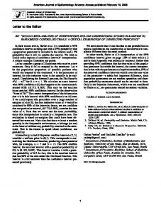
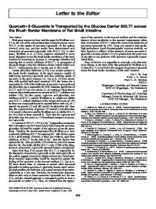
Figure 1. A 1 H-15 N HSQC spectrum of CheY2 from S. meliloti recorded at 298 K. 15 N labeled protein was dissolved in 320 µl 95% 2 H O/5% H O, 20 mM NaP and 5 mM MgCl at pH 6.9 to give a 2 2 i 2 concentration of 1.2 mM. The well resolved peaks are labeled with the corresponding assignment, peaks with no label arise from side chain amide groups. Arrows signify peaks that stem from a minor conformation for Gly 125 and Phe 125 at the C- and Ile 7 at the N-terminus.
point in 1 H, 23 Hz/data point in 13 C and 36 Hz/data point in 15 N. Data were referenced indirectly using the 1 H chemical shift of the methyl group in DSS and multiplying this value by 0.25144953 for 13 C and 0.101329118 for 15 N (Markley et al., 1998). Sequential backbone assignment was accomplished utilising the HNCO, HCACO, HNCA, CBCA(CO)NH, HBHA(CO)NH and 15 N separated NOESY spectra, the side chain assignment was done using the HCCH-TOCSY experiment (for a review, see Sattler et al., 1999). Data were processed in XWINNMR (Bruker, Karlsruhe) and evaluated in AURELIA (Bruker) (Neidig et al., 1995).
Extent of assignments and data deposition All backbone 1 H, 15 N and 13 C resonances and 65% of the side chain chemical shifts were assigned.
Signals in the postulated active site encompassing residues 51 to 61 were comparatively weaker than those of the other regions of CheY2, indicating structural heterogeneity. Also, Gly 126 and Phe 125 at the C- and Ile 7 at the N-terminus show evidence of a minor conformer. The chemical shift values of the carbonyl-carbon, α-proton and α-carbon were used for the secondary structure prediction with the CSI (Wishart and Sykes, 1994) and indicate a well defined globular fold with an (a/b)5 globular structure. Figure 1 shows the 15 N HSQC spectrum with the assignments. The chemical shifts for CheY2 have been deposited in the BMRB database (http://www.bmrb.wisc.edu) with accession code 4896.
Acknowledgements This work was supported by the Deutsche Forschungsgemeinschaft Schm68/34-1.
References Barkhuijsen, H., de Beer, R., Bovee, W.M., Creyghton, J.H. and van Ormondt, D. (1985) Magn. Reson. Med., 2, 86–89. Marion, D. and Wüthrich, K. (1983) Biochem. Biophys. Res. Commun., 113, 967–974. Santoro, J., Bruix, M., Pascual, J., Lopez, E., Serrano, L. and Rico, M. (1995) J. Mol. Biol., 247, 717–725. Markley, J.L., Bax, A., Arata, Y., Hilbers, C.W., Kaptein, R., Sykes, B.D., Wright, P.E. and Wüthrich, K. (1998) Pure Appl. Chem., 70, 117–142. Neidig, K.-P., Geyer, M., Görler, A., Antz, C., Saffrich, R., Beneicke, W. and Kalbitzer, H.R. (1995) J. Biomol. NMR, 6, 255–270. Sattler, M., Schleucher, J. and Griesinger, C. (1999) Prog. NMR Spectrosc., 34, 93–158. Schleucher, J., Schwendinger, M., Sattler, M., Schmidt, P., Schedletzky, O., Glaser, S.J., Sørensen, O.W. and Griesinger, C. (1994) J. Biomol. NMR, 4, 301–306. Sourjik, V. and Schmitt, R. (1998) Biochemistry, 37, 2327–2335. Stock, A.M., Mottonen, J.M., Stock, J.B. and Schutt, C.E. (1989) Nature, 337, 745–749. Wishart, D. and Sykes, B.D. (1994) J. Biomol. NMR, 4, 171–180.