Journal of Biomolecular NMR, 18: 369–370, 2000. KLUWER/ESCOM © 2000 Kluwer Academic Publishers. Printed in the Netherlands.
369
Letter to the Editor: Sequential resonance assignments of the extracellular ligand binding domain of the human TGF-β type II receptor Andrew P. Hincka,∗ , Kerfoot P. Walker, IIIa , Nathan R. Martina, Shashank Deepa , Cynthia S. Hincka & Dar´on I. Freedbergb a Center
for Biomolecular Structure Analysis, Department of Biochemistry, University of Texas Health Science Center at San Antonio, San Antonio, TX 78229–3900, U.S.A. b Center for Biologics Evaluation and Research, Food and Drug Administration, Bethesda, MD, U.S.A. Received 12 July 2000; Accepted 2 October 2000
Key words: ligand, NMR assignments, receptor, TGF, Transforming growth factor-beta, type II receptor 1 H, 13 C,
and 15 N chemical shift assignments for the extracellular ligand binding domain of human TβR2.
Biological context Transforming growth factor beta (TGF-β) is a potent growth suppressor of many different normal cell types (Massague, 1998). Significantly, many cancer cell types show a diminished sensitivity to TGF-β mediated growth inhibition. In addition to its important role in growth regulation, TGF-β also affects the adhesive properties of cells by regulating key components involved in cell adhesion. TGF-β signaling is initiated by binding of growth factor ligand to two related singlepass transmembrane receptor serine/threonine kinases, known as the TGF-β type I (TβR1) and type II (TβR2) receptors. In the sequential model for TGF-β receptor activation, growth factor ligand first binds to TβR2. TβR1, which is incapable of binding ligand in the absence of TβR2, associates with the TGF-β•TβR2 complex and is activated by the constitutive kinase activity of TβR2 (Massague, 1998). Presently, the three-dimensional structure of each of the major TGF-β isoforms (TGF-β1, -β2, and -β3) is known (reviewed by Massague, 1998). There is no experimentally determined three-dimensional structural information available regarding the ligand binding domains of the TGF-β receptors, although Xray crystal structures of extracellular ligand binding domain of the activin type II receptor and the extracellular domain of the bone morphogenic protein (BMP) receptor 1A bound to the TGF-β superfamily member BMP-2 have recently been reported (Greenwald et al., 1999; Kirsch et al., 2000). Herein, we report backbone ∗ To whom correspondence should be addressed.
[email protected]
E-mail:
Methods and results The coding region for the extracellular ligand binding domain of the human TβR2 was inserted into plasmid pET32a (Novagen). This construct lacked the coding region for the first 14 residues and included an asparagine- to alanine-substitution at position 19. The latter was important in eliminating problems associated with deamidation of the naturally occurring asparagine at this position (Lin et al., 1992). Recombinant soluble TβR2, herein designated sTβR2, was obtained by overexpressing the protein in E. coli strain BL21(DE3), and by isolating the protein from the insoluble fraction. The protein was then subjected to repeated dialysis against 20 mM acetic acid (pH 3.55), and then folded by slowly transferring the acid-soluble protein to an equal or greater volume of buffer containing 200 mM Tris-Cl, 2 mM reduced glutathione, 0.5 mM oxidized glutathione, pH 8.0. The properly folded protein was separated from misfolded protein and other impurities using anion-exchange chromatography, and was found to exhibit identical TGF-β binding properties relative to that of the full-length human sTβR2 domain as produced in a cultured mouse myeloma cell line (R&D Systems) (Qian et al., 1996). Isotopically enriched forms of the sTβR2 domain were prepared for NMR spectroscopy by culturing the cells on M9 minimal medium containing 0.1% 15 NH4 Cl and 0.3% uniformly 13 C-labeled D-glucose.
370 Extent of assignments and data deposition
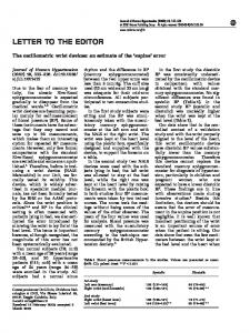
Figure 1. A summary of the backbone assignments and secondary structure of sTβR2. (A) The 2D 1 H-15 N HSQC spectrum of E. coli recombinant sTβR2 with peaks labeled according to their assignments. D170 , N180 , and G200 correspond to peaks arising from a minor form due to incomplete removal of the N-terminal methionine. S1330 and N1340 correspond to a minor form detected at the C-terminus. (B) The predicted secondary structure of the sTβR2 domain as deduced by the consensus chemical shift index (Wishart and Sykes, 1994).
The sequential backbone assignments of the sTβR2 domain were made using triple-resonance methodology with 1–2 mM 15 N/13 C samples dissolved in 25 mM sodium acetate, 25 mM NaCl, 5% 2 H O at pH 5.5. The sample temperature was 27 ◦ C. 2 The NMR data was collected using a Bruker AMX2500 spectrometer and 1 H, 13 C and 15 N chemical shifts were referenced to DSS (Markley et al., 1998). The sequential connectivities were made using the CBCA(CO)NH and HNCACB pair of experiments, and were confirmed using the HNCO and HN(CA)CO pair. The C(CO)NH experiment facilitated the identification of complete sidechain 13 C assignments. Backbone Hα and sidechain Hβ resonances for the majority of residues were obtained using the HBHA(CO)NH experiment. The remainder of the 1 H resonances were assigned using the HCCH-TOCSY experiment.
Essentially complete backbone and side-chain sequential resonance assignments were obtained for 112 of the 122 amino acid residues in the sTβR2 domain (Figure 1A). Amongst these residues, the only missing assignments include the 1 Hε/13 Cε resonances for two lysines and the phenylalanine aromatic ring resonances. Among the 10 unassigned residues, partial assignments are available for four of these (C78, K101,S114, and I125). The unassigned residues cluster to four regions of the amino acid sequence: C78, C98–K101, M112–S114, and I124–I125. It appears likely that the NMR signals in these regions of the protein are missing owing to conformational broadening since all of the amides in the 1 H/15 N HSQC which yield detectable correlations in the triple-resonance experiments have already been assigned. The secondary structure of the sTβR2 domain, as deduced by the consensus chemical shift index (Figure 1B), is comprised exclusively of β-strand, and is consistent with our expectations based on the X-ray structure of the homologous activin type II receptor ligand binding domain (Greenwald et al., 1999). The assignments have been deposited with BioMagResBank under accession 4779.
Acknowledgements The authors acknowledge Andrzej Krezel who suggested deamidation as the source of heterogeneity in the sTβR2 samples. This work was supported by NIHGMS (GM58670) and the Robert A. Welch Foundation (AQ1431).
References Greenwald, J., Fischer, W.H., Vale, W.W. and Choe, S. (1999) Nat. Struct. Biol., 6, 18–22. Kirsch, T., Sebald, W. and Dreyer, M.K. (2000) Nat. Struct. Biol., 7, 492–496. Lin, H.Y., Wang, X.-F., Elinor, N.-E., Weinberg, R.A., and Lodish, H.F. (1992) Cell, 68, 775–785. Markley, J.L., Bax, A., Arata, Y., Hilbers, C.W., Kaptein, R., Sykes, B.D., Wright, P.E., and Wüthrich, K. (1998) Pure Applied Chem., 70, 117–142. Massague, J. (1998) Annu. Rev. Biochem., 67, 753–791. Qian, S.W., Burmester, J.K., Tsang, M.L.-S., Weatherbee, J.A., Hinck, A.P., Ohlsen, D.J., Sporn, M.B. and Roberts, A. (1996) J. Biol. Chem., 271, 30656–30662 Wishart, D.S. and Sykes, B.D. (1994) J. Biomol. NMR, 4, 171–180.