The Plant Journal (2001) 28(3), 361±369
Local expression of enzymatically active class I b-1, 3-glucanase enhances symptoms of TMV infection in tobacco Gregor L. Bucher²,³, Corina Tarina², Manfred Heinlein, Francesco Di Serio§, Fred Meins, Jr.*, Victor Alejandro Iglesias¶ Friedrich Miescher Institute, A Branch of the Novartis Research Foundation, Maulbeerstrasse 66, CH-4058 Basel, Switzerland Received 11 July 2001; revised 5 September 2001; accepted 5 September 2001. * For correspondence (fax +41 (0)61 697 3976; e-mail
[email protected]). ³ Present address: Max-Planck-Institut fuÈr ZuÈchtungsforschung, Carl-von-Linne Weg 10, D-50829 KoÈln, Germany. § Present address: Centro di Studio del CNR sui Virus e le Virosi delle Colture Mediterranee, via G. Amendola 165/A, I-70126 Bari, Italy. ¶ Present address: Hoffmann-La Roche AG, Grenzacherstrasse 124, CH-4070 Basel, Switzerland. ² These authors contributed equally to this publication.
Summary Mutant tobacco plants de®cient for class I b-1,3-glucanase (GLU I) are decreased in their susceptibility to virus infection. This is correlated with delayed virus spread, a reduction in the size exclusion limit of plasmodesmata and increased cell-wall deposition of the b-1,3-glucan callose. To further investigate a role of GLU I during cell-to-cell movement of virus infection, we inserted the GLU I coding sequence into TMV for overexpression in infected cells. Compared with the size of local lesions produced on plants infected with virus expressing either an enzymatically inactive GLU I or a frameshift mutant of the gene, the size of local lesions caused by infection with virus expressing active GLU I was consistently increased. Viruses expressing antisense GLU I constructs led to lesions of decreased size. Similar effects were obtained for virus spread using plants grown at 32°C to block the hypersensitive response. Together, these results indicate that enzymatically active GLU I expressed in cells containing replicating virus can increase cell-to-cell movement of virus. This supports the view that GLU I induced locally during infection helps to promote cell-to-cell movement of virus by hydrolyzing callose. Moreover, our results provide the ®rst direct evidence that a biological function of a plant b-1,3-glucanase depends on its catalytic activity. Keywords: callose, b-1, 3-glucanase, plasmodesmata, TMV, tobacco, virus spread.
Introduction b-1,3-Glucanases are induced during the hypersensitive response of plants to viral, bacterial and fungal pathogens. Considerable evidence suggests that the class I isoform (GLU I) has an important role in the resistance of plants to infection. For example, GLU I has anti-fungal activity in vitro and helps protect plants against infection with some pathogenic fungi (Kombrink and Somssich, 1997; Leubner-Metzger and Meins, 1999). Studies of GLU Ide®cient mutants generated by antisense transformation have shown that GLU I can also act as a susceptibility factor in viral pathogenesis (Beffa et al., 1996). Following infection with TMV and tobacco necrosis virus, GLU Ide®cient mutants of tobacco and Nicotiana sylvestris show ã 2001 Blackwell Science Ltd
greatly reduced virus symptoms in both the local-lesion and systemic response. Viruses such as TMV spread from cell to cell via plasmodesmata in processes mediated by viral movement proteins (MP) (Deom et al., 1992). GLU-I de®cient mutants exhibit delayed spread of TMV and a recombinant potato virus X; delayed cell-to-cell movement of a cucumber mosaic virus MP fusion with green ¯uorescent protein; and a substantial decrease in the size exclusion limit (SEL) of plasmodesmata (Iglesias and Meins, 2000). This suggests that reduced virus susceptibility of the GLU Ide®cient mutants results from downregulation of plasmodesmatal traf®cking. 361
362
Gregor L. Bucher et al.
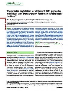
Local deposition of callose, which is a substrate for b1,3-glucanases (Stone and Clarke, 1992), may constrict the neck region of plasmodesmata, thus reducing plasmodesmatal traf®cking and virus spread (Botha and Cross, 2000; Robards and Lucas, 1990). Hypersensitive GLU I-de®cient mutants show greatly increased callose deposition in sieve tubes elevated temperatures and locally at discrete sites in response to elicitors, wounding and virus infection (Beffa et al., 1996; Iglesias and Meins, 2000). Together, this has led to the hypothesis that the induction of GLU I in response to infection may support MP-driven virus spread by degrading callose. In the present experiments we used GLU I-expressing TMV vectors to examine the role of GLU I during viral infection of the hypersensitive, local-lesion host N. tabacum cv. Xanthi-nc. Our results establish that increased expression of GLU I in virus-infected cells can increase the size of local lesions and, in the absence of the hypersensitive response, can promote virus spread. We also show that these biological effects of b-1,3-glucanase depend on the enzymatic activity of the protein. Results Construction of TMV vectors The TMV-based expression vector 4 GD-Pl (Karrer et al., 1998) was used to express recombinant GLU I coding sequences precisely in those cells infected with virus. The vector and constructs are summarized in Figure 1. The vector contains a modi®ed cDNA of TMV that allows the insertion of foreign sequences immediately downstream of the TMV coat-protein (CP) promoter. A CP subgenomic promoter derived from the related tobacco mild green mosaic virus directs expression of the coat protein, allowing expression of the virion. The vectors SGLU and ASGLU carry GLU I cDNA in sense and antisense orientation, respectively. FSGLU encodes a transcript almost identical to that of SGLU, but is unable to produce the GLU I polypeptide due to a frameshift mutation. MGLU encodes an enzymatically inactive mutant of GLU I in which E262 and E318, essential for hydrolytic activity, have been replaced by glutamine residues (Chen et al., 1995). The empty vector 4 GD-PI and the vector LUC, which carries a luciferase reporter gene, were used as controls. Capped, infectious transcripts generated by in vitro transcription with T7 polymerase were ampli®ed and then encapsidated in BY-2 tobacco protoplasts. The TMV constructs replicate in protoplasts and are stable in planta To detect possible effects of the inserted sequences on virus replication, infectious transcripts of the constructs
Figure 1. Partial Maps of TMV expression vectors. The open reading frames of the TMV vector 4 GD-Pl (VEC) (Karrer et al., 1998) are indicated. GLU I and luciferase genes were cloned into the SalI site and are under control of the TMV coat-protein (CP) sub-genomic promoter. Arrows represent the bacteriophage T7 RNA polymerase promoter (pT7), and the TMV movement protein (pMP), TMV coat protein (pTMVCP), tobacco mild green mosaic virus coat protein (pTMGMVCP) subgenomic promoters. Amino acid substitutions in the inactive GLU I insert and the position of the one-base pair deletion in the frameshift mutant (vertical arrow) are indicated. The 1150 bp GLU I inserts, the 1689 bp luciferase insert, and open reading frames in the vector are drawn roughly to scale.
were electroporated into tobacco BY-2 protoplasts. Total RNA was isolated from the protoplasts 0, 16, 28, and 42 h after electroporation. Accumulation of virus was assayed by RNA-blot hybridization of total RNA using a probe for TMV replicase sequences. Figure 2(a,b) shows that the recombinant SGLU, FSGLU and MGLU RNAs accumulated at nearly the same rate, but more slowly than empty vector RNA, and more rapidly than ASGLU. No intact viral RNA was detected in any of the zero time samples indicating that the in vitro-transcribed RNA incorporated into protoplasts is below the limit of detection. These results veri®ed that the recombinant GLU I viruses were capable of replication, and showed that the replication rate of the virus is in¯uenced by the presence and nature of the GLU I insert. Foreign genes inserted into TMV are often deleted during replication of the virus (Dawson et al., 1989). To con®rm that the recombinant TMV constructs were stable in planta, at least 10 individual local lesions per construct were sampled 7 days post inoculation (dpi) from leaves of infected plants and analyzed by RT±PCR. Two pairs of primers were used in separate ampli®cation reactions. The ®rst pair consisted of an insert-speci®c primer and a primer speci®c for TMV sequences encoding the MP. The second pair consisted of primers complementary to the 3¢-nontranslated region of the TMV genome (3¢-UTR) and to the MP coding region. Figure 2(c) shows that the insertspeci®c primers ampli®ed the expected 452 bp SGLU, MGLU and FSGLU fragments and the expected 572 bp ASGLU fragment con®rming that the inserts were still present after viral replication in planta. PCR with the ã Blackwell Science Ltd, The Plant Journal, (2001), 28, 361±369
b-1,3-glucanase enhances virus symptoms 363 second pair of primers ampli®ed the 2.16 kbp fragment expected for the virus with inserts. No 1.00 kbp fragment expected for the empty vector was ampli®ed indicating that the concentration of revertant viruses was below the limit of detection. TMV-encoded GLU I is expressed in protoplasts Expression of TMV-vector encoded GLU I was determined by infection of BY-2 protoplasts and analysis of protein extracts for the presence of GLU I antigen. Comparison of the immunoblots in Figure 3 shows that similar amounts of GLU I antigen accumulated over a period of 40 h in protoplasts infected with SGLU and MGLU. No accumulation above background was detected in either uninoculated protoplasts or in protoplasts infected with the empty vector. Similarly, no accumulation was detected in protoplasts infected with the FSGLU and LUC constructs, and protoplasts infected with the ASGLU construct showed a small decline in accumulation (data not shown). Production of virus under the conditions of the experiment was con®rmed by probing for TMV coat protein, which only accumulated in virus infected protoplasts. The high level of GLU I detected in zero-time samples is due to hostencoded GLU I, which is known to be induced by the procedure used to prepare protoplasts (Grosset et al., 1990). Together, these results indicate that additional GLU I antigen accumulating in SGLU and MGLU infected cells is due to expression of the viral GLU I genes and con®rm that the frameshift mutant FSGLU is translationally inactive. Expression of luciferase encoded by the LUC vector used as an insert control was con®rmed by measurements of enzyme activity (data not shown). GLU I encoded by MGLU is enzymatically inactive To verify that GLU I proteins encoded by SGLU and MGLU differ in enzymatic activity, both proteins were expressed in E. coli with a C-terminal (His)6-tag. Figure 4(a) shows that puri®ed His-tagged GLU I (SGLUh) and the His-tagged mutant GLU I (MGLUh) were recognized by anti-GLU I polyclonal antibodies. Both SGLUh and MGLUh were also recognized by anti-His monoclonal antibody, which did not
Figure 2. Replication of 4 GD-Pl and its derivatives in protoplasts and in planta. (a,b) RNA-blot hybridization analysis of equal amounts (5 mg) of total RNA from BY-2 protoplasts inoculated with the TMV constructs shown in Figure 1. Blots were hybridized with a probe for the TMV replicase. The 6.65 and 7.80 kb bands corresponding to the full-length vector genomes indicated on the left were quanti®ed and are expressed as ng TMV mg-1 total RNA. T, wild type TMV; C, mock inoculated BY-2 protoplasts. (c) RT±PCR analysis of total RNA extracted from lesions on tobacco leaves 7 dpi using the primers indicated. Sizes in bp of the ampli®ed fragments are shown on the left.
ã Blackwell Science Ltd, The Plant Journal, (2001), 28, 361±369
react with either unmodi®ed tobacco GLU I or a crude extract of untransformed E. coli (Figure 4b). Enzyme-
364
Gregor L. Bucher et al.
Figure 3. Expression of GLU I protein in protoplasts infected with SGLU and MGLU Immunoblot analyses of extracts prepared from virus-infected BY-2 protoplasts sampled immediately after inoculation (0 h) and after 40 h. The probes used were antibodies directed against GLU I (GLU) and TMV coat protein (CP). Control, mock-inoculated protoplasts.
activity tests using the b-1,3-glucan laminarin as substrate showed that SGLUh and wild-type GLU I puri®ed from tobacco exhibited very similar speci®c activities; whereas the His-tagged active-site mutant MGLUh was completely inactive (Figure 4c).
Virus-encoded GLU I expression increases lesion size and promotes virus spread We measured the areas of local lesions produced on leaves of Xanthi-nc tobacco plants 7 dpi maintained at 22°C after infection with the different viruses. Two fully expanded leaves of each plant were inoculated with coated viruses of each construct. Table 1 shows the average lesion areas obtained in four independent experiments. Using the t-test of means (P < 0.0005) as a criterion, local lesions caused by the empty vector construct were signi®cantly larger than those caused by TMV constructs with inserts. The lesions caused by SGLU were signi®cantly larger (approximately 2-fold) and the lesions caused by ASGLU were signi®cantly smaller (approximately 1.23fold) in area than those caused by LUC used as an insert control. In contrast, the lesions caused by the inactive constructs FSGLU and MGLU did not differ signi®cantly in area from those caused by LUC. The effect of local GLU I expression on virus spread in the absence of the hypersensitive response was examined by incubating inoculated plants at 32°C. At this temperature, tobacco carrying the N gene does not exhibit a hypersensitive response, TMV spreads from cell to cell and GLU I is not induced (VoÈgeli-Lange et al., 1988). After 3 days, the plants were transferred to 22°C to permit lesion formation by virus-infected cells. The size of these lesions was then measured after 20 h when lesions were ®rst detectable. Thus, the size of the necrotic regions re¯ects the extent of virus spread at 32°C in the absence of the
Figure 4. Expression of His-tagged SGLU and MGLU inserts in E. coli. (a,b) Immunoblot analyses of puri®ed his-tagged GLU I (SGLUh) and inactive mutant GLU I (MGLUh) expressed in E. coli. Equal amounts of protein were applied to each lane and blots were probed with antibodies directed against GLU I (a) and the His tag (b). Note that SGLUh and MGLUh are slightly larger in size than GLU I as expected for His tagged proteins. (c) b-1,3-Glucanase activity assays of puri®ed His-tagged proteins.Control, extract of empty-vector transformed E. coli; GLU I, puri®ed GLU I standard.
hypersensitive response. Table 2 shows the average areas of necrotic regions obtained in three independent experiments. Necrotic regions caused by SGLU were signi®cantly larger (T-test of means, P < 0.0005) and the necrotic regions caused by ASGLU were signi®cantly smaller (P < 0.0005) than those obtained after inoculation with MGLU virus encoding enzymatically inactive GLU I.
Discussion Enhanced local expression of GLU I in virus infected cells increases lesion size Previous studies have shown that a reduced susceptibility of GLU I-de®cient hypersensitive plants for virus disease, reduced plasmodesmatal traf®cking and increased callose deposition in local lesions are correlated (Beffa et al., 1996; Iglesias and Meins, 2000). Although these ®ndings implicated GLU I in virus pathology and callose metabolism, direct evidence that GLU I helps to promote virus infection in wild-type plants was lacking. For example, it could be argued that decreased virus susceptibility resulted from ã Blackwell Science Ltd, The Plant Journal, (2001), 28, 361±369
b-1,3-glucanase enhances virus symptoms 365 Table 1. Effect of GLU I expression on the size of lesions produced by TMV expression vectors in tobacco leaves at 22°C Lesion area (mm2) Exp.
Vector
SGLU
1 2 3 4 Average
4.03 3.93 3.87 ± 3.94
2.97 2.67 2.71 2.55 2.73
6 0.15 (110) 6 0.20 (110) 6 0.15 (59) 6 0.03 (3)***
6 6 6 6 6
ASGLU 0.10 0.09 0.09 0.10 0.04
(98) (104) (48) (81) (4)***
1.18 1.12 0.98 1.25 1.13
6 6 6 6 6
0.08 0.07 0.05 0.06 0.03
FSGLU (80) (86) (35) (81) (4)***
1.53 1.47 1.40 1.43 1.46
6 6 6 6 6
MGLU 0.08 0.10 0.05 0.05 0.01
(103) (109) (51) (80) (4)
1.46 1.49 1.50 1.41 1.47
6 6 6 6 6
LUC 0.09 0.10 0.05 0.05 0.01
(99) (113) (50) (80) (4)
1.49 1.41 1.39 1.42 1.43
6 6 6 6 6
0.07 0.09 0.04 0.07 0.01
(56) (108) (36) (80) (4)
Lesions were scored on at least two infected leaves of the same plant 7 dpi at 22°C. Data for individual experiments are expressed as the mean area 6 SD for the number of lesions measured indicated in parentheses. The average lesion areas obtained in independent experiments are expressed 6 SEM for the number of experiments indicated in parentheses; ***signi®cantly different (P < 0.0005, t-test of means) from LUC used as a control.
Table 2. Virus spread at 32°C in tobacco leaves infected with TMV expression vectors Necrotic region area (mm2) Exp.
ASGLU
1 2 3 Average
6.12 4.71 5.25 5.25
6 6 6 6
1.31 1.45 1.07 0.75
SGLU (12) (19) (17) (48)
12.5 6 13.2 6 9.11 6 11.6 6
MGLU 1.51 (27) 1.53 (43) 0.69 (40) 0.76 (110)
7.18 6 8.10 6 7.14 6 7.52 6
0.89 (26) 0.89 (42) 0.68 (40) 0.48 (108)
Plants were infected with virus, incubated for 3 days at 32°C and shifted to 22°C. Necrotic regions on at least two infected leaves of two plants were scored 20 h later. Data for individual experiments are expressed as the average area of necrotic regions 6 SEM for the number of lesions scored in parenthesis. The average area of necrotic regions obtained from three independent experiments is expressed 6 SEM for the total number of regions scored in parenthesis. Based on data pooled from the independent experiments, the average area of necrotic lesions relative to MGLU was signi®cantly smaller for ASGLU (P < 0.0005, T-test of means) and signi®cantly larger for SGLU (P < 0.0005).
various indirect effects of GLU I-de®ciency including the induction of compensatory b-1,3-glucanases with anti-viral functions (Beffa et al., 1993), subtle morphological alterations affecting the ef®ciency of experimental infection and the induction of systemic resistance (Ryals et al., 1996). In the present study, we expressed GLU I sequences during virus infection of a wild-type host using the infecting TMV itself as expression vector. This method restricts ectopic GLU I over-expression locally and temporally to cells containing replicating virus, and, hence, is likely to have negligible effects on the development and physiology of the plant. Our most important ®nding was that the expression of GLU I in infected cells led to an increased size of necrotic lesions in a local-lesion tobacco host. Experiments were performed to ensure that this effect was due to expression of GLU I. Lesion size depends on the nature of the virus, the genotype of the host and environmental factors including temperature, light and leaf age (Helms and McIntyre, 1962; Matthews, 1981). ã Blackwell Science Ltd, The Plant Journal, (2001), 28, 361±369
First, we established that the increased lesion size obtained with the sense GLU I construct relative to the insert control LUC was highly signi®cant and consistent in four independent experiments. Earlier studies (Hilf and Dawson, 1993) and the present work show that the presence of inserts in TMV vectors decreased replication ef®ciency and lesion size. We showed that the vectors SGLU, MGLU, FSGLU and LUC replicated at comparable rates in protoplasts, con®rmed that the virus preparations did not contain detectable empty-vector virus, and found that empty-vector revertants did not arise in plants during the 7-day period of the experiment. We also veri®ed by expression in E. coli that the SGLU insert encodes GLU I, con®rmed that this GLU I is expressed in tobacco protoplasts and showed using a frame-shift mutant that production of GLU I is required for the effect on lesion size. Together, these results lead us to suggest that increased local expression of GLU I in cells containing replicating TMV could facilitate the cell-to-cell spread of the virus in a local-lesion host.
366
Gregor L. Bucher et al. Nevertheless, the results are consistent with the ®nding of an approximately 50% reduction in lesion size reported for GLU I-de®cient plants (Beffa et al., 1996) and thus are likely to be due to antisense inhibition of endogenous GLU I expression in infected cells. Local expression of GLU I promotes virus spread in the absence of the hypersensitive response
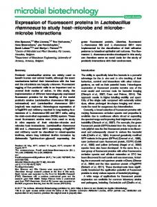
Figure 5. A heuristic model emphasizing the proposed roles of GLU I and movement protein in regulating plasmodesmatal traf®cking and virus spread.
Enzymatically active GLU I is required Infection with MGLU encoding an enzymatically inactive form of GLU I increased the accumulation of GLU I antigen in protoplasts but did not increase lesion size in plants. Although the His-tagged mutant GLU I expressed in E. coli was still recognized by antibody directed against enzymatically active GLU I, it was devoid of enzymatic activity. Therefore, an enzymatically active GLU I is required for the effect on lesion size. It is commonly believed that hydrolytic activity is required for the biological activity of GLU I (Fritig et al., 1998; Leubner-Metzger and Meins, 1999). Our experiments provide the ®rst direct evidence for this important assumption. Infection with antisense GLU I virus constructs reduces lesion size Within infection sites of TMV, the MP increases the SEL of plasmodesmata only at or near the leading front of infection (Oparka et al., 1997) in a region where host GLU I genes are induced early in the response to TMV infection (VoÈgeli-Lange et al., 1988; VoÈgeli-Lange et al., 1994). Relative to the LUC insert control, infection with the antisense construct ASGLU led to a reduced lesion size. This effect was modest, approximately 20%, possibly owing to the substantially lower replication rate of ASGLU relative to the other viral constructs.
Local GLU I expression could increase lesion size by promoting movement of virus from cell to cell, by indirect effects on the hypersensitive response, or by a combination of both mechanisms. To distinguish between these possibilities we compared the effect of SGLU, ASGLU and MGLU infection at 32°C, which blocks the hypersensitive response but permits virus spread. Relative to the MGLU control, local GLU I expression encoded by SGLU increased virus spread, whereas ASGLU, which downregulates host GLU I expression decreased virus spread. These ®ndings, together with earlier studies demonstrating that TMV movement is delayed in the GLU I-de®cient mutant (Iglesias and Meins, 2000), indicate that GLU I can promote cell-to-cell movement in the absence of a hypersensitive response. Although we cannot rule out additional GLU I effects on local-lesion size formation, this seems unlikely since lesion size in a local-lesion host as well as rates of virus spread in a susceptible host are strictly correlated with GLU I content in a graded series of antisense transformants (Beffa et al., 1996). A model linking GLU I induction, callose deposition and virus spread Early studies on the wound response in plants identi®ed plasmodesmata as a site for callose deposition (Olesen and Robards, 1990). Other studies have shown that treatment of onion tips with 2-deoxy-D-glucose, an inhibitor of callose deposition, results in a more open con®guration of plasmodesmata (Overall and Blackman, 1996). Moreover, recent studies indicate that conformational changes in plasmodesmatal ultrastructure triggered by osmotic forces or changes in hormonal balance are correlated with structural changes in the neck and collar regions of plasmodesmata and with the closure of the plasmodesmatal ori®ce. Callose deposition in the plasmodesmatal neck region thus may contribute to the regulation of plasmodesmatal permeability by down-regulating of cell-to-cell traf®cking via plasmodesmata (Botha and Cross, 2000). Studies of pea seedborne mosaic virusinfected pea cotyledons showed that a transient reduction in callose occurs in the vicinity of plasmodesmata at the advancing edge of the infected region (Roberts et al., 1998). This suggests that viruses can promote the local degradation of callose. Our observation that expression of ã Blackwell Science Ltd, The Plant Journal, (2001), 28, 361±369
b-1,3-glucanase enhances virus symptoms 367 enzymatically active GLU I in virus-infected cells increases the size of necrotic lesions supports a role of callose degrading enzyme as a virus susceptibility factor in plants. Oparka et al. (1997) demonstrated that within an expanding infection site, plasmodesmatal gating is limited to the leading edge of the infection front, although MP is present in plasmodesmata throughout the infection site. Therefore, in principle, one could speculate that the induction of host-encoded, callose-degrading enzymes an important factor in plasmodesmatal gating at the leading edge of infection. Based on these considerations, we propose a heuristic model, which emphasizes the role of GLU I and viral MP in cell-to-cell movement of viruses. According to the model shown in Figure 5, host defense functions impede virus traf®cking at least in part by increasing the rate of callose synthesis, resulting in increased callose deposition and a decreased plasmodesmatal SEL. Viral functions, on the other hand, promote callose degradation mediated by GLU I, and elaborate MPs that increase SEL and facilitate intercellular movement of infectious virus via plasmodesmata (Deom et al., 1992). A role for GLU I in plasmodesmatal traf®cking and viral pathogenesis requires that the enzyme is targeted to the cell wall. Because GLU I is usually localized in the vacuole, alternative targeting of the enzyme to the extracellular compartment is proposed. Although direct evidence is still lacking, secretion of GLU I has been reported for tobacco cells in suspension (Kunze et al., 1998) and for certain other vacuolar proteins in plants (Kjemtrup et al., 1995). Our model also does not rule out the possiblity that class II and III b-1,3-glucanases, which are secreted isoforms induced locally by virus infection (Kauffmann et al., 1987), also contribute to callose degradation. Experimental procedures Plasmid construction, in vitro transcription, and encapsidation Unless indicated otherwise, standard methods for nucleic acid analysis and molecular cloning were used (Sambrook et al., 1989). Foreign genes were cloned into the polylinker of the TMV expression vector 4 GD-PI (Karrer et al., 1998). A wild-type, chimeric tobacco class I b-1,3-glucanase gene (SGL6) with Sna BI sites upstream of the 5¢-untranslated region of chitinase gene CHN48 and downstream of the stop codon of the hybrid GLU36 and GLU31 cDNA (Holtorf et al., 1999) was generated by PCR ampli®cation using the primers Sn5 (5¢-TTCCTCTATATACGTAAGTTCATTT-3¢) and Sn3-Rev (5¢-CCATCCAAGGAATACGTATGTAA-3¢). To obtain the frameshift mutant FSGLU, a single C in codon 36 (CCA) was deleted by ampli®cation of SGL6 using the overlapping primers 5¢-ATGCTAGGCAACAACTTGCAAATC-3¢ and 5¢-GATTTGCAAGTTGTTGCCTAGCAT-3¢, which eliminated a unique Bst XI restriction site. Site-directed mutagenesis of SGL6 with the primer pairs 5¢-GTTGTGTCCCAGAGTGGCTGGCC-3¢ and 5¢-GGCCAGCCACCTGGGACACAAC-3¢, and 5¢-CCTGAACTGCAGAAACATTTTGG-3¢ and 5¢-CCAAAATGTTTCTGCAGTTCAGG-3¢ ã Blackwell Science Ltd, The Plant Journal, (2001), 28, 361±369
was used to obtain the mutant MGLU with E262Q and E318Q. The mutants were then ampli®ed with the Sn5 and Sn3-rev primers. After cutting with Sna BI, the mutants and wild-type SGL6 were cloned into Sal I-cut and Klenow-®lled 4 GD-Pl to give FSGLU, MGLU and SGLU and ASGLU, with wild-type SGL6 genes in sense and antisense orientation, respectively. To construct the luciferase expression vector LUC, the luc + gene was excised from vector pSP-luc + (Promega, Madison, WI, USA) with Eco RV and Bgl II, Klenow-treated and cloned into linearized 4 GD-Pl. Ngo AIV-linearized vectors were transcribed in vitro using a MEGAscript T7 kit (Ambion, Austin, TX, USA) and a 10 : 1 cap analogue to GTP ratio. Capped transcripts introduced by electroporation into BY-2 protoplasts as indicated below, and coated viruses were harvested by sonication 16 h later. After removal of cellular debris by low-speed centrifugation, viruses were suspended in 10 mM phosphate buffer, pH 7.0 and stored at ±80°C. Virion yield was determined from measurements of TMV coat protein obtained with an ELISA kit (Adgen Diagnostic Systems, Auchincruit, UK) according to the manufacturer's instructions.
Protoplast preparation and electroporation Suspensions of BY-2 cells were maintained, protoplasts were isolated and electroporation was performed as described by Watanabe et al. (1987). For each TMV construct, two aliquots of 6 3 105 protoplasts were inoculated with infectious RNA by electroporation and incubated in 10 ml of protoplast culture medium at 28°C. Samples of 1.0 ml were used for RNA-blot hybridization and immunoblot analyses.
Plant infection experiments Similar-sized leaves of hypersensitive Nicotiana tabacum cv. Xanthi-nc plants with 4±6 leaves were mechanically inoculated with coated virus using carborundum as described by Holt and Beachy (1991). Inoculated plants were raised in a growth chamber with 16 : 8 h light/dark regimen and a light intensity of 15 000 lux at 22°C and 70% relative humidity. Infected leaves were scanned 7 dpi and the surface area of individual local lesions measured by using PHOTOSHOP software (Adobe, San Jose, CA, USA). After scanning, discs 5-mm in diameter surrounding individual lesions were excised and used to isolate total RNA. To detect virus spread in susceptible plants, the inoculated plants were incubated at 32°C for 3 days, after which they were shifted to 22°C for 20 h and the area of necrotic regions measured.
RNA analyses Total RNA was isolated by the method of Chomczynski (1993) using TRIzol reagent (GibcoBRL Life Technologies, Rockville, MD, USA). RNA-blot hybridization was performed with 5 mg aliqouts of total RNA using the 1676 bp Nde I fragment of 4 GD-Pl representing the TMV replicase gene as the DNA probe. Signals were quanti®ed with a PhosphorImager (Molecular Dynamics, Sunnyvale, CA, USA) and expressed as ng TMV RNA mg-1 of total RNA using known amounts of TMV RNA as a standard. The identity and stability of virus constructs introduced into plants was con®rmed by RT±PCR, using `Ready To Go' beads (Amersham Pharmacia Biotech, Uppsala, Sweden). The primers used represented vector MP sequences (MP: 5¢-CGGTCAGTGCCGAACAAGAACTATAGAAAT-3¢), the vector 3¢-untranslated region (3¢UTR: 5¢-GCGGATGTATATGAACCATATACATTTGACCC-3¢) and
368
Gregor L. Bucher et al.
GLU I inserts in sense (SGLU: 5¢-CCGGAAGCAATGTGCTTCACATCTGAATT-3¢) and antisense (ASGLU: 5¢-GCCAATATAGGAACTTATTTGATGCAATGCTGG-3¢) orientation.
Protein analyses Protein extracts were prepared as described by Beffa et al. (1993), and protein content was measured by the method of Bradford (1976) using bovine g-globulin as a standard. Immunoblot analysis was performed using polyclonal anti-GLU I as described by Keefe et al. (1990) or using anti-His monoclonal antibodies (PentaHis Antibody, Qiagen, Basel, Switzerland). Signals were quanti®ed using a PhosphorImager. Luciferase activity was measured with a 1250 Luminometer (LKB, Wallac, Perkin Elmer Life Sciences, Wallac Oy., Turku, Finland) and is expressed in arbitrary units mg-1 protein. b-1,3-Glucanase activity was determined radiometrically (Keefe et al., 1990) and is expressed as pKat mg-1 protein using authentic tobacco GLU I as the standard.
Expression of GLU I inserts in E. coli Inserts of TMV vectors encoding GLU I were cloned into the bacterial expression vector pET-3a (Novagen, Madison, WI, USA) with the addition of a 3¢-His6 tag by ampli®cation with the primers 5¢-ATTGACATAGCATTAATGCAATCGATAGGT-3¢ and 5¢-AGTTTCGGATCCTCATCAATGATGATGATGATGATGCCCAAAGTTGATATTATATTTGGG-3¢. Proteins were expressed under standard conditions (37°C, induction with 1 mM IPTG) in E. coli strain BL21 (Novagen) transformants, and puri®ed in the native state using a Ni-NTA Agarose column as recommended by the manufacturer (Qiagen).
Acknowledgements We thank Erik Karrer (The Scripps Research Institute, La Jolla, USA) for providing the 4 GD-Pl expression vector, Sjoend van Eeden, Monique Thomas, Jacqueline Ferralli and Irina Petruska for technical assistance, and our colleagues Thomas Hohn and Andreas Gisel for critical comments.
References Beffa, R.S., Hofer, R.-M., Thomas, M. and Meins, F., Jr (1996) Decreased susceptibility to virus disease of b-1,3-glucanasede®cient plants generated by antisense transformation. Plant Cell, 8, 1001±1011. Beffa, R.S., Neuhaus, J.-M. and Meins, Jr., F. (1993) Physiological compensation in antisense transformants: Speci®c induction of an ersatz glucan endo-1,3-bglucosidase in plants infected with necrotizing viruses. Proc. Natl Acad. Sci. USA, 90, 8792±8796. Botha, C.E.J. and Cross, R.H.M. (2000) Toward reconciliation of structure with function in plasmodesmata: Who is the gatekeeper? Micron, 31, 713±721. Bradford, M.M. (1976) A rapid and sensitive method for the quantitation of microgram quantities of protein utilizing the principle of protein-dye binding. Analyt. Biochem. 72, 248±254. Chen, L., Garrett, T.P.J., Fincher, G.B. and Hoj, P.B. (1995) A tetrad of ionizable amino acids is important for catalysis in barley bglucanases. J. Biol. Chem. 270, 8093±8101. Chomczynski, P. (1993) A reagent for the single-step
simultaneous isolation of RNA, DNA and proteins from cell and tissue samples. Biotechniques, 15, 532±535. Dawson, W.O., Lewndowki, D.J., Hilf, M.E., Bubrick, P., Raffo, J., Shaw, J.J., Grantham, G.L. and Desjardins, P.R. (1989) A tobacco mosaic virus hybrid expresses and loses an added gene. Virol. 172, 285±292. Deom, C.M., Lapidot, M. and Beachy, R.N. (1992) Plant virus movement proteins. Cell, 69, 221±224. Fritig, B., Heitz, T. and Legrand, M. (1998) Antimicrobial proteins in induced plant defense. Curr. Opin. Immunol. 10, 16±22. Grosset, J., Meyer, Y., Chartier, Y., Kauffmann, S., Legrand, M. and Fritig, B. (1990) Tobacco mesophyll protoplasts synthesize 1,3-b-glucanase, chitinases, and `osmotins' during in vitro culture. Plant Physiol. 92, 520±527. Helms, K. and McIntyre, G.A. (1962) Studies on size of lesions of tobacco mosaic virus on pinto bean. Virol. 18, 535±545. Hilf, M.E. and Dawson, W.O. (1993) The tobavirus capsid protein functions as a host-speci®c determinant in long-distance movement. Virol. 193, 106±114. Holt, C.A. and Beachy, R.N. (1991) In vivo complementation of infectious transcripts from mutant tobacco mosaic virus cDNAs in transgenic plants. Virol. 181, 108±117. Holtorf, H., SchoÈb, H., Kunz, C., Waldvogel, R. and Meins, F., Jr (1999) Stochastic and nonstochastic post-transcriptional silencing of chitinase and b-1,3-glucanase genes involves increased RNA turnover. A possible role for ribosome independent RNA degradation. Plant Cell, 11, 471±484. Iglesias, V.A. and Meins, Jr., F. (2000) Movement of plant viruses is delayed in a b-1,3-glucanase-de®cient mutant showing a reduced plasmodesmatal size exclusion limit and enhanced callose deposition. Plant J. 21, 157±166. Karrer, E.E., Beachy, R.N. and Holt, C.A. (1998) Cloning of tobacco genes that elicit the hypersensitive response. Plant Mol. Biol. 36, 681±690. Kauffmann, S., Legrand, M., Geoffroy, P. and Fritig, B. (1987) Biological function of `pathogenesis-related' proteins: four PR proteins of tobacco have 1,3-b-glucanase activity. EMBO J. 6, 3209±3212. Keefe, D., Hinz, U. and Meins, Jr., F. (1990) The effect of ethylene on the cell-type-speci®c and intracellular localization of b-1,3-glucanase and chitinase in tobacco leaves. Planta, 182, 43±51. Kjemtrup, S., Borkhsenious, O., Raikhel, N.V. and Chrispeels, M.J. (1995) Targeting and release of phytohemagglutinin from the roots of bean seedlings. Plant Physiol. 109, 603±610. Kombrink, E. and Somssich, I.E. (1997) Pathogenesis-related proteins and plant defense. In: The Mycota V, Part A (Carroll, G. and Tudzynski, P. eds). Berlin-Heidelberg, Germany: Springer Verlag, pp. 107±128. Kunze, I., Kunze, G., BroÈker, M., Manteuffel, R., Meins, F., Jr and MuÈntz, K. (1998) Evidence for secretion of vacuolar amannosidase, class I chitinase, and class I b-1,3-glucanase in suspension cultures of tobacco cells. Planta, 205, 92±99. Leubner-Metzger, G. and Meins, Jr., F. (1999) Function and regulation of plant b-1,3-glucanases (PR-2). In: Pathogenesis Related Proteins in Plants (Datta, S.K. and Muthukrishnan, S. eds). Boca Raton, FL, USA: CRC Press, pp. 49±76. Matthews, R.E.F. (1981). Plant Virology. New York, USA:, Academic Press. Olesen, P. and Robards, A.W. (1990) The neck region of plasmodesmata. In: Parallels in Cell to Cell Junctions in Plants and Animals (Robards, A.W., Lucas, W.J., Pitts, J.D., Jongsma, H.J. and Spray, D.C. eds). Berlin, Germany: Springer Verlag, pp. 145±170. ã Blackwell Science Ltd, The Plant Journal, (2001), 28, 361±369
b-1,3-glucanase enhances virus symptoms 369 Oparka, K.J., Prior, D.A.M., Santa Cruz, S., Padgett, H. and Beachy, R.N. (1997) Gating of epidermal plasmodesmata is restricted to the leading edge of expanding infection sites of tobacco mosaic virus (TMV). Plant J. 12, 781±789. Overall, R.L. and Blackman, L.M. (1996) A model of the macromolecular structure of plasmodesmata. Trends Plant Sci. 1, 307±311. Robards, A. and Lucas, W. (1990) Plasmodesmata. Annu. Rev. Plant Physiol. Plant Mol. Biol. 41, 369±419. Roberts, I.M., Wang, D., Findlay, K. and Maule, A.J. (1998) Ultrastructural and temporal observations of the potyvirus cylindrical inclusions (CIs) show that the CI protein acts transiently in aiding virus movement. Virol. 245, 173±181. Ryals, J.A., Neuenschwander, U.H., Willits, M.G., Molina, A., Steiner, H.-Y. and Hunt, M.D. (1996) Systemic acquired resistance. Plant Cell, 8, 1809±1819. Sambrook, J., Fritsch, E.F. and Maniatis, T. (1989). Molecular
ã Blackwell Science Ltd, The Plant Journal, (2001), 28, 361±369
Cloning. A Laboratory Manual. New York, USA: Cold Spring Harbor Laboratory Press. Stone, B.A. and Clarke, A.E. (1992). Chemistry and Biology of (1®3) -b-Glucans. Victoria, Australia: La Trobe University Press. VoÈgeli-Lange, R., FruÈndt, C., Hart, C.M., Nagy, F. and Meins, Jr., F. (1994) Developmental, hormonal, and pathogenesis-related regulation of the tobacco class I b-1,3-glucanase B promoter. Plant Mol. Biol. 25, 299±311. VoÈgeli-Lange, R., Hansen-Gehri, A., Boller, T. and Meins, F., Jr (1988) Induction of the defense-related glucanohydrolases, b1,3-glucanase and chitinase, by tobacco mosaic virus infection of tobacco leaves. Plant Sci. 54, 171±176. Watanabe, Y., Meshi, T. and Okada, Y. (1987) Infection of tobacco protoplasts with in-vitro transcribed tobacco mosaic virus RNA using an improved electroporation method. FEBS Lett. 219, 65±69.