DEVELOPMENTAL DYNAMICS 236:2918 –2924, 2007
PATTERNS & PHENOTYPES
Expression of Multiple Class Three Semaphorins in the Retina and Along the Path of Zebrafish Retinal Axons Davon C. Callander,1 Ryan E. Lamont,2 Sarah J. Childs,2 and Sarah McFarlane1*
Retinal ganglion cells (RGCs) extend axons that exit the eye, cross the midline at the optic chiasm, and synapse on target cells in the optic tectum. Class three semaphorins (Sema3s) are a family of molecules known to direct axon growth. We undertook an expression screen to identify sema3s expressed in the retina and/or brain close to in-growing RGC axons, which might therefore influence retinal-tectal pathfinding. We find that sema3Aa, 3Fa, 3Ga, and 3Gb are expressed in the retina, although only sema3Fa is present during the time window when the axons extend. Also, we show that sema3Aa and sema3E are present near or at the optic chiasm. Furthermore, sema3C, 3Fa, 3Ga, and 3Gb are expressed in regions of the diencephalon near the path taken by RGC axons. Finally, the optic tectum expresses sema3Aa, 3Fa, 3Fb, and 3Gb. Thus, sema3s are spatiotemporally placed to influence RGC axon growth. Developmental Dynamics 236:2918 –2924, 2007. © 2007 Wiley-Liss, Inc. Key words: Sema3; visual system; diencephalon; optic tectum; axon guidance Accepted 1 August 2007
INTRODUCTION Semaphorins (Semas) are a large family of molecules characterized by a highly conserved extracellular SEMA domain of approximately 500 amino acids (Semaphorin Nomenclature Committee, 1999). The Semas are most well known for their role in axon guidance. There are eight different classes of Semas; class 1 and 2 are encoded by invertebrates; class 3–7 are present in vertebrates; and the eighth class, V, is found in viruses (Yazdani and Terman, 2006). Of the vertebrate Semas, classes 4 –7 are membrane bound, through either glycophosphatidyl inositol linkage or
transmembrane regions, while members of class 3 are secreted. Nine plexin molecules (A1–A4, B1–B3, C1, D1), which also contain a conserved SEMA domain, form the functional receptors of Semas (Tamagnone et al., 1999). The secreted class 3 Semas (Sema3A-3H) require a holoreceptor complex consisting of a neuropilin (Nrp1 or Nrp2) and a plexin for activity, with the exception of Sema3E, which only requires a plexin (Gu et al., 2005). The extracellular portion of neuropilin serves as the Sema binding subunit, while the plexin subunit conveys the signal to the cytoplasm (Nakamura et al., 1998).
1
Class 3 Semas have been investigated as repulsive axon guidance molecules (Kolodkin et al., 1992; Luo et al., 1993; Kruger et al., 2005). In this regard, Sema3A has been most well studied. For example, in Sema3A null mice sensory axons grow aberrantly into regions of the spinal cord that normally express Sema3A and neuronal processes of pyramidal neurons are oriented randomly rather than towards the brain surface (Behar et al., 1996). There are also severe defects in cranial nerve axon fasciculation in these mice (Taniguchi et al., 1997; Catalano et al., 1998). Furthermore, perturbation of Sema3Aa signalling in
Hotchkiss Brain Institute, University of Calgary, Calgary, Canada Department of Biochemistry and Molecular Biology, University of Calgary, Calgary, Canada Grant sponsor: Canadian Institutes of Health Research (CIHR); Grant numbers: MOP-14138 and MOP-53230; Grant sponsor: Heart and Stroke Foundation of Canada. *Correspondence to: Dr. Sarah McFarlane, University of Calgary, Hotchkiss Brain Institute, HSC Rm 2207, 3330 Hospital Dr., NW, Calgary, AB, T2N 4N1, Canada. E-mail:
[email protected] 2
DOI 10.1002/dvdy.21315 Published online 14 September 2007 in Wiley InterScience (www.interscience.wiley.com).
© 2007 Wiley-Liss, Inc.
SEMA3S IN THE DEVELOPING VISUAL SYSTEM 2919
zebrafish causes the axons of ventral spinal motoneurons, which normally grow along the medial portion of the somite, to extend aberrantly into anterior or posterior regions (Shoji et al., 1998; Sato-Maeda et al., 2006). It is now known that Sema3s serve a wide range of functions in addition to axon guidance, including angiogenesis and organogenesis (Ito et al., 2000; Yazdani and Terman, 2006). In the developing zebrafish visual system, neuropilin receptors are expressed in retinal ganglion cells (RGCs) at the time their axons project from the eye to the brain (Liu et al., 2004), which suggests that class 3 Semas may be important for the development of the optic projection. Indeed, in zebrafish, Sema3D is required at the midline for RGC axons to cross correctly at the optic chiasm, as well as at the optic tectum for appropriate topographic mapping (Liu et al., 2004; Sakai and Halloran, 2006). Furthermore, Sema3A repels embryonic Xenopus laevis RGC growth cones in vitro (Campbell et al., 2001). There is little known about the expression of the class 3 Semas in the embryonic zebrafish visual system. Here, we use in situ hybridization to characterize the expression profiles of the majority of the class 3 sema gene family members during the entire period of zebrafish RGC axon outgrowth and target recognition. Our study provides the first comprehensive examination of sema3 expression in the developing visual system and will be key for future studies that address the involvement of Sema3 family members in the formation of connections between visual neurons.
RESULTS There are eight members of the class 3 Sema family, and in zebrafish, due to a whole genome duplication (Amores et al., 1998), many of these genes have two related family members. We examined the expression patterns of sema3Aa, 3C, 3E, 3Fa, 3Fb, 3Ga and 3Gb mRNA from early proliferative to later differentiated stages of the embryonic zebrafish visual system. Visual system expression of sema3D and 3H has been described previously (Stevens and Halloran, 2005, 2006; Sakai and Halloran, 2006), and
sema3B has not yet been identified in zebrafish. RGCs first differentiate in the ventronasal portion of the retina at approximately 28 hr post fertilization (hpf) (Laessing and Stuermer, 1996). Soon after differentiation, RGCs distributed throughout the cup-shaped retina become polarized and project an axon toward the vitreal surface of the retina. The axons travel in the optic fiber layer from their point of origin towards the optic disc. Once there, they make a 90° turn, dive through the outer layers of the retina, and exit the eye at the optic nerve head (Fig. 1A). The axons of the first RGCs exit the eye at 32 hpf and cross the optic chiasm at 36 –38 hpf (Fig. 1A,B; Burrill and Easter, 1994, Stuermer, 1988). After they cross the midline, the axons turn dorsally, travel along the pial surface of the diencephalon, and at 48 hpf begin to reach their main midbrain target, the optic tectum (Fig. 1A,B; Stuermer, 1988). By 68 hpf, a startle response to changes in light and dark has developed and shortly after, at 73 hpf, embryos can focus on and follow moving objects (Easter and Nicola, 1996).
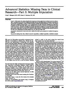
sema3Aa Expression In the optic pathway, sema3Aa is expressed at 30 hpf in bilateral nuclei in the dorsal posterior tuberculum and in the pretectal/dorsal thalamus area of the diencephalon (Fig. 1C,D) that lies just anterior and ventral to the optic tectum. These domains are close to the path taken by RGC axons as they travel towards the optic tectum (Fig. 1B). The dorsal posterior tuberculum expression persists until at least 72 hpf (Fig. 1G). In situ label is also seen in the optic tectum from 30 – 60 hpf (Fig. 1C,E, data not shown). sema3Aa is not expressed in the retina until 72 hpf, when it is turned on by cells in the outermost part of the inner nuclear layer (INL; Fig. 1G,H). Additionally, expression is evident in a number of other regions of the head, including the telencephalon (Fig. 1C, asterisk), ventral mesenchyme, and hindbrain (Fig. 1C,E).
sema3C Expression The sema3C in situ probe labels a pair of bilateral nuclei in the dorsal poste-
rior tuberculum from 30 –50 hpf (Fig. 2A,B), but this expression is lost by 60 hpf. We found no evidence for sema3C expression within the retina at these time points. In the head region, the early pectoral fin buds, pharyngeal arches, and the jaw express sema3C (data not shown).
sema3E Expression sema3E is found in the visual pathway at the optic chiasm (Fig. 1A,B) between 30 and 72 hpf (Fig. 3B,C,D,F), with the first RGC axons crossing the midline at this decision point between 34 and 36 hpf (Burrill and Easter, 1994). At 30 hpf, in situ label is present in the posterior tectum (Fig. 3A). Similar to sema3C, no retinal expression is evident. sema3E is also seen in the hindbrain, jaw, ventral mesenchyme, and in the hypothalamus (Fig. 3B,F).
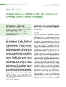
sema3Fa Expression Low levels of sema3Fa are seen in the ventral retina at 30 –50 hpf (Fig. 4B,D arrowheads). At 30 hpf, there are several different domains of sema3Fa expression in and around the optic pathway (Fig. 1A,B), including strong expression in the ventral posterior tuberculum and optic tectum, with weaker label in the dorsal posterior tuberculum (Fig. 4B). As development proceeds, sema3Fa becomes restricted to the ventral posterior tuberculum and to a laterally located pair of bilateral nuclei of the dorsal posterior tuberculum (Fig. 4D,F). This expression continues until 60 hpf. By 72 hpf, only diffuse label is observed in the anterior brain (Fig. 4H). Label is also seen in neural tissues distant from ingrowing RGC axons. The cerebellum begins to show sema3Fa at 30 hpf (Fig. 4A), with expression gradually weakening by 72 hpf (Fig. 4G). At 60 –72 hpf, sema3Fa is evident in the jaw, pharyngeal arches, and pectoral fin buds (Fig. 4E,G,H).
sema3Fb Expression From 30 hpf onwards, punctate sema3Fb expression is found in the pretectal region of the diencephalon (Fig. 5B,D). Additionally, there is expression in the optic tectum ventral to
2920 CALLANDER ET AL.
Fig. 2. Expression of sema3C. A, B: A dorsal view of a whole-mount embryo is shown in A, and a transverse section through the eyes and brain is shown in B. sema3C transcripts are seen in the dorsal posterior tuberculum at 40 hpf. PTd, dorsal posterior tuberculum. Scale bar in B ⫽ 50 m. Fig. 1. Expression of sema3Aa. A: Schematic diagram of a transverse section of the optic pathway. A RGC resides in the RGC layer (1) and sends out an axon that exits the retina at the optic nerve head (2), crosses the midline at the optic chiasm (3), extends along the pial surface of the diencephalon (4), and enters the main target, the optic tectum (5). B: Transverse section of a 72-hpf brain with an HRP-labelled RGC optic projection (brown). The eyes and skin have been removed. The optic chiasm, pathway, and tectum are labelled 3, 4, and 5, respectively. C and E are lateral views of whole-mount embryos and D and F are stage-matched transverse sections. C, D: sema3Aa transcripts appear in the pretectum/dorsal thalamus and the posterior tectum at 30 hpf. Bilateral nuclei in the dorsal posterior tuberculum also show expression. From 30 –72 hpf, sema3Aa transcripts are seen in the telencephalon (asterisk), the hindbrain, and the ventral mesenchymal tissue. E, F: At 50 hpf, signal is maintained in the dorsal posterior tuberculum (F) and the posterior tectum (E). G, H: At 72 hpf, in transverse sections the outer region of the inner nuclear layer begins to express sema3Aa. The boxed area in G is shown at a higher magnification in H. hb, hindbrain; INL, inner nuclear layer; ONL, outer nuclear layer; ptec, posterior tectum; pt/DT, pretectum/dorsal thalamus; PTd, dorsal posterior tuberculum; RGC, retinal ganglion cell layer; vm, ventral mesenchyme. Scale bars ⫽ 50m (B,D,F,G), 10 m (H).
Fig. 4. Expression of sema3Fa. A,C,E, and G are lateral views of whole-mount embryos and B,D,F, and H are stage-matched transverse sections. A,B: At 30 hpf, sema3Fa transcript is seen in the ventral retina (arrowhead), in the optic tectum, and in the ventral and dorsal regions of the posterior tuberculum. There is also label in the cerebellum. C,D: At 50 hpf, sema3Fa is expressed in the ventral posterior tuberculum and in bilateral nuclei in the dorsal posterior tuberculum. Expression in the ventral retina (arrowhead) continues. E,F: At 60 hpf, expression in the dorsal posterior tuberculum and cerebellum is maintained. G,H: At 72 hpf, there is diffuse label in the anterior brain (H). There is also expression in the jaw (E,G,H), pharyngeal arches (E,G). and the pectoral fin buds (E). cb, cerebellum; FB, fin bud; j, jaw; PA, pharyngeal aches; PTd, dorsal posterior tuberculum; PTv, ventral posterior tuberculum; tec, tectum. Scale bars in B,D,F, H ⫽ 50 m.
the axons at 60 and 72 hpf (compare Fig. 1A,B and 5D; data not shown). There is no obvious retinal expression of sema3Fb (Fig. 5). Several other structures express sema3Fb. From 30 –50 hpf, there are weak and strong signals located in rhombomeres (r) 2 and r4, respectively (Fig. 5A, data not
shown). Furthermore, sema3Fb is found in the telencephalon (Fig. 5A,C, arrowhead), jaw (Fig. 5D), and ventral mesenchymal tissue (Fig. 5A,C).
Fig. 3. Expression of sema3E. Lateral (A,E) and ventral views (C) of whole-mount embryos, and stage-matched transverse sections (B,D,F). A: sema3E label is seen in the posterior tectum at 30 hpf. B,D,F: sema3E transcripts appear in the optic chiasm at 30 hpf and persist until 72 hpf. sema3E is seen in the ventral mesenchymal tissue (B), the hypothalamus (B), and the jaw (F). tec, tectum; hb, hindbrain; H, hypothalamus; j, jaw; oc, optic chiasm; ptec, posterior tectum; vm, ventral mesenchyme. Scale bars in B,D,F ⫽ 50 m.
their axons arborize in the optic tectum. In the brain, sema3Ga is present in discrete lateral regions of the dorsal posterior tuberculum (Fig. 6B,D,F). In other regions, the dorsal hindbrain strongly expresses sema3Ga at 30 and 40 hpf and mRNA becomes restricted to two ventrolateral regions at 72 hpf (Fig. 6A,C,E). Additionally, the pharyngeal arches show expression at 72 hpf.
sema3Ga Expression
sema3Gb Expression
sema3Ga is first found in RGCs in the neural retina at 72 hpf (Fig. 6F), when
sema3Gb is found in the peripheral retina at 40 hpf (Fig. 7B, asterisk) and
SEMA3S IN THE DEVELOPING VISUAL SYSTEM 2921
Fig. 5. Expression of sema3Fb. A,C: Lateral views of whole-mount embryos. B,D: Stagematched transverse sections. B,D: At 40 – 60 hpf, the pretectal region of the diencephalon expresses sema3Fb transcripts. C,D: At 60 hpf, the region of the tectum just ventral to where the RGC axons terminate expresses sema3Fb. The jaw (D), ventral mesenchyme (A,C), telencephalon (arrowhead; A,C), and rhombomeres 2 and 4 (A) also show expression. j, jaw; pt, pretectum; r, rhombomere; tec, tectum; vm, ventral mesenchyme. Scale bars in B,D ⫽ 50 m.
also has restricted expression in several areas of the diencephalon (Fig. 7B) including in the dorsolateral region of the dorsal posterior tuberculum, along the dorsoventral axis of the posterior tuberculum and in the pretectal area. The posterior tectum expresses sema3Gb at 40 hpf and the expression becomes punctate throughout the tectum at 50 hpf and onwards (Fig. 7). In other regions of the head, sema3Gb is restricted to the rhombomere boundaries of the hindbrain at 40 hpf (Fig. 7A, arrowheads). While sema3Gb is still present in the hindbrain at 50 –72 hpf (Fig. 7C, data not shown), the boundary expression disappears as the rhombomeres become less well defined.
DISCUSSION Sema3s have complex expression patterns in the embryonic zebrafish brain and visual system. Two genes, sema3Fa and 3Gb, show restricted expression in the eye as RGC axons grow through the retina, whereas sema3Aa and 3Ga are evident in the eye only after RGC axons have reached the optic tectum. A number of sema3s are found in the diencephalon and optic tectum, before, during, and after RGC axonal migration. Furthermore, their receptors, the neuropilins (NRP), are
Fig. 6. Expression of sema3Ga. A,C,E: Lateral views of whole-mount embryos. B,D,F: Stagematched transverse sections. A,B: At 40 hpf, sema3Ga expression is seen in the dorsal posterior tuberculum as well as in the dorsal hindbrain. Blood vessels bordering the lens are also stained at 40 (B) and 60 (D) hpf. C,D: The dorsal posterior tuberculum expression persists at 60 hpf. E,F: Expression is seen in the RGC layer at 72 hpf (E, F), whereas signal in the dorsal hindbrain has switched to two ventrolateral regions (compare C and E). sema3Ga transcripts are present in the dorsal posterior tuberculum from 40 – 60 hpf (B,D,F) and in the pharyngeal arches at 72 hpf (E). hb, hindbrain; PA, pharyngeal arches; PTd, dorsal posterior tuberculum; RGC, retinal ganglion cell layer. Scale bars in B,D,F ⫽ 50 m.
expressed by differentiating RGCs (Nrp1a, 1b, 2a) and cells of the inner nuclear layer (INL; Nrp2b) (Liu et al., 2004). As such, RGCs and INL cells likely respond to class 3 Semas. Here, we consider the expression of sema3s in light of their well-recognized function as repulsive guidance molecules (Pasterkamp and Kolodkin, 2003), conscious of the fact that they likely have other roles as well.
Retinal Expression of sema3s There are multiple factors known to be involved in intraretinal axon pathfinding, including netrin-1, slit, and sonic hedgehog (Deiner et al., 1997; Kolpak et al., 2005; Thompson et al., 2006). These factors comprise an expanding group of molecular cues known to be responsible for the guidance of RGC axons along the optic fiber layer, toward the optic disc and
Fig. 7. Expression of sema3Gb. A,C: Lateral views of whole-mount embryos. B,D: Stagematched transverse sections. A: At 40 hpf, in situ signal is evident in the posterior tectum and the rhombomeric boundaries (arrowheads). B: At 40 hpf, sema3Gb is expressed in the peripheral retina (asterisk). Several areas of the diencephalon express sema3Gb: dorsolateral region of the dorsal posterior tuberculum, the dorsoventral axis of the posterior tuberculum, and the pretectal area. C,D: At 50 hpf, a subset of cells dispersed throughout the tectum express sema3Gb, which is evident in the wholemount embryo (dashed line outlines the tectum) and in the section. pt, pretectum; ptec, posterior tectum; tec, tectum; PT, posterior tuberculum; dlPTd, dorsolateral region of the dorsal posterior tuberculum; Scale bars in B,D ⫽ 50 m.
out of the retina. Whether Sema3s play a role in the retina to guide RGC axons is unknown. sema3Fa, expressed in the ventral retina, is the only Sema3 that is found early enough and in the appropriate location to potentially influence zebrafish intraretinal RGC axon guidance. It may help direct RGC axons away from the ventral retina. Two other Sema3s, sema3Aa and 3Ga, are found in distinct cell layers within the retina well after RGC axons have left the eye, with expression in the INL and the RGCs, respectively. In addition to its function as a repellent, Sema3A attracts the dendrites of pyramidal neurons towards the marginal zone during mouse cortical development (Polleux et al., 2000). Thus, although it is expressed too late to guide RGC axons, Sema3Aa could still attract or repel the neural processes of cells in the inner and outer nuclear layers towards the synaptic outer plexiform layer. Alternatively, Sema3Aa may control the complexity of the dendritic arbors of RGCs as it does with murine cortical neurons (Fenstermaker et al., 2004). Similarly, Sema3Ga in the RGCs could guide the growth of either RGC den-
2922 CALLANDER ET AL.
drites or INL cell neural processes towards the synaptic inner plexiform layer between the RGC layer and the INL. The expression of sema3s in the zebrafish retina appears to differ from that reported in other species. For instance, while sema3Aa is expressed in a select population of cells in the INL of the zebrafish retina well after the optic projection has formed, in both the chick and mouse retinas Sema3A is found throughout the embryonic eye (Taniguchi et al., 1997; Chilton and Guthrie, 2003). Moreover, we found no sema3E label in the zebrafish retina, but sema3E is expressed in the outer layers of the embryonic mouse retina (Steinbach et al., 2002). Finally, whereas sema3Fa is present in the ventral zebrafish retina, in rat it is expressed in the outer retinal layers (Giger et al., 1998).
Sema3s in, and Adjacent to, the Optic Pathway Sema3s are found alongside the optic projection from its entry into the ventral diencephalon to its innervation of the optic tectum. First, sema3s are expressed at the zebrafish optic chiasm, where RGC axons cross the midline and extend into the contralateral brain (Burrill and Easter, 1994). It is known that Sema3D is required at this decision point because in Sema3D morphants approximately half the RGC axons project in an aberrant ipsilateral fashion (Sakai and Halloran, 2006). The partial penetrance of the Sema3D phenotype suggests that there may be additional guidance molecules important at this decision point. Indeed, we found several sema3s at or near the optic chiasm when RGC axons cross the midline. For example, sema3E, which is repellent to chick RGC growth cones (Steinbach et al., 2002), is expressed at the optic chiasm. Furthermore, sema3Fa and sema3Gb are expressed dorsal to the optic chiasm, and are placed such that they could prevent the axons from straying dorsally at the midline. Thus, multiple Sema3s could act together to ensure proper guidance of RGC axons, as they do for zebrafish cranial neural crest cell migration (Yu and Moens, 2005). Little is known about the factors
that regulate the guidance of RGC axons within the diencephalon after they have crossed the midline. One of the few molecules to be implicated is Robo2 (Karlstrom et al., 1996; Fricke et al., 2001). The astray/robo2 mutant zebrafish exhibit some RGC axon pathfinding errors in the posterior tuberculum en route to the optic tectum. We show that several sema3s are expressed within the diencephalon in regions adjacent to the optic projection, where as repellents they could serve to restrict RGC axons to the optic tract. Sema3A is found in the telencephalon of several species including zebrafish, Xenopus, mouse, and chick (Campbell et al., 2001; Melendez-Herrera and Varela-Echavarria, 2006). In Xenopus, the telencephalic expression is just anterior to the optic projection and Sema3A is repellent to RGC growth cones in vitro (Campbell et al., 2001). Yet, the telencephalic sema3Aa expression domain appears to be too anterior to the optic tract to suggest a function for Sema3Aa in zebrafish RGC axon guidance. Other sema3s, however, are expressed close to the optic projection. In the dorsal posterior tuberculum, there are discrete and unique domains of expression of several sema3s (sema3Aa, 3C, 3Fa, 3Ga, and 3Gb), whose temporal patterns also differ somewhat. For instance, in this region sema3C is only found from 30 –50 hpf, while sema3Aa is expressed throughout the 30 –72-hpf period. Presumably, these genes play important roles in the differentiation of the dorsal part of the posterior tuberculum, and, moreover, based on proximity to the optic pathway, could restrict the RGC axons to the pial neuropil of the diencephalon. Future studies to address the importance of Sema3 family members in RGC axon guidance in the diencephalon will have to take into account the multiple players. Next, the pretectal/dorsal thalamus region expresses sema3Aa, 3Fb, and 3Gb, which could function to prevent the RGC axons from straying away from the optic tract. Finally, three sema3s are expressed in distinct expression domains in the optic tectum at 50 hpf and beyond, when RGC axons are innervating this midbrain target. sema3Gb is evident in cells dispersed throughout the anterior
tectum and sema3Fb is present just below the RGC axons in this same region. As such, they could control the termination domain of RGC axons. In contrast, sema3Aa and 3E are found in the posterior optic tectum, where they may prevent axons from extending straight through the target into the hindbrain, similar to the role that has been described for ephrinA5 (Frisen et al., 1998). In summary, sema3s have highly localized expression in the developing zebrafish brain and retina. Our expression data will help guide future functional analyses of the role of Sema3s in zebrafish visual system development.
EXPERIMENTAL PROCEDURES Animals Zebrafish embryos from the TL strain were raised at 28.5°C according to Westerfield (1995) and staged by hours post fertilization (Kimmel et al., 1995; Westerfield, 1995). Embryos were raised in 0.1 ⫻ Danieau’s solution. To block pigment formation in embryos, 0.003% phenylthiourea (PTU) was added to the media at 24 hpf.
Visualization of the Optic Projection Briefly, 72-hpf embryos were anaesthetized in Modified Barth’s Saline (8.8 mM NaCl, 0.1 KCl, 0.7 mM MgSO4, 5 mM HEPES (pH 7.8), 25 mM NaHCO3) supplemented with 0.4 mg/ml tricaine (ethyl 3-aminobenzoic ethyl ester, methanesulfonate salt; Sigma), 50 mg/ml gentamicin sulphate (Sigma), and 10 mg/ml Phenol Red (Sigma). Embryos were pinned in a Sylgard dish (Dow Corning) and the lens of one eye was removed with an electrolytically sharpened tungsten needle. Two boluses of horseradish peroxidase (HRP) dissolved in 1% lysolecithin (Sigma) were placed in the lens cavity. The embryos were left undisturbed for 20 min to allow the RGC axons to anterogradely transport the HRP, and then fixed in 4% paraformaldehyde (PFA) for 2 hr. After rinsing in Phosphate Buffered Saline (PBS), the eyes and the skin covering
SEMA3S IN THE DEVELOPING VISUAL SYSTEM 2923
the head were removed and the embryos were permeabilized in 0.5% Triton X-100. The HRP was then visualized by performing a diaminobenzidine (DAB; Sigma) reaction. The fish were then post fixed in 1% glutaraldehyde for 1 hr and washed with PBS.
Probe Synthesis and WholeMount In Situ Hybridization Digoxygenin (DIG) labelled antisense RNA probes were generated for class 3 semas as follows. For sema3Aa, AL919018 was digested with Xbal (Invitrogen) and transcribed with T7 polymerase (Promega), and AL923380 was digested with XhoI (Invitrogen) and transcribed with T3 polymerase (Promega) to generate a probe for sema3C. A probe template for sema3E was generated by nested RACE PCR amplification from a 50-hpf RACE library (Clonetech) using the primers 5⬘ – GGA GCT GAA CAG GAC GTG GGT GTT CCA GG-3⬘ (forward) and 5⬘ – CCA TCC TAA TAC GAC TCA CTA TAG GGC – 3⬘ followed by 5⬘ – GGA GAG GGC CGT CTG CAG CAA TAC ACC ATG -3⬘ and 5⬘- ACT CAC TAT AGG GCT CGA GCG GC – 3⬘ and the Advantage 2 PCR system (Clonetech). PCR products were cloned into pCRII (Invitrogen) and a 2.7-kb fragment was identified by sequencing to contain 2.1 kb of sema3E coding sequence and 0.6 kb of 3⬘ untranslated region. This sequence has been deposited in GenBank under accession no. EF656360. To make DIG-labelled RNA probes against sema3E, the above plasmid was digested with XbaI (Invitrogen) and transcribed with SP6 polymerase (Promega). Additional probes, which include sema3Fa, 3Fb, 3Ga, and 3Gb, have been described previously (Yu et al., 2004). Then, 30 –72-hpf embryos were fixed and processed as whole-mounts by in situ hybridization with digoxygenin (DIG) labelled antisense RNA probes. Whole-mount embryos were fixed in 4% paraformaldehyde in PBS. RNA in situ hybridization was carried out as described previously (Thisse et al., 1993) with slight modifications: the rinse in glycine was omitted and the embryos were incubated at 65°C. Transferring the embryos into 100% methanol for 5 min stopped the co-
louration reaction. This was followed by several washes in PBS and 0.1% Tween-20 (PBT), refixation with 4% PFA, and storage in PBT at 4°C. Multiple independent in situ hybridization procedures were carried out to ensure specificity and reproducibility. Whole-mount embryos were photographed in 3% methylcellulose using a Zeiss Axiocam HRc. To obtain sections, embryos were dehydrated through a series of ethanol/water washes to 100% ethanol, embryos were then infiltrated with catalyzed JB4 medium (Polysciences), and 5-m sections were cut on a Leica microtome with glass knives. Digital images of sections were taken with an Optronics camera. All images were processed for brightness and contrast with Adobe Photoshop CS.
ACKNOWLEDGMENTS The authors thank Dr. Cecelia Moens for supplying probes for sema3Fa, Fb, Ga, and Gb. D.C.C. is supported by the Canadian Institutes of Health Research (CIHR) Training Grant in Genetics, Child Health and Development and R.E.L. is supported by the Heart and Stroke Foundation of Canada. S.C. is an Alberta Heritage Foundation for Medical Research (AHFMR) scholar and a Tier II Canada Research Chair (CRC). S.M. is an AHFMR senior scholar and a Tier II CRC Chair. This work was supported by operating grants from CIHR to S.M. and S.J.C.
REFERENCES Amores A, Force A, Yan YL, Joly L, Amemiya C, Fritz A, Ho RK, Langeland J, Prince V, Wang YL, Westerfield M, Ekker M, Postlethwait JH. 1998. Zebrafish hox clusters and vertebrate genome evolution. Science 282:1711–1714. Behar O, Golden JA, Mashimo H, Schoen FJ, Fishman MC. 1996. Semaphorin III is needed for normal patterning and growth of nerves, bones and heart. Nature 383:525–528. Burrill JD, Easter SS, Jr. 1994. Development of the retinofugal projections in the embryonic and larval zebrafish (Brachydanio rerio). J Comp Neurol 346:583– 600. Campbell DS, Regan AG, Lopez JS, Tannahill D, Harris WA, Holt CE. 2001. Semaphorin 3A elicits stage-dependent collapse, turning, and branching in Xenopus retinal growth cones. J Neurosci 21:8538 –8547. Catalano SM, Messersmith EK, Goodman CS, Shatz CJ, Chedotal A. 1998. Many
major CNS axon projections develop normally in the absence of semaphorin III. Mol Cell Neurosci 11:173–182. Chilton JK, Guthrie S. 2003. Cranial expression of class 3 secreted semaphorins and their neuropilin receptors. Dev Dyn 228:726 –733. Deiner MS, Kennedy TE, Fazeli A, Serafini T, Tessier-Lavigne M, Sretavan DW. 1997. Netrin-1 and DCC mediate axon guidance locally at the optic disc: loss of function leads to optic nerve hypoplasia. Neuron 19:575–589. Easter SS, Jr., Nicola GN. 1996. The development of vision in the zebrafish (Danio rerio). Dev Biol 180:646 –663. Fenstermaker V, Chen Y, Ghosh A, Yuste R. 2004. Regulation of dendritic length and branching by semaphorin 3A. J Neurobiol 58:403–412. Fricke C, Lee JS, Geiger-Rudolph S, Bonhoeffer F, Chien CB. 2001. astray, a zebrafish roundabout homolog required for retinal axon guidance. Science 292:507– 510. Frisen J, Yates PA, McLaughlin T, Friedman GC, O’Leary DD, Barbacid M. 1998. Ephrin-A5 (AL-1/RAGS) is essential for proper retinal axon guidance and topographic mapping in the mammalian visual system. Neuron 20:235–243. Giger RJ, Urquhart ER, Gillespie SK, Levengood DV, Ginty DD, Kolodkin AL. 1998. Neuropilin-2 is a receptor for semaphorin IV: insight into the structural basis of receptor function and specificity. Neuron 21:1079 –1092. Gu C, Yoshida Y, Livet J, Reimert DV, Mann F, Merte J, Henderson CE, Jessell TM, Kolodkin AL, Ginty DD. 2005. Semaphorin 3E and plexin-D1 control vascular pattern independently of neuropilins. Science 307:265–268. Ito T, Kagoshima M, Sasaki Y, Li C, Udaka N, Kitsukawa T, Fujisawa H, Taniguchi M, Yagi T, Kitamura H, Goshima Y. 2000. Repulsive axon guidance molecule Sema3A inhibits branching morphogenesis of fetal mouse lung. Mech Dev 97: 35–45. Karlstrom RO, Trowe T, Klostermann S, Baier H, Brand M, Crawford AD, Grunewald B, Haffter P, Hoffmann H, Meyer SU, Muller BK, Richter S, van Eeden FJ, Nusslein-Volhard C, Bonhoeffer F. 1996. Zebrafish mutations affecting retinotectal axon pathfinding. Development 123:427–438. Kimmel CB, Ballard WW, Kimmel SR, Ullmann B, Schilling TF. 1995. Stages of embryonic development of the zebrafish. Dev Dyn 203:253–310. Kolodkin AL, Matthes DJ, O’Connor TP, Patel NH, Admon A, Bentley D, Goodman CS. 1992. Fasciclin IV: sequence, expression, and function during growth cone guidance in the grasshopper embryo. Neuron 9:831–845. Kolpak A, Zhang J, Bao ZZ. 2005. Sonic hedgehog has a dual effect on the growth of retinal ganglion axons depending on its concentration. J Neurosci 25:3432– 3441.
2924 CALLANDER ET AL.
Kruger RP, Aurandt J, Guan KL. 2005. Semaphorins command cells to move. Nat Rev Mol Cell Biol 6:789 –800. Laessing U, Stuermer CA. 1996. Spatiotemporal pattern of retinal ganglion cell differentiation revealed by the expression of neurolin in embryonic zebrafish. J Neurobiol 29:65–74. Liu Y, Berndt J, Su F, Tawarayama H, Shoji W, Kuwada JY, Halloran MC. 2004. Semaphorin3D guides retinal axons along the dorsoventral axis of the tectum. J Neurosci 24:310 –318. Luo Y, Raible D, Raper JA. 1993. Collapsin: a protein in brain that induces the collapse and paralysis of neuronal growth cones. Cell 75:217–227. Melendez-Herrera E, Varela-Echavarria A. 2006. Expression of secreted semaphorins and their receptors in specific neuromeres, boundaries, and neuronal groups in the developing mouse and chick brain. Brain Res 1067:126 –137. Nakamura F, Tanaka M, Takahashi T, Kalb RG, Strittmatter SM. 1998. Neuropilin-1 extracellular domains mediate semaphorin D/III-induced growth cone collapse. Neuron 21:1093–1100. Pasterkamp RJ, Kolodkin AL. 2003. Semaphorin junction: making tracks toward neural connectivity. Curr Opin Neurobiol 13:79 –89. Polleux F, Morrow T, Ghosh A. 2000. Semaphorin 3A is a chemoattractant for cortical apical dendrites. Nature 404:567– 573.
Sakai JA, Halloran MC. 2006. Semaphorin 3d guides laterality of retinal ganglion cell projections in zebrafish. Development 133:1035–1044. Sato-Maeda M, Tawarayama H, Obinata M, Kuwada JY, Shoji W. 2006. Sema3a1 guides spinal motor axons in a cell- and stage-specific manner in zebrafish. Development 133:937–947. Semaphorin Nomenclature Committee. 1999. Unified nomenclature for the semaphorins/collapsins. Cell 97:551– 552. Shoji W, Yee CS, Kuwada JY. 1998. Zebrafish semaphorin Z1a collapses specific growth cones and alters their pathway in vivo. Development 125:1275– 1283. Steinbach K, Volkmer H, Schlosshauer B. 2002. Semaphorin 3E/collapsin-5 inhibits growing retinal axons. Exp Cell Res 279:52–61. Stevens CB, Halloran MC. 2005. Developmental expression of sema3G, a novel zebrafish semaphorin. Gene Expr Patterns 5:647–653. Stevens CB, Halloran MC. 2006. Corrigendum to “Developmental expression of sema3G, a novel zebrafish semaphorin” [Gene Expression Patterns 5 (2005) 647– 653]. Gene Expr Patterns 6:331. Stuermer CA. 1988. Retinotopic organization of the developing retinotectal projection in the zebrafish embryo. J Neurosci 8:4513–4530. Tamagnone L, Artigiani S, Chen H, He Z, Ming GI, Song H, Chedotal A, Winberg
ML, Goodman CS, Poo M, Tessier-Lavigne M, Comoglio PM. 1999. Plexins are a large family of receptors for transmembrane, secreted, and GPI-anchored semaphorins in vertebrates. Cell 99:71– 80. Taniguchi M, Yuasa S, Fujisawa H, Naruse I, Saga S, Mishina M, Yagi T. 1997. Disruption of semaphorin III/D gene causes severe abnormality in peripheral nerve projection. Neuron 19:519 –530. Thisse C, Thisse B, Schilling TF, Postlethwait JH. 1993. Structure of the zebrafish snail1 gene and its expression in wild-type, spadetail and no tail mutant embryos. Development 119:1203–1215. Thompson H, Camand O, Barker D, Erskine L. 2006. Slit proteins regulate distinct aspects of retinal ganglion cell axon guidance within dorsal and ventral retina. J Neurosci 26:8082–8091. Westerfield M. 1995. The zebrafish book: a guide for the laboratory use of zebrafish (Danio rerio). Eugene: University of Oregon Press. Yazdani U, Terman JR. 2006. The semaphorins. Genome Biol 7:211. Yu HH, Moens CB. 2005. Semaphorin signaling guides cranial neural crest cell migration in zebrafish. Dev Biol 280:373– 385. Yu HH, Houart C, Moens CB. 2004. Cloning and embryonic expression of zebrafish neuropilin genes. Gene Expr Patterns 4:371–378.