mitochondrial protein. mM patoms xmin-' xg protein-'. A: Pre-extracted mitochondria Acetyl-CoA. nL-carnitine. Acetyl-CoA and nL-carnitine. DL- Acetyl-carnitine.
European J. Biochem. 11 (1969) 234-240
Localization and Function of External and Internal Carnitine Acetyltransferases in Mitochondria of Rat Liver and Pig Kidney D. BRDICZKA,K. GERBITZ,and D. PETTE Fachbereich Biologie der Universitat Konstanz (Received July 9, 1969)
Subfractionation of rat liver mitochondria by the “digitonin method” revealed that carnitine acetyltransferase is present in two different compartments. The major part of the mitochondrial activity of this enzyme is found to be located in the inner mitochondrial compartment which contains also the soluble enzymes of the citric acid cycle, fatty acid oxidation and amino metabolism. At least 25 O/, of the total mitochondrial activity of carnitine acetyltransferase can be assigned to the outer mitochondrial compartment. The localization of this “external” carnitine acetyltransferase corresponds thus to that of mitochondrial adenylate kinase. Carnitine dependent oxidation of acetyl-CoA was studied in pig kidney mitochondria after treatment with low digitonin concentrations. It was found that oxidation of acetyl-CoA in the presence of carnitine depends on the amount of carnitine acetyltransferase present in the outer mitochondrial compartment. Extraction of external carnitine acetyltransferase decreases the oxidation rate of acetyl-CoA carnitine. This may be partly restored by re-addition of extracted external carnitine acetyltransferase. Relatively high digitonin concentrations (0.75 mg/lO mg mitochondrial protein) render inner mitochondrial membrane permeable to acetyl-CoA. It is concluded that the inner membrane is a barrier to acetyl-CoA and that transport of acetyl-CoA across this membrane is mediated by the function of external and internal carnitine acetyltransferase.
+
As shown by Fritz and Yue [i], acetyl CoA is unable to penetrate mitochondrial membranes. On the other hand, transport of acetyl CoA both into and out of mitochondria occurs in thc presence of carnitine [I -31. Because of these findings, carnitine acetyltransferase which catalyses the reversible acetylation of carnitine by acetyl-CoA, is believed t o mediate transport of acetyl groups across the mitochondrial membranes. This mechanism of acetyl transfer should be linked to the presence of carnitine acetyltransferases both outside and inside the mitochondrial membrane which is impermeable t o acetylEnzymes. Adenylate kinase, or ATP: AMP phosphotransferase (EC 2.7.4.3); ATP citrate lyase, or ATP: citrate oxaloacetate-lyase (CoA-acetylating and ATP dephosphorylating) (EC 4.1.3.8) ; carnitine acetyltransferase, or acetylCoA: carnitine 0-acetyltransferase (EC 2.3.1.7) ; citrate synthase, or citrate oxaloacetate lyase (CoA-acetylating) (EC 4.1.3.7) ; glutamate dehydrogenase, or L-glutaniate:NAD(P) oxidoreductase (deaminating) (EC 1.4.1.3) ; 3-hydroxyacylCoA dehydrogenase, or L-3-hydroxyacyl-CoA:NAD oxidoreductase (EC 1.1.1.35); kynurenine hydroxylase, or Lkynurenine, reduced-NADP: oxygen oxidoreductase (3hydroxylating) (EC 1.14.1.2) ; monoamine oxidase, or monoamine :oxygen oxidoreductase (deaminating) (EC 1.4.3.4) ; (NADP) malate dehydrogenase, or L-ma1ate:NADP oxidoreductase (decarboxylating) (EC 1.1.1.40) ; succinate dehydrogenase, or succinate: (acceptor) oxidoreductase (EC 1.3.99.1).
CoA. I n earlier investigations, small amounts of carnitine acetyltransferase were found in the cytoplasmic fraction, and higher amounts of the enzyme were shown to be firmly bound to the mitochondrial fraction [a]. Findings of Beenakkers et al. [5,6] and Barker et al. [7] suggest the whole cellular activity of carnitine acetyltransferase is located exclusively within the mitochondria. Barker et al. concluded that little or no carnitine acetyltransferase is available, in vivo, t o acetyl-CoA outside the mitochondria of liver or mammary gland of goat, guinea pig, and rat [ 7 ] . On the other hand, Beenakkers and Henderson [6] showed that flight muscle mitochondria of the locust contain two forms of carnitine acetyltransferase, one easily extractable, the other one more tightly attached to the mitochondrial membranes. These findings suggest that the enzyme occurs in two different compartments of the mitochondrion. The present study was undertaken in order to determine whether carnitine acetyltransferase is really localized within two different mitochondrial compartments. For this reason, a comparison was made between intact mitochondria and those with ruptured outer membranes after treatment with digitonin according to the method of Schnaitman and Greenawalt [S].
Vol.11, No.2, 1969
D. BRDICZKA, K. GERBITZ,and D. PETTE
235
MATERIALS AND METHODS
Extraction of outer mitochondrial compartment
DL-Kynurenine was obtained from Fluka AG. (Buchs, Switzerland), L-malic acid and digitonin from CAL Biochem. Digitonin was twice recrystallized from ethanol. Bovine serum albumin was obtained from Behring-Werke (Marburg). All other chemicals used were products of C. F. Boehringer & Soehne (Mannheim). Pig kidneys were obtained from the slaughter house about half a n hour after killing the animals. For experiments with rat liver mitochondria, male Sprague-Dawley rats (170-230 g weight) starved for 24 hours, were used.
Rat liver mitochondria incubated for 20 min with 2.1 mg digitonin110 mg protein I Centrifugation, 15 min a t 9500 x g
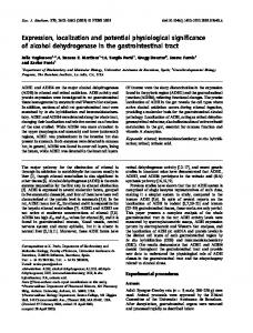
Subfractionation of Mitochondria [8,9] Suspensions of rat liver mitochondria in 0.44 M sucrose (buffered with 10 mM triethanolamine, 2 mM EDTA, pH 7.4) were diluted with the same medium to a final concentration of 30mg of mitochondrial protein per ml. Two samples each containing 2 ml of this suspension, were prepared. One sample was incubated with digitonin (2.1mg/lO mg protein), the other serving as control, was treated identically but without addition of digitonin. After 20 min of incubation, mitochondria were recovered by centrifugation a t 9500 x g for 10 min. The supernatants were re-centrifuged a t high speed (60 rnin a t 145000 xg) in order to remove membrane fragments (pellet PI). The remaining supernatant was called S,. Mitochondria collected in the first pellet after low speed centrifugation were washed once by resuspension in sucrose medium and subsequent centrifugation a t 9500 x g. Supernatant of this washing procedure was called S,. I n a subsequent step of preparation, mit,ochondria collected in the low speed pellet after washing were resuspended in 0.1 M phosphate buffer p H 7.2, and subjected to ultrasonic treatment (Branson Sonifier 5 x 10 see) in order to open the inner mitochondrial membrane. The homogenate was centrifuged a t 145000 x g for 15 min to give supernatant S, and pellet Pz.A scheme of this preparation procedure is given in Fig. 1. Measurements of Oxygen Uptake Pig kidney mitochondria isolated in 0.3 M sucrose were suspended, to a concentration of 50-60 mg proteinlml, in a solution containing 0.3 M sucrose, 2 mM EDTA, and 1 O/, albumin, buffered with 10 mM triethanolamine p H 7.4. After addition of 0.50 mg and 0.75 mg digitonin/lO mg of protein respectively, the suspensions were incubated for 20 min a t + 4". Subsequently the samples were diluted four-fold with sucrose medium and centrifuged for 15 min a t 9500 x g . Control mitochondria were treated identically but without addition of digitonin. Sedimented mitochondria were gently resuspended in the incubation medium. The supernatants were concentrated 16*
-r
Sediment resuspended in 0.44 M sucrose medium and centrifuged 15 rnin a t 9500 x g
Supernatant centrifuged 60 rnin a t 145000 x g
r2
I
m
Supernatant Supernatant
s1
Pellet Pi
Extraction of inner mitochondrial compartment I Sediment treated with sonifier 5 x 10 sec in 0.1 M phosphate buffer pH 7.2 I Centrifugation, 15 rnin a t 145000x g Pellet P2
Supernatant 53
Fig. 1. Schematic representation of subfractionation procedure. Abbreviations are explained in text
to a small volume by vacuum dialysis against 0.15 M sucrose (Sartorius-Membranfilter GmbH., Gottingen). Oxygen uptake was measured polarographically with a ''Clark" type platinum electrode in a test volume of 1ml a t 25" and pH7.4. The assay mixture contained: 0.3 M sucrose, 2 mM EDTA, I0 miK triethanolamine, 2 mM phosphate, 2 mM MgC1, , 1 mM ADP, 1 mM ATP. Mitochondria were added in a concentration of 8 to 9 mglprotein ml. The test mixture was gently stirred during measurement. Assay of Enzyme Activities Monoamine oxidase, adenylate kinase, succinate dehydrogenase, and glutamate dehydrogenase were assayed as described previously [lo]. Kynurenine hydroxylase was determined according to Saito et al. [ i l l . Activity of carnitine acetyltransferase was measured by following the reduction of NAD in a Beckman spectrophotometer at 340 nm. Concentrations of the assay mixture were as follows : 100 mM Tris-C1buffer, pH 8.0,2.5 mM EDTA, 5 mM NAD, 50 mM L-malate, 0.3 mM CoA-SH, 20 U/ml malate dehydrogenase, 28 U/ml condensing enzyme, 2 mM DL-acetylcarnitine. The reaction was started by adding DL-acetylcarnitine. RESULTS
Intramitochondrial Localization of Carnitine Acetyltransferase Localization of carnitine acetyltransferase within the mitochondria was studied by subfractionation of mitochondria. This method used in two recent com-
Localization of Mitochondria1 Carnitine Acetyltransferase
236
European J. Biochem.
Table 1. Subfractionation of rat liver mitochondria Mitochondria suspended in sucrose medium were incubated with 2.1 mg digitonin/lO mg mitochondrial protein. Centrifugation for 15 min a t 9500 x g resulted in a supernatant containing soluble proteins of the outer mitochondrial compartment and fragments of the outer mitochondrial membrane. These membrane fragments were separated from soluble proteins by a high speed centrifugation (145000xg for 60 min) to give pellet Pi and supernatant S1. A small part of soluble proteins of the outer mitochondrial Compartment remained within the mitochondrial pellet obtained after first l o v ~speed centrifugation. When mitochondria were washed once in sucrose medium these proteins were collected in supernatant S2. By sonification of washed mitochondria in 0.1 Bf phosphate buffer inner membranes were ruptured. Soluble proteins of the inner mitochondrial compartment were separated in supernatant 53 from fragments of the inner mitochondrial membrane as collected in the pellet P2 after centrifugation a t 145000X g for 15 min. Control experiments were performed identically without addition of digitonin. Enzyme activity was calculated as units per fraction (monoamine oxidase as mu), and as percentages of total measured activities. Mean values of five experiments are given ~
Braction
~~
~~
Glutamate dehydrogenase
with digitonin
“0
~
Xonoamine oxidase
U
‘lo
mu
“0
~~
Succinate dehydrogenase
u
Carnitine acetyltransferase
“0
U
“0
Xynurenine hydroxylase
U
OIo
Protein mg
Olio
38.5 5.7 8.0 183.8 13.0
15.4 2.3 3.2 73.9 5.2
70.5 0.6 1.7 0.2 0.2
96.7 0.7 2.2 0.2 0.2
24.9 135.6 109.4 0.0 183.2
5.7 30.4 23.9 0.0 40.0
0.03 0.87 0.80 0.27 16.39
0.2 4.3 4.0 1.5 90.0
0.083 0.026 0.007 0.124 0.014
32.5 10.1 2.6 49.6 5.2
0.014 0.021 0.039 0.005 0.023
14.5 20.6 38.1 4.6 22.2
11.2 3.3 2.5 19.5 13.5
22.8 5.6 5.1 42.6 26.9
Tota1249.0
100.0
73.2
100.0
453.1
100.0
18.40
100.0
0.254
100.0
0.102
100.0
50.0
100.0
1.8 2.2 6.5 175.1 44.2
0.8 1.0 2.9 77.8 17.5
1.9 0.7 4.6 33.6 7.2
3.9 1.4 9.1 71.0 14.6
0.0 26.1 25.3 15.3 379.0
0.0 6.1 5.6 3.4 84.9
0.0 0.14 0.17 0.20 18.0
0.0 0.7 0.9 1.1 97.4
0.004 0.006 0.010 0.166 0.018
1.9 2.7 4.7 82.0 8.7
0.0 0.008 0.008 0.008 0.040
0.0 13.8 13.6 11.4 61.2
1.4 1.8 2.8 20.6 23.4
2.7 3.5 5.7 41.0 45.1
Tota1229.8
100.0
48.0
100.0
445.7
100.0
18.51
100.0
0.204
100.0
0.064
100.0
50.0
100.0
S1 Pi 82 S3 P2
without digitonin
Sl P1 S2 S3 P2
munications [lo, 1.21, allows separate extraction of two mitochondrial compartments. By treating rat liver mitochondria with low concentrations of digitonin, the outer membrane is ruptured. Practically 100 of the mitochondrial adenylate kinase is found in the supernatants of such preparations. Another mitochondrial compartment is opened when digitonin treated mitochondria are exposed to sonification. The supernatants of these preparations contain the soluble citric acid cycle enzymes, 3-hydroxyacyl-CoA dehydrogenase, and glutamate dehydrogenase. These results are in good agreement with recent findings of other authors [8,9,13- 151. Data summarized in Table 1 are mean values of 5 experiments performed as described above. Total activities of the listed enzymes were determined in each fraction and expressed both as units per fraction and percentages of total activities in all fractions. Monoamine oxidase and kynurenine hydroxylase, both enzymes bound to the external mitochondrial membrane [S, 161, are recovered after digitonin treatment with a high percentage of their total activity in the particulate fraction P, which predominantly contains ruptured external mitochondrial membranes. Relatively high activities of these enzymes are also found in the particulate fraction P, , thus indicating that this fraction is contaminated with outer mitochondrial membranes. On the other hand, fraction P,
contains the bulk of fragmented inner mitochondrial membranes, as is demonstrated by its high activity of succinate dehydrogenase. I n Table 1 adenylate kinase and glutamate dehydrogenase represent “soluble” mitochondrial enzymes. Adenylate kinase being localized within the outer mitochondrial compartment, is detected with almost 1000/, of its total activity in supernatant S, as obtained after mild digitonin treatment of the mitochondria. Glutamate dehydrogenase which is a soluble enzyme of the inner mitochondrial compartment [lo, 14,171, is extracted with the bulk of its activity after sonification of digitonin treated mitochondria. 74O/, of its total activity is found in the respective supernatant S,. It may be noted, however, that 20°/, of glutamate dehydrogenase activity is already set free before sonification, i. e. during digitonin treatment and subsequent washing (S, , S, , Pl). As has been pointed out in previous communications [lo, 1.21, the fragility of the inner mitochondrial membrane increases markedly after the outer membrane has been ruptured by digitonin. Therefore, a slight contamination with enzymes actually localized in the inner compartment occurs in the first three fractions (Sl, S,,P,). This contamination is not observed in control experiments. The distribution of carnitine acetyltransferase in Table I shows that most enzyme activity is concen-
Vol.11, N0.2, 1969
D. BRDICZKA, K. GERBITZ,and D. PETTE
237
Table 2. Carnitine dependent oxidation of acetyl-CoA by mitochondria of pig kidney Respiration of isolated mitochondria (8-9 mg protein/ml) was measured polarographically with the platinum electrode. Two samples of mitochondrial preparations were pre-incubated with 0.5 mg and 0.75 mg digitonin/lO mg mitochondrial protein respectively. Control mitochondria were treated identically, but without digitonin. After incubation, mitochondria were separated by centrifugation. The remaining supernatants containing carnitine acetyltransferase activity were concentrated by vacuum dialysis and added t o the test mixture (part B of this Table). For each substrate oxygen consumption was corrected by subtracting the utilization attributable to malate. The given data are mean values of 5 experiments Oxygen consumption a t digitonin concn. in ore-incubation of Substrate in assay mixture
Concn.
0.75 mg per 10 mg
0 (control)
mM
A: Pre-extracted mitochondria Acetyl-CoA nL-carnitine Acetyl-CoA and nL-carnitine DL- Acetyl-carnitine Substrate in assay mixture
0.2 2.5 0.2 2.5 1.0 Concn.
B: Pre-extracted mitochondria Acetyl-CoA
+ extracted enzymes
and DL-carnitine
trated in fractions containing soluble proteins (S, and SJ. Obviously, the enzyme is not firmly bound to mitochondrial membranes. Approximately 55 of the total activity of carnitine acetyltransferase remains unextracted as long as the inner membrane is intact. This part of carnitine acetyltransferase activity shows extractability similar t o that of glutamate dehydrogenase. It is suggested, therefore, that this portion of carnitine acetyltransferase activity is located in the same compartment as glutamate dehydrogenase (inner mitochondrial compartment). On the other hand, 4501, of the activity of carnitine acetyltransferase is detected in the first three fractions, which contain only 20 of the total activity of glutamate dehydrogenase. Glutamate dehydrogenase activity found in these fractions may be chosen as a measure of contamination with soluble enzyme from the inner mitochondrial compartment [lo]. The possibility that carnitine acetyltransferase activity in fractions S,, S,,P,, may also be due to leakage from the inner compartment should be considered. However, comparing the proportion of carnitine acetyltransferase activities with that of glutamate dehydrogenase activities in the fractions S,, S,,PI, a surplus of about 26 O/, of carnitine acetyltransferase activity is found. At least this amount of carnitine acetyltransferase activity is therefore located in the same compartment as adenylate kinase. The data in Table I may thus be interpreted as indicating the presence of two differently extractable carnitine acetyltransferases in rat liver mitochondria. At least 25 Olio of the total mitochondrial activity of carnitine acetyltransferase is probably located in the
0.2 2.5
mitochondrial protein
patoms xmin-’ x g protein-’
0 0
0 0
3.1 0
8.4 14.0
3.6 14.4
5.0 13.2
Oxygen consumption with a total activity (mujml) of carnitine acetyltransferase added to the assay of: 2.6
mM
0.5 mS!
8.0
-
patoms xmin-’ x g protein-’
10.0
12.0
-
outer mitochondrial compartment (“external” caror even less, nitine acetyltransferase) whereas 75 of the activity is present in the inner mitochondrial compartment (“internal” carnitine acetyltransferase) . With regard to external carnitine acetyltransferase, it is obvious that a relatively high proportion of the enzyme is found in the high speed pellet P,. This observation indicates that external carnitine acetyltransferase may be adsorbed to the outer membrane.
Function of Carnitine Acetyltransferase in the Transport of Acetyl-CoA Localization of carnitine acetyltransferase on both sides of the inner mitochondrial membrane which is assumed to be impermeable to acetyl-CoA [l,61, suggests that both enzymes mediate transport of acetylCoA across the mitochondrial membrane by reversible formation of acetyl-carnitine in the presence of carnitine. I n order to investigate whether carnitine acetyltransferase activity within the outer mitochondrial compartment has the ability to produce acetylcarnitine from added acetyl-CoA and carnitine, another series of experiments was performed on digitonin-treated and untreated mitochondria (Table 2). Pig kidney mitochondria were used because of their higher content of carnitine acetyltransferase activity as compared with rat liver mitochondria. By pre-incubation with digitonin (0.5 and 0.75mg digitonin110 mg mitochondrial protein respectively), mitochondria were partially depleted of carnitine acetyltransferase activity located in the outer mitochondrial compartment. Oxygen consumption was
238
Localization of Mitochondria1 Carnitine Acetyltransferase
observed in the presence of L-malate after addition of acetyl-CoA, DL-carnitine, and DL-acetylcarnitine by the polarographic method. Mitochondria treated with 0.5 mg digitonin110 mg protein are able to oxidize L-malate as well as untreated mitochondria (20 patoms 0 x min-I x g protein-l). The respiration of mitochondria treated with 0.75 mg digitonin110 mg protein is much lower (13 patoms xmin-lxg protein-I). As shown in Table2, acetyl-CoA and DLcarnitine added separately failed t o increase the oxygen consumption of control mitochondria and of mitochondria treated with 0.5 mg digitonin/iO mg protein. Mitochondria treated with a higher digitonin concentration (0.75 mg/lO mg protein), however, are able to oxidize added acetyl-CoA to some extent. Treatment of mitochondria with this and higher digitonin concentrations probably results in a permeability of the inner mitochondrial membrane to acetylCoA. On the other hand, simultaneous addition of acetyl-CoA and m-carnitine increases the respiration of intact and digitonin-treated mitochondria. A significantly higher respiration is observed in the control. When DL-acetylcarnitine is used as substrate, oxygen consumption is approximately 14 patoms 0 x min-l x g protein-l in all preparations and thus is much higher than the values obtained when acetyl-CoA and DLcarnitine are added simultaneously. This shows that the capacity of the acetyl transferring systems to produce acetylcarnitine is less than the maximum capacity of mitochondria to oxidize it. Digitonin treatment appears to decrease the ability of mitochondria to produce acetylcarnitine. I n digitonintreated (0.5mg) mitochondria, oxygen consumption in the presence of acetyl-CoA and DL-carnitineis only one fourth of that obtained when DL-acetylcarnitine is used as substrate. The somewhat higher oxygen consumption of mitochondria treated with 0.75 mg digitonin/lO mg protein, may be attributed to the increased permeability of the inner membrane to acetyl-CoA. Assuming that digitonin-treated mitochondria have lost some carnitine acetyltransferase from the outer compartment (see first section), these results support the proposed role of carnitine acetyltransferase in mediating the transport of acetyl-CoA across the inner mitochondrial membrane. From the data presented in Table 2, it appears that the acetylating step is limiting also in control mitochondria, since DL-acetylcarnitine is oxidized more rapidly than simultaneously added acetyl-CoA and carnitine. The results given in section B of Table 2 and in Fig.2, however lead to another explanation. It is obvious that control mitochondria also lose some of their carnitine acetyltransferase activity by leakage. This leakage is certainly due t o mechanical influence during preparation. The data in Fig.2 make it clear that the supernatant of control mitochondria also contains carnitine acetyltransferase activity, although
0
European J. Bioehem.
05 0.75 Digitonin (rngilOmg protein)
+
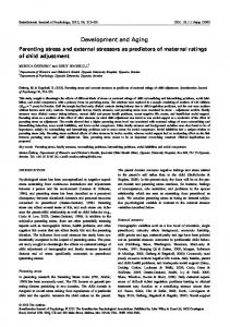
Fig. 2. Mitochondrial oxidation of acetyl-CoA carnitine related to amount of extracted external carnitine acetyltransferase. Pig kidney mitochondria were incubated for 20 min with 0, 0.5 mg and 0.75 mg digitonin110 mg mitochondrial protein respectively. After incubation the samples were centrifuged for 15 min at 95OOxg. Activity of carnitine acetyltransferase set free from the mitochondria into the supernatants was determined and is given as pmolesx min-l x g protein-l. Oxygen consumption of the mitochondria recovered in the pellet was measured polarographically with L-malate and acetyl-CoA in the presence of D,L-carnitine. Values were corrected by substracting oxidation rates attributable t o malate and are expressed as patoms x min-l x g protein-1
only one fourth of the activity which is found in the supernatant from mitochondria treated with 0.5 mg digitonin. There is a higher amount ofcarnitine acetyltransferase activity in the supernatant following incubation with higher concentrations of digitonin (0.75 mg). The right columns in Fig.2 represent the capacity of mitochondria to oxidize added acetyl-CoA in the presence of DL-carnitine. It can be seen, that the higher extraction of carnitine acetyltransferase activity from mitochondria treated with digitonin is followed by a decrease of the oxidation rate of acetylCoA in the presence of carnitine. This is less pronounced in mitochondria treated with the greater digitonin concentration, probably as a result of acetyl-CoA penetrating the inner membrane. As is shown in the lower part of Table 2, re-addition of supernatant fractions to the mitochondrial suspension, increases the oxidation rate of acetyl-CoA in the presence of carnitine. The measured oxygen consumption is then close to the values obtained when DL-acetylcarnitine is oxidized. DISCUSSION
The current hypothesis for explaining carnitinemediated transport of acetyl groups across the mito-
D. BRDICZKA, K. GERBITZ,and D. PETTE
Vol.11, No.2, 1969
chondrial membranes, postulates two forms of carnitine acetyltransferase located on both sides of the membrane which is a barrier to acetyl-CoA [6]. Fig.3 gives a scheme of the postulated model. The outer membrane is permeable to acetyl-CoA and between this membrane and the inner membrane carnitine acetyltransferase activity is present. Acetylcarnitine generated in this space (“external” carnitine acetyltransferase) enters the inner mitochondrial compartment where the acetyl group is transferred again to CoA (“internal” carnitine acetyltransferase). A first condition for this hypothesis is the location of carnitine acetyltransferase activity in both the outer and inner mitochondrial compartment. This is supported by the data presented in this paper. The second condition is that the outer mitochondrial membrane is permeable to acetyl-CoA whereas the
outer membrane
outer compartment
inner membrane
inner compartment
Fig. 3. Proposed scheme of localization and function of external ( C A T I ) and internal ( C A T IT) carnitine acetyltransferase in mitochondria[6]
inner membrane is not. This is supported by the finding that mitochondria with ruptured outer membranes (after digitonin treatment, 0.5 mg/iO mg mitochondrial protein) are still unable to oxidize external acetyl-CoA. Beattie [I91 reported similar results for acyl-CoAin the case of digitonin-treated mitochondria from rat liver. Relatively small digitonin concentrations (0.5 mg/iO mg mitochondrial protein) are sufficient t o open the outer mitochondrial membrane, as is proved by extraction of enzymes located between outer and inner mitochondrial membranes. A similar conclusion may be drawn from the finding that [14C]sucrose accumulated in the outer compartment of rat liver mitochondria can be released by treatment of the mitochondria with low digitonin concentration [20]. As has been shown already by Schnaitman et al. [9], the outer mitochondrial membrane is ruptured by low digitonin concentrations and is fragmented totally by higher digitonin concentrations. A similar dependence on concentration may also be proposed for the action of digitonin on the inner membrane which becomes permeable to acetyl-CoA after treatment with digitonin concentrations above 0.5 mg/ 10 mg protein. Thus, acetyl-CoA penetrates mitochondria from pig kidney as shown in this communi-
239
cation, and also the inner membrane of rat liver mitochondria is no longer impermeable to [14C]sucrose after treatment with higher digitonin concentrations [20]. Digitonin concentrations which lead to a complete extraction of soluble enzymes of the outer mitochondrial compartment, inevitably also lead to a higher permeability of the inner membrane. Therefore, digitonin concentrations were applied in a series of experiments which should not affect the inner mitochondrial membrane. Under these conditions extraction of carnitine acetyltransferase activity from the outer mitochondrial compartment, is incomplete. Nevertheless, a sibpifieant decrease of acctyl-CoA oxidation in the presence of carnitine is observed (cf.Table 2). However, the capability of mitochondria to oxidize acetyl-CoA in the presence of carnitine can be partly restored by the addition of carnitine acetyltransferase to the incubation medium. A certain discrepancy appears to exist with the findings of Barker et al. [7]. These authors measured activities of carnitine acetyltransferase in intact and lysed mitochondria (Liver and mammary gland) and found little or no enzyme activity available t o external acetyl-CoA in intact mitochondria. Glutamate dehydrogenase activities were related to carnitine acetyltransferase activities in the experiments of these authors. It has been shown, however, that glutamate dehydrogenase is located within the inner mitochondrial compartment [lo, 14,171 and may therefore only be related to the activity of carnitine acetyltransferase within this compartment. Our results indicate that carnitine acetyltransferase of the external mitochondrial compartment is easily extractable and may be set free even by gentle mechanical treatment.Intactnessof the external mitochondrial compartment may be judged from the activity level of adenylate kinase, and this enzyme was not included into the studies of Barker et al. [7]. Another possible explanation is, that in lipogenetic tissues mitochondrial carnitine acetyltransferase functions mainly with respect to carnitine-dependent transport of acetyl-CoA into the extramitochondrial compartment. This suggestion is in agreement with Bressler and Katz [a] who reported results favouring the thesis that function of mitochondrial carnitine acetyltransferase in liver and fat tissue is mainly related to lipogenesis. It is noteworthy, however, that no significant increase of carnitine acetyltransferase activity could bc detected in rat liver, when the animals were held on a diet inducing lipogenesis and increasing i0-fold the activity levels of (NADP) malate dehydrogcnase and ATP citrate lyase [Zi]. These results are not necessarily inconsistent with the hypothesis of Bressler and Katz. Differences in the catalytic properties of the external and internal mitochondrial carnitine acetyltransferase may be expected independent of the discussed functions of carnitine acetyltransferase.
240
D. BRDICZKA et al. : Localization of Mitochondrial Carnitbe Acetyltransferase
Purification of both enzymes is in progress at present, in order to determine whether catalytic differences do really exist. This work was generously supported by the Deutsche Porschungsgemeinschaft. REFERENCES 1. Fritz, J. B., and Yuc, K. T. N., J . Lipid Res. 4 (1963) 279. 2. Bressler, R., and Katz, R. J., J . Biol. Chem. 240 (1965) 622. 3. Bremer, J., J . Biol. Chem. 237 (1962) 3628. 4. Marquis, N. R., and Fritz, J. B., J . Biol. Chem. 240 (1965) 2193. 5. Beenakkers, A. M. T., and Klingenberg, M., Biochem. Biophys. Acta, 84 (1964) 205. 6. Beenakkers, A. M. T., and Henderson, P. T., European J . Biochem. 1 (1967) 187. 7. Barker, P. J., N. J. Fincham, and D. C. Hardwick, Biochem. J . 110 (1968) 739. 8. Schnaitnian, C. A., Erwin, V. G., and Greenawalt, J. W., J . Cell Biol. 32 (1967) 719. 9. Schnaitman, C. A., and Greenawalt, J. W., J . Cell Biol. 38 (1968) 158. 10. Brdiczka, D., Pette, D., Brunner, G., and Miller, F., European J . Biochem. 5 (1968) 294. 11. Saito, Y . , Hayaishi, O., and Rothberg, S., J . Biol. Chem. 229 (1957) 921.
European J. Biochem.
12. Bottger, I., Wieland, O., Brdiczka, D., and Pette, D., European J . Biochem. 8 (1969) 113. 13. Beattie, D. S., Biochem. Biophys. Res. Commun. 31 (1968) 901. 14. Sottocasa, Gl., Kuylenstierna, B., Ernster, L., and Bergstrand, A., in Methods i n Enzymology (edited by P. Colowick and N. 0. Kaplan), Academic Press, New York 1967, Vol. X, p. 448. 15. Parsons, D. I?., Williams, G. R., Thompson, W., Wilson, D., and Chance, B., Mitochondrial Structure an Compartmentation (edited by E. Quagliariello, S. Papa, E. C. Slater, and J. M. Tager), Adriatica Editrice, Bari 1967, p. 29. 16. Okamoto, H., Yamamoto, S., Nozaki, M., and 0. Hayaishi, Biochem. Biophys. Res. Commun. 26 (1967) 309. 17. Norum, K. R., Farstad, &I., and Bremer, J., Biochem. Biophys. Res. Commun. 24 (1966) 797. 18. Norum, K. R., and Bremer, J., J . Biol. Chem. 242 (1967) 407. 19. Beattie, D. S., Biochem. Biophys. Res. Commun. 30 (1968) 57. 20. Gerbitz, K. D., and Brdiczka, D., unpublished results. 21. Brdiczka, D., and Pette, D., unpublished results.
s.
D. Brdiczka, K. Gerbitz, and D. Pette Fachbereich Biologie der Universitat BRD-775 Konstanz, Postfach 733, Germany