Mating induces an immune response and developmental switch in the Drosophila oviduct Anat Kapelnikov*, Einat Zelinger*, Yuval Gottlieb*†, Kahn Rhrissorrakrai‡, Kristin C. Gunsalus‡, and Yael Heifetz*§ *Department of Entomology, Hebrew University, Rehovot 76100, Israel; and ‡Center for Comparative Functional Genomics, Department of Biology, New York University, New York, NY 10003 Edited by Bruce S. Baker, Stanford University, Stanford, CA, and approved June 9, 2008 (received for review November 20, 2007)
Mating triggers physiological and behavioral changes in females. To understand how females effect these changes, we used microarray, proteomic, and comparative analyses to characterize gene expression in oviducts of mated and unmated Drosophila females. The transition from non-egg laying to egg laying elicits a distinct molecular profile in the oviduct. Immune-related transcripts and proteins involved in muscle and polarized epithelial function increase, whereas cell growth and differentiation-related genes are down-regulated. Our combined results indicate that mating triggers molecular and biochemical changes that mediate progression from a ‘‘poised’’ state to a mature, functional stage. antimicrobial peptides 兩 network 兩 reproduction
S
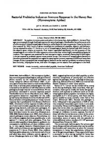
uccessful fertilization is the culmination of concerted interactions between oocyte and sperm. For many animals, the microenvironment of the female reproductive tract (RT) plays an important role in mediating the interaction between gametes (1). The oviducts secrete a variety of molecules that generate the correct osmolarity for supporting the production, maintenance, and modification of gametes and that protect the oviduct and the gametes/fetus from microbial infection and other stressors (2). In mammals, the oviducts secrete glycoproteins, thought to enhance sperm binding to the zona pellucida of oocytes and decrease polyspermy; protease inhibitors, which regulate proteolytic activity to protect the integrity of the zona pellucida, blastomeres, and oviductal tissues; and growth factors, which may enhance embryonic development (2). The microenvironment of the female RT may also influence fertility in insects. Female insects store sperm in specialized organs called the spermatheca and seminal receptacle, which allows the female to fertilize eggs for days after mating. In Drosophila melanogaster, the spermathecal ducts secrete glucose dehydrogenase, which influences sperm motility as well as sperm storage and release (3). Secretory glands in the spermatheca produce lipoproteins, phospholipids, carbohydrates, and proteins that may help maintain sperm viability and maximal fertilization potential. In Drosophila, mating may induce molecular and biochemical changes in the female RT that allow it to support a high rate of ovulation, fertilization, and oviposition (4). Shortly after mating, females begin ovulating (5). Mature oocytes become activated in the oviduct (5) in transit from the ovary to the uterus (Fig. 1A), where sperm released from the sperm storage organs enter the egg through an aperture in the eggshell called the micropyle (6). Mating induces specific physiological changes in the oviduct. In nonlaying, unmated females, a hydrated matrix is detected between the intima and the microvillar surface of the oviduct; after egg laying begins, the intima lies close to the oviduct epithelium, suggesting changes in epithelial cell activity (7). This might affect the osmolarity of the extracellular fluid in the oviduct, which is necessary to support high ovulation and egg laying rates. Major changes also occur in the peptidergic nerve termini innervating different parts of the RT, including distinct domains within the lateral and common oviducts (4). These observations suggest that the oviducts are not ‘‘passive’’ con13912–13917 兩 PNAS 兩 September 16, 2008 兩 vol. 105 兩 no. 37
duits, and that each domain in the female RT (ovary, sperm storage organs, female accessory glands, and uterus) may be regulated locally and possibly in synchrony with other domains. Heifetz and Wolfner (4) hypothesized that, before mating, the female RT of Drosophila does not possess maximal biosynthetic capacity and secretory activity but is ‘‘poised’’ and waiting for a signal provided by mating to continue development. Molecular profiling of female whole-body and lower RT (soma that store sperm) suggests that mating does induce physiological changes (8, 9). To further understand how the female reproductive system achieves maximal functionality to support a high fertility rate, we examined the effect of mating on the oviduct (soma in which eggs are activated). We hypothesize that mating directly or indirectly induces transcriptional and translational changes, transforming the oviduct from a resting state to a physiological state that can sustain a high rate of ovulation of properly activated fertilizable eggs. Here, we provide molecular evidence for such a developmental switch within the oviduct. Results Mating Induces Up-Regulation of Immune-Related Transcripts in the Female Oviduct. Mating in Drosophila triggers profound changes
in female behavior and physiology (5). Females begin to oviposit 3 h after mating, suggesting that the female oviduct undergoes significant physiological changes during this period to prepare for supporting massive egg activation and movement. To identify genes whose expression changes after mating, we analyzed RNA extracted from the oviduct (Fig. 1 A) of unmated and mated females at 3 h after mating. The expression profile of unmated oviducts revealed 5,011 transcripts as ‘‘present’’ [Fig. 1B; see supporting information (SI) Methods]. This set is enriched for genes involved in structural constituents of the ribosome, nucleotide binding, protein binding, transporter activity, translation regulator activity, and actin binding (for P values see Table S1). We next examined differences between unmated and mated female oviducts. From the mated group, 5,411 transcripts were present. Of all transcripts in either group (5,615 in total), 0.95% (53/5,615) were detected only in the oviduct of unmated females (Figs. 1B and S1 A). These transcripts showed overrepresentation of genes involved in serine-type, endopeptidase activity (Table S1), whose down-regulation may eliminate enzymatic activity that could interfere with oviduct maturation or seminal Author contributions: E.Z. and Y.H. designed research; A.K., E.Z., Y.G., and Y.H. performed research; A.K., E.Z., K.C.G., and Y.H. contributed new reagents/analytic tools; A.K., E.Z., K.R., K.C.G., and Y.H. analyzed data; and A.K., E.Z., K.C.G., and Y.H. wrote the paper. The authors declare no conflict of interest. This article is a PNAS Direct Submission. Data deposition: The data reported in this paper have been deposited in the Gene Expression Omnibus (GEO) database, www.ncbi.nlm.nih.gov/geo (accession no. GSE12332). †Present §To
address: Department of Entomology, Volcani Center, Bet Dagan 50250, Israel.
whom correspondence should be addressed. E-mail:
[email protected].
This article contains supporting information online at www.pnas.org/cgi/content/full/ 0710997105/DCSupplemental. © 2008 by The National Academy of Sciences of the USA
www.pnas.org兾cgi兾doi兾10.1073兾pnas.0710997105
C CO SSO AG
5615 total oviduct transcripts Unmated 5011
53
198
differentially expressed
E
M CecA1 CG15745 CecA2 AttA AttB CG9080 CG14527 Jhamt
LO
UT
B
UM
Lower RT
Oviduct
OV
(P