Banda et al. Particle and Fibre Toxicology 2014, 11:64 http://www.particleandfibretoxicology.com/content/11/1/64
RESEARCH
Open Access
Mechanisms of complement activation by dextran-coated superparamagnetic iron oxide (SPIO) nanoworms in mouse versus human serum Nirmal K Banda1, Gaurav Mehta1, Ying Chao2, Guankui Wang3, Swetha Inturi3, Liliane Fossati-Jimack4, Marina Botto4, LinPing Wu5, Seyed Moein Moghimi5,6 and Dmitri Simberg3*
Abstract Background: The complement system is a key component of innate immunity implicated in the neutralization and clearance of invading pathogens. Dextran coated superparamagnetic iron oxide (SPIO) nanoparticle is a promising magnetic resonance imaging (MRI) contrast agent. However, dextran SPIO has been associated with significant number of complement-related side effects in patients and some agents have been discontinued from clinical use (e.g., Feridex™). In order to improve the safety of these materials, the mechanisms of complement activation by dextran-coated SPIO and the differences between mice and humans need to be fully understood. Methods: 20 kDa dextran coated SPIO nanoworms (SPIO NW) were synthesized using Molday precipitation procedure. In vitro measurements of C3 deposition on SPIO NW using sera genetically deficient for various components of the classical pathway (CP), lectin pathway (LP) or alternative pathway (AP) components were used to study mechanisms of mouse complement activation. In vitro measurements of fluid phase markers of complement activation C4d and Bb and the terminal pathway marker SC5b-C9 in normal and genetically deficient sera were used to study the mechanisms of human complement activation. Mouse data were analyzed by non-paired t-test, human data were analyzed by ANOVA followed by multiple comparisons with Student-Newman-Keuls test. Results: In mouse sera, SPIO NW triggered the complement activation via the LP, whereas the AP contributes via the amplification loop. No involvement of the CP was observed. In human sera the LP together with the direct enhancement of the AP turnover was responsible for the complement activation. In two samples out of six healthy donors there was also a binding of anti-dextran antibodies and C1q, suggesting activation via the CP, but that did not affect the total level of C3 deposition on the particles. Conclusions: There were important differences and similarities in the complement activation by SPIO NW in mouse versus human sera. Understanding the mechanisms of immune recognition of nanoparticles in mouse and human systems has important preclinical and clinical implications and could help design more efficient and safe nano-formulations.
Introduction Superparamagnetic iron oxide (SPIO) is one of the most widely cited metal oxide nanoparticle that has been used as magnetic resonance imaging (MRI) contrast agent alone and as a component of multifunctional nanomedicines [1]. Dextran SPIO consists of magnetite-maghemite * Correspondence:
[email protected] 3 The Skaggs School of Pharmacy and Pharmaceutical Sciences, University of Colorado Anschutz Medical Campus, 12850 East Montview Blvd., Aurora, CO 80045, USA Full list of author information is available at the end of the article
(Fe3O4 and γ-Fe2O3) crystalline cores of 3–10 nm size coated with dextran or carboxymethyl dextran [2]. Despite the tremendous medical need in efficient MRI contrast agents [3], several dextran SPIO formulations have been withdrawn from the clinical use due to hypersensitivity in patients (Sinerem, Combidex, Feridex). Another problem of these nanomaterials is the propensity of dextran SPIO for liver and spleen clearance, which limits imaging to macrophage-rich organs. In order to design contrast agents with reduced toxicity and improved pharmacokinetics, a basic understanding of immune recognition of these
© 2014 Banda et al.; licensee BioMed Central Ltd. This is an Open Access article distributed under the terms of the Creative Commons Attribution License (http://creativecommons.org/licenses/by/4.0), which permits unrestricted use, distribution, and reproduction in any medium, provided the original work is properly credited. The Creative Commons Public Domain Dedication waiver (http://creativecommons.org/publicdomain/zero/1.0/) applies to the data made available in this article, unless otherwise stated.
Banda et al. Particle and Fibre Toxicology 2014, 11:64 http://www.particleandfibretoxicology.com/content/11/1/64
materials in both mouse (preclinical) and human (clinical) systems of paramount importance. The complement system accounts for about 5% of globulins in serum and is responsible for recognition, elimination and destruction of pathogens [4]. Activation of the complement on the foreign surface takes place via either the classical pathway (CP), the lectin pathway (LP) or the alternative pathway (AP). The CP activation is triggered via initial binding of IgG or IgM to the pathogen surface, followed by binding and activation of C1q component and formation of C4bC2a, a C3 convertase. C4bC2a cleaves C3 into C3a and C3b, and the latter covalently attaches via highly reactive thioester group to hydroxyls and amines on the foreign surface [5]. More C3b is formed through the alternative pathway (AP) via the formation of alternative C3 convertase C3bBb. Lectin pathway (LP) is somewhat different in mice vs. humans. In mice, the activation is primarily triggered via initial binding of mannose-binding lectin -A and -C or ficolin A to carbohydrates on the pathogen surface, leading to activation of MBL-associated serum protease MASP-2 and formation of C4bC2a, the C3 convertase. In humans, five different sugar recognition molecules have been identified that are able to initiate the LP: MBL, M-, L-, and H-ficolins; and collectin 11 (CL11 or CL-K1), but the downstream activation of the classical C3 convertase is believed to be similar in mice and humans [6]. Activation of the complement plays a major role in the immune recognition of nanoparticles and pathogens [7]. Opsonization by C3b and its cleavage products (e.g., iC3b) triggers recognition by complement receptors CR3 (also known as CD11b/CD18 or Mac-1), complement receptor CR4 (CD11c/CD18), and complement receptor immunoglobulin (CRIg) [8,9], leading to particle uptake by macrophages. Complement cleavage byproducts C3a and C5a are among the most potent anaphylatoxins and proinflammatory molecules with low nanomolar affinity [10]. Many nanoparticulate systems including iron oxides exhibit signs of the complement activation in vivo [11-20]. At the same time, despite the accumulating evidence on the involvement of complement in acute and often life threatening reactions observed in some patients infused with dextran SPIO, the mechanisms of complement activation are not clear. Our earlier report using shotgun proteomics demonstrated the absorption of the LP components MBL-A/C and MASP-1/2 from mouse plasma on the SPIO surface [21]. The involvement of the LP in the complement activation would be a logical assumption, since dextran is a polysaccharide and as such may be recognizable via the LP [22]. This contrasts the reported mechanisms of activation in human sera. Pedersen et al. demonstrated that iron oxide nanoparticles of large curvature activate the CP in human plasma [13] due to the presence of specific anti-dextran IgM
Page 2 of 10
antibodies in certain individuals. In view of the abovementioned similarities and differences between mouse and human complement systems, we set out to systematically study the mechanisms and pathways of the complement activation in mouse versus human sera. For the study below we used our previously described 20 kDa dextran-coated SPIO nanoworms (SPIO NW) that have physicochemical and biological properties similar to Feridex [23,24]. Our data suggest that SPIO NW activate complement in mouse and human sera, but the mechanisms of activation are different, which could bear important implications on preclinical and clinical studies of these materials.
Results and discussion Mechanisms of complement activation by SPIO in mouse sera
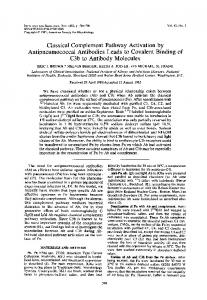
Dextran SPIO contrast agents Feridex I.V.™ (Feridex) and Sinerem™ have been discontinued due to safety issues and are no longer available on the market. Although we used Feridex in our earlier studies [25], the remaining amount was not sufficient for a full scale complement study, and therefore we used our previously described dextrancoated SPIO nanoworms [23]. These nanoparticles are prepared by precipitation of 20 kDa dextran with FeCl2/FeCl3 using the established method of Molday and MacKenzie [26]. The same method was used for preparation of Feridex and other dextran SPIO [27,28], with the difference being that for Feridex manufacturing 10 kDa (T-10) dextran was used, whereas we used 20 kDa dextran. SPIO NW (Figure 1a and Additional file 1: Figure S1) have a worm-like shape with multiple crystalline cores (~6-7 nm each crystal) embedded in the dextran meshwork and with an average hydrodynamic diameter of 169 ± 77.43 nm and zeta potential of −6.05 ± 8.29 mV (Figure 1a). Our previous studies showed that Feridex and SPIO NW have similar physicochemical and immunological properties [24]. To verify the complement activation by SPIO NW and Feridex in mouse serum, we incubated the particles in normal mouse serum at the concentration similar to the concentrations used in vivo (100 μg/mL serum, or 4 mg/kg body weight), washed multiple times by ultracentrifugation and analyzed for the presence of complement fragments in serum supernatant and on the purified particles. The binding and activation of a complement leads to C3 cleavage and covalent deposition of C3b via active thioester bond on the pathogen surface. Main C3 fragments are schematically shown in Figure 1b. The C3 deposition and the pattern of C3 fragments on the surface of Feridex (Additional file 2: Figure S2) were similar to those of SPIO NW. Western blot analysis of fragments deposited on SPIO NW revealed presence of C3b α1’ and α2’ chains (Figure 1b lane 1), suggesting complement activation with subsequent cleavage of C3b to iC3b by Factor I
Banda et al. Particle and Fibre Toxicology 2014, 11:64 http://www.particleandfibretoxicology.com/content/11/1/64
Page 3 of 10
Figure 1 SPIO-mediated complement activation in knockout mouse sera. (a) Model of worm-like polycrystalline SPIO NW shows magnetite/ maghemite crystals (10 nm, brown color) randomly coated with 20 kDa dextran chains (arrow). Size bar for transmission electron microscopy image: 100 nm; (b) scheme of C3 chains and cleaved fragments; (c) nanoparticles were incubated with normal mouse sera (C57BL/6) and C3 fragments were detected on purified SPIO NW or in whole serum by western blotting. Fragments of C3 are clearly detected in purified SPIO NW sample. Zymosan particles (1 mg/ml) also showed strong complement activation; (d) deposition of C3 fragments on SPIO NW surface as a function of normal serum concentration as detected with dot blot immunoassay (see Methods); (e) deposition of C3 fragments in sera deficient for the LP components shows complete dependency on the MBL-MASP-2 but not on FcnA. All experiments were repeated at least three times based on n = 3; (f) deposition of C3 fragments in normal mouse sera pre-incubated with different concentrations of the LP inhibitor mannose; (g) deposition of C3 fragments in sera deficient for the AP. N = 3 for each bar, repeated at least 3 times. ***p < 0.0001, and **p < 0.01.
[29]. Nanoparticle-treated serum showed an increased concentration of C3 fragments, compared to the nontreated sera (Figure 1c, lane 3). Zymosan (1 mg/mL) caused complete disappearance of α chain in serum, suggesting a much more potent activation of the complement than SPIO NW (Figure 1c, lane 4). The reason for the shifted position of α1’ chain eluted from SPIO NW could be due to binding of α1’ chain via thioester to high molecular weight components on the surface of SPIO NW (e.g., dextran). The reason for lack of detection of C3 β chain (~70 kDa) on washed SPIO NW and zymosan treated serum is not clear (albeit consistent), but could be due to a weaker immunoreactivity of the antibody toward the eluted β chain compared to the α chain fragments. Next, SPIO NW were incubated with different dilutions of sera (80%, 40%, 20%) and washed using ultracentrifugation. Dot blotting of washed SPIO NW on nitrocellulose membrane
and immunodetection of C3 fragments showed linear decrease in C3 fragment deposition with decrease in serum concentrations (Figure 1d). There was no detectable C3 in the supernatant from the last wash of the nanoparticles, suggesting that dot blot assay is detecting the nanoparticle bound C3 fragments (shown in Figure 1c) and not the carryover protein. In the subsequent experiments, we used dot blot as the main method to quantify and compare the complement activation in mouse sera and will refer to all C3 fragments as “C3”. In order to further investigate the pathways by which SPIO NW trigger mouse complement we examined C3 fragment deposition after incubation of SPIO NW with sera genetically lacking specific complement components. The serum was mixed with SPIO NW at a final iron concentration of 0.15 mg/ml and serum concentration of 75% v/v. There was a 97% decrease in C3 deposition in
Banda et al. Particle and Fibre Toxicology 2014, 11:64 http://www.particleandfibretoxicology.com/content/11/1/64
serum from wild type (WT) mice supplemented with 5 mM ethylenediamine tetraacetic acid (EDTA) (Figure 1e), since complement activation via all pathways requires both Ca2+ and Mg2+ ions. Also, there was not detectable C3 deposition in C3−/− serum. Activation of the LP proceeds following the binding of serum MBL or the binding of serum Ficolin A (FcnA) to a LP-activating surface [30]. There was 95% less C3 deposition in MBL-A/C−/− mouse serum compared to normal mouse serum (Figure 1e). There was a similar decrease in C3 deposition in the sera lacking both MBL-A/C and FcnA (MBL-A/C−/−FcnA−/−), and no decrease in FcnA−/− serum (Figure 1e), confirming that complement activation in mouse serum depends on MBL-A/C and not on FcnA. Three different types of mannose-associated serine proteases (MASPs), i.e. MASP1, MASP-2 and MASP-3 have been reported to be associated with MBL or ficolins in mouse sera [30,31]. In order to confirm the role of the LP in the complement activation, we measured C3 deposition using mouse serum deficient for MASP-2. In MASP-2−/− mouse serum there was a significant (p