Assistant Professor, Department of Computer Science & Engineering, ... Abstract: To provide an efficient image compression method for medical imaging , an ..... Conference on Biomedical Engineering, Medical & Biological Engineering &.
IJARCCE
ISSN (Online) 2278-1021 ISSN (Print) 2319-5940
International Journal of Advanced Research in Computer and Communication Engineering ISO 3297:2007 Certified
Vol. 7, Issue 3, March 2018
Medical Image Compression using Block Processing with DCT Himani1, Pawan Kumar Mishra2 M.Tech Scholar, Department of Computer Science & Engineering, Uttarakhand Technical University, Dehradun, India1 Assistant Professor, Department of Computer Science & Engineering, Uttarakhand Technical University, Dehradun, India 2 Abstract: To provide an efficient image compression method for medical imaging , an effective approach Discrete Cosine Transform (DCT) with block processing is presented in this paper. To stimulate image transformation, discrete cosine transformation is used first. The discrete cosine transformation transform image into several parts by keeping image visual quality. DCT is most commonly used transformation based on fourier transformation. After successful transformation using DCT method block processing is applied on transformed image. In block processing operations are performed on blocks of image instead processing whole image. In our proposed paper block processing on image is done with 8X8 block. Experimental results shows that block processing with dct compress images faster compared with conventional DCT and older variant of other variant of DCT. Keywords: Image Compression, Medical Imaging, PSNR, MSE, Compression Ratio (CR), Compression size, Block processing. CT, MRI, PET. I. INTRODUCTION Medical imaging is an application area of imaging which is used to generate images of the human body (or parts and function thereof) for medical or clinical purposes. Medical imaging is used for generate images of internal body part of human body. Medical image is an essential part of modern healthcare, it provides a large set of data that is used in research and treatment of diseases, it provides a wealth of information that is increasingly relied upon in the clinical management of patients and conduct planning. Medical imaging data is collected from community and human resource with respect to time involved in these activities. Once acquired, such data is not removed lightly because it is needed for future research and for repetition of test, these things will be minimize risk of research data and maximize productivity. Advances in technology have created the opportunity for radiology systems to use complex. Compression algorithms to reduce the file size of each image in an effort to partially offset the increase in data volume created by new or more complex modalities. In field of medical imaging many types of images are used, some type are CT, MRI, and the combination of positron emission CT and CT (PET-CT), these all are becoming standard in health care industry. All treatments of a patient is totally depended in these images. Format of Medical images is Digital Imaging and Communications in Medicine (DICOM) format. Defiantly DICOM is a non-compressed raw format. The large amount of data creates demand for compression for storage and data transmission purpose. Image compression is a process of data compression and it is used for reducing redundancies in image. Compression is divided in Lossy and Lossless. Lossless techniques, gives a correct reconstruction of the original image, whereas lossy techniques achieve higher compression ratios by allowing some acceptable degradation. The selection of method is depend on application area. In general, for medical imaging, we cannot afford the loss of information and hence lossless compression is preferred. For telemedicine transmission, to exploit greater data compression and hence faster transmission speed, lossy compression is acceptable as long as the required diagnostic data is preserved. This paper introduced a lossless image compression algorithm for possible telemedicine transmission use. JPEG image compression is based on DCT. Among compression techniques the discrete cosine transformation is a fast transformation. It is powerful technique for image compression and widely used for compression.
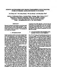
Figure 1 Block Diagram of DCT Compression
For the compression of medical images, the DCT algorithm has achieved good performance and many variants have been developed to for different purpose. Yen-YuChen et al. [1] used DCT-based subband decomposition and modified Copyright to IJARCCE
DOI 10.17148/IJARCCE.2018.7330
164
IJARCCE
ISSN (Online) 2278-1021 ISSN (Print) 2319-5940
International Journal of Advanced Research in Computer and Communication Engineering ISO 3297:2007 Certified
Vol. 7, Issue 3, March 2018
SPIHT data organization for medical image compression. Yung-Gi Wu et al.[2] Medical image compression by sampling DCT coefficients. Manisha Gaonkar and Anuja P Parameswaran [3] proposed Medical image compression by sampling DCT coefficients. V. Naga Prudhvi Raj et al. [4] A Novel Approach to Medical Image Compression Using Sequential 3D DCT. Singh S1, Kumar V, Verma HK. [5]proposed a DWT-DCT hybrid scheme for medical image compression.Marian Kazubek, Artur Przelaskowski, Tomasz Jamrógiewicz et al. [6] proposed an application of Medical Image Data Characteristics for constructing DCT-based compression algorithm. II. RELATED WORK Andrew B. Watson [7] NASA Ames Research Center has proposed DCT technique for converting a signal into elementary frequency components. It is widely used in image compression and develops some simple functions to compute the DCT and to compress image. These functions illustrate the power of Mathematic in the prototyping of image processing algorithms. Prachi Natub, H. B. Kekrea, and Tanuja Sarode [8] proposed color image compression using vector quantization and hybrid wavelet transform. Many image compression technologies are being developed. Wavelet transformation is used for image compression. After advancement in this compression technique hybrid wavelet transformation was introduced. Compression result was very good of hybrid wavelet compare to other compression. Wavelet transformation with vector quantization is applied to increase compression ratio. Prabhpreet Kaur, Navpreet Saroya[9] presents Discrete Cosine Transform (DCT) and Discrete Wavelet transform (DWT) implementation because these are lossy techniques. Archana Deshlahra, G. S. Shirnevar [10] proposed comparative study of 3 transform techniques, which are Discrete Cosine Transform (DCT), Discrete Wavelet Transform (DWT) & Hybrid (DCT+DWT) Transform. Khan Wahid and Suchitra Shrestha [11] proposed a hybrid algorithm that performs a Discrete Cosine Transform (DWT) on Discrete Wavelet Transform coefficients. In this paper, presented algorithm gives good result much better in term of peak-signal-to-noise-ratio with a higher compression ratio to standalone DCT and DWT algorithms. Dr. K. Kuppusamy, D. Malarvizhi, [12] introduced a new technique for simultaneous image acquisition and compression called adaptive compressed sampling. It creates demand for image reconstruction on compressed images. Reconstruction is a challenging work after decompression of image, because loss in quality and data makes no use of image after compression. III. PROPOSED METHODOLOGY An image compression process for medical images is generally defines a specific set of images which are containing sensitive information of human being, loss of information from these images means that no use of images. After image compression, compressed image is compared with original image. For comparing two images several parameters are required such as compression ratio, PSNR, MSE and compressed size.
Figure 2 Architecture of proposed methodology Copyright to IJARCCE
DOI 10.17148/IJARCCE.2018.7330
165
IJARCCE
ISSN (Online) 2278-1021 ISSN (Print) 2319-5940
International Journal of Advanced Research in Computer and Communication Engineering ISO 3297:2007 Certified
Vol. 7, Issue 3, March 2018
The details about the steps adopted in the methodology are described in the following subsections. DCT compression occurs in following steps as: The image is subdivided into small blocks. If the total size of the image is N x N then each of the blocks has the size of (N/n) X (N/n). Mostly we use N/n=8. The main purpose of having such non overlapping blocks is that we can perform parallel processing on each of the blocks. We are able to find correlation existing in the same block and can exploit more redundancy in much better way. In our approach first original image is loaded in the system which is main thing of whole process. Image can be in any extension. After loading of image original image size will be display on user window, by checking this size user can check image size. In second step user start compression process by pressing compress image button. This process will take few seconds to load compress image and perform required calculation. Basically in case of image compression PSNR, MSE, compressed size, compression ratios are calculated. PROPOSED ALGORITHM
Proposed Algorithm for Medical Image Compression Setup Initialize required variables Create Forms to Collect Data Start Step1. R read original image Step2. R get red channel Step3. TGenerate 8X8 discrete cosine transform Matrix Step4. BPerform 8X8 block processing over R using Matrix generated in step 3 Step5. mask create a mask [1 1 1 1 0 0 0 0 1 1 1 0 0 0 0 0 1 1 0 0 0 0 0 0 1 0 0 0 0 0 0 0 0 0 0 0 0 0 0 0 0 0 0 0 0 0 0 0 0 0 0 0 0 0 0 0 0 0 0 0 0 0 0 0] Step6. B2 block process over B using mask Step7. I2 block process over B2 using T, and inverse of T Step8. Repeat steps 3 through 6 for Green and Blue channel STEP9. L CONCATENATE R, G AND B TO GET A COMPRESSED IMAGE IV. PERFORMANCE Compression Ratio Compression ratio is defined as ratio between original size and compressed size. If x 1 and x2 represents the number of information- carrying units in two sets that represents the same information, the relative data redundancy R D of the first data set can be defined as RD = 1- 1/CR Where CR, is commonly called the compression ratio. CR = x1/ x2 MSE The MSE is a measure of the quality of an estimator—it is always non-negative, and values closer to zero are better. MSE is defined as following:-
In this equation compressed version and original version of image is used for calculation. I(x,y) is the original image, and I'(x,y) is the approximated version and M,N are the dimensions of the images. A lower value for MSE means less error.
Copyright to IJARCCE
DOI 10.17148/IJARCCE.2018.7330
166
IJARCCE
ISSN (Online) 2278-1021 ISSN (Print) 2319-5940
International Journal of Advanced Research in Computer and Communication Engineering ISO 3297:2007 Certified
Vol. 7, Issue 3, March 2018
PSNR It is designed to measure the difference in pixels value between of two images. PSNR is calculated via the mean squared error (MSE). PSNR can be defined as:PSNR = 20 * log10 (255 / √(MSE)) Logically, a higher value of PSNR is good because it means that the ratio of Signal to Noise is higher. Signal is used for original image and noise is used for error in reconstruction. So, if compression scheme having a lower MSE (and a high PSNR), you can recognise that it is a better one. V. RESULT AND DISCUSSIONS In the proposed work, medical image are compressed using block processing with DCT method. DCT is popular method for image compression because of its performance. In our work first images are transformed using DCT and then block processing is used on transformed image. After image compression is done first MSE of image is calculated because it is needed in calculation of PSNR. Based on these two parameters compression ratio is calculated between original image and compressed image. The algorithms are implemented on MATLAB 2016a version. SYSTEM IMAGE
Proposed System Image compression
Figure 3 Proposed Paper Image Compression Window To load an image in system user has to click on read image button. After clicking on this button following window will appear where user has to select image for compression.
Figure 4 Reading an Image Window After loading an image user will able to see image for compression in below figure 5.
Figure 5 Original Image Loaded Window Copyright to IJARCCE
DOI 10.17148/IJARCCE.2018.7330
167
IJARCCE
ISSN (Online) 2278-1021 ISSN (Print) 2319-5940
International Journal of Advanced Research in Computer and Communication Engineering ISO 3297:2007 Certified
Vol. 7, Issue 3, March 2018
After image loading user has to click on compress image button to get compressed image, after this step user will get result of compression which is shown in next figure.
Figure 6 Proposed Paper Result Window In above figure 6, two images are loaded in system, Image first (right side) is original image and second image (left side) is compressed image. All Calculation result for MSE, PSNR and compression ratio are below.
Figure 7 Medical Image sequence Table 1 Comparison of PSNR is following:
Sr. 1 2 3 4 5 6 7 8 9 10 11 12 13 14 Copyright to IJARCCE
3DSPIHT COMPRESSION SIZE 80.5592 78.0232 52.1846 91.418 78.8301 50.4844 73.7559 61.002 58.5225 83.374 69.1318 61.887 77.002 60.5615
BLOCK PROCESSING WITH DCT COMPRESSION SIZE 50.5206 40.4854 34.2314 58.29 43 33.3076 51.541 38.7031 40.1357 55.7227 40.7979 41.6855 51.6797 37.791
DOI 10.17148/IJARCCE.2018.7330
168
ISSN (Online) 2278-1021 ISSN (Print) 2319-5940
IJARCCE
International Journal of Advanced Research in Computer and Communication Engineering ISO 3297:2007 Certified
Vol. 7, Issue 3, March 2018
15 16 17
62.0898 105.775 83.334
40.9746 60.8066 43.2383
Table 2 Comparison of image size is following:
Sr. 1 2 3 4 5 6 7 8 9 10 11 12 13 14 15 16 17
3DSPIHT MSE
BLOCK PROCESSING WITH DCT MSE
0.052296 0.0581552 0.0527614 0.0852592 0.0932309 0.0516024 0.0502525 0.0541364 0.0515927 0.0646349 0.0725783 0.0621307 0.0546344 0.0556946 0.0652244 0.14161 0.112097
0.0123321 0.00961908 0.0113714 0.0116339 0.00847277 0.0106825 0.0111207 0.00931134 0.0119299 0.0124505 0.00930575 0.0116474 0.0116975 0.0086066 0.0110861 0.0112609 0.0095756
Table 3 Comparison of MSE is following:
Sr. 1 2 3 4 5 6 7 8 9 10 11 12 13 14 15 16 17
Copyright to IJARCCE
3DSPIHT PSNR 61.2204 62.2995 61.5727 61.4636 62.8505 61.8441 61.6695 62.4407 61.3644 61.1789 62.4433 61.4685 61.4499 62.7825 61.683 61.6151 62.3191
BLOCK PROCESSING WITH DCT PSNR 67.2204 68.2995 67.5727 67.4636 68.8505 67.8441 67.6695 68.4407 67.3644 67.1789 68.4433 67.4685 67.4499 68.7825 67.683 67.6151 68.3191
DOI 10.17148/IJARCCE.2018.7330
169
IJARCCE
ISSN (Online) 2278-1021 ISSN (Print) 2319-5940
International Journal of Advanced Research in Computer and Communication Engineering ISO 3297:2007 Certified
Vol. 7, Issue 3, March 2018
Table 4 Comparison of Compression ratio is following:
Sr. 1 2 3 4 5 6 7 8 9 10 11 12 13 14 15 16 17
3DSPIHT Compression Ratio 80.5592 78.0232 77.5093 80.827 75.3379 77.5774 80.7119 77.9232 78.54 81.1933 76.7153 80.1363 80.599 77.2328 78.292 79.2655 74.4633
BLOCK PROCESSING WITH DCT Compression Ratio 86.9107 86.0056 85.2468 87.7749 86.5474 85.2064 86.5214 85.9932 85.2824 87.4306 86.2587 86.6207 86.9791 85.7931 85.6744 88.0804 86.7501
Figure 8 Compression size comparison
Figure 9 MSE comparison
Copyright to IJARCCE
DOI 10.17148/IJARCCE.2018.7330
170
IJARCCE
ISSN (Online) 2278-1021 ISSN (Print) 2319-5940
International Journal of Advanced Research in Computer and Communication Engineering ISO 3297:2007 Certified
Vol. 7, Issue 3, March 2018
Figure 10 PSNR comparison
Figure 11 Compression Ratio comparison
CONCLUSION AND FUTURE SCOPE In this paper, a fast algorithm based on block processing with DCT is proposed for 3D medical image compression. This work has been done on real medical images, where lossless compression is needed. In medical science lossless compression is preferred because loss of data can make diagnosis result incorrect and doctor can feel difficulty during treatment. The proposed algorithm significantly reduced the compression size, maximize the peak signal noise ratio 6%, and improve compression ratio and MSE. Our proposed algorithm is general and it will be applied to any kind of high resolution images (in field of medical science for e.g. MRI, CT scan) in our future work. The field of medical image compression is ripe for explosive growth. In future work, the study can be enhanced by combining others technique with our proposed algorithm. Also, the work can be carried out with other modern techniques to get better result. REFERENCES [1] Yen-YuChen.: „Medical image compression using DCT-based subband decomposition and modified SPIHT data organization‟, International Journal of Medical Informatics Volume 76, Issue 10, October 2007, Pages 717-725. [2] Yung-Gi Wu, W.A.: „Medical image compression by sampling DCT coefficients‟, IEEE Transactions on Information Technology in Biomedicine ( Volume: 6, Issue: 1, March 2002 ) [3] Anuja P Parameswaran & Manisha Gaonkar: „DCT and DWT in Medical Image Compression‟, International Journal on Advanced Computer Theory and Engineering (IJACTE), IET Image Process., 2017, Vol. 11 Iss. 1, pp. 80-87. [4] V. Naga Prudhvi Raj. „A Novel Approach to Medical Image Compression Using Sequential 3D DCT‟, IEEE Transactions on Information Technology in Biomedicine ( Volume: 4, Issue: 13, Dec 2005). [5] Singh S1, Kumar V, Verma HK. „DWT-DCT hybrid scheme for medical image compression‟, Journal of Medical Engineering & Technology 2007 Mar-Apr;31(2):109-22. [6] Artur Przelaskowski, Marian Kazubek, Tomasz Jamrógiewicz. „Application of Medical Image Data Characteristics for constructing DCT-based compression algorithm.‟, proceedings of the 10th Nordic-Baltic Conference on Biomedical Engineering, Medical & Biological Engineering & Computing, vol. 34 , Supplement I, part I, pp.243-244, 1996. [7] Andrew B. Watson, “Image Compression Using the Discrete Cosine Transform”, 1994.
Copyright to IJARCCE
DOI 10.17148/IJARCCE.2018.7330
171
IJARCCE
ISSN (Online) 2278-1021 ISSN (Print) 2319-5940
International Journal of Advanced Research in Computer and Communication Engineering ISO 3297:2007 Certified
Vol. 7, Issue 3, March 2018 [8] H. B. Kekrea, Prachi Natub, and Tanuja Sarode, "Color Image Compression using Vector Quantization and Hybrid Wavelet Transform", Procedia Computer Science 89, 2016. [9] Navpreet Saroya , Prabhpreet Kaur ,"Analysis of Image Compression Algorithm Using DCT and DWT Transforms”, ISSN: 2277 128X, Issue 2, Volume 4, February 2014. [10] Archana Deshlahra, G. S. Shirnevar, Dr. A. K. Sahoo ,"A Comparative Study of DCT, DWT & Hybrid (DCT-DWT) Transform", 2016. [11] Suchitra Shrestha and Khan Wahid,"Hybrid DWT-DCT Algorithm for Biomedical Image and Video Compression Applications", 2016. [12] Malarvizhi , Dr. K. Kuppusamy ,"A New Entropy Algorithm for Image Compression Using DCT", International Journal of Engineering Trends and Technology- Issue3, Volume3, 2012.
BIOGRAPHIES Ms. Himani completed Bachelor of Technology in computer science from Uttarakhand technical university Dehradun in 2015 and she is currently pursuing Master of technology under Uttarakhand technical university Dehradun, India.
Mr Pawan Kumar Mishra is working as an Assistant Professor and Head of the Department, Computer Science and Engineering, Faculty of Technology, Uttarakhand Technical University, Dehradun, India.
Copyright to IJARCCE
DOI 10.17148/IJARCCE.2018.7330
172