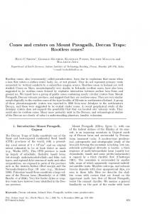
AHMAD MAJD, SAEED IRIAN, PARISA JONOUBI, FARIDE ASGARI J. Plant Develop. 21(2014): 83–93
MERISTEM STRUCTURE, DEVELOPMENT OF CONES AND MICROSPOROGENESIS OF TEHRAN PINE (PINUS ELDARICA Medw.) Ahmad MAJD1, Saeed IRIAN1, Parisa JONOUBI1, Faride ASGARI1* Abstract:
Tehran pine (Pinus eldarica Medw.) belongs to Pinaceae family with significant economic and ecological benefits. To gain further insight into anatomical-developmental structure of P. eldarica, both the vegetative and generative meristematic tissues and microsporogenesis were been studied during certain stages of development. To do this, meristematic tissues and male cones were initially fixed in FAA solution (37% formaldehyde, 96% ethanol and glacial acetic acid with a 2:7:1 ratio, respectively). They were then embedded in paraffin and sectioned using a rotary microtome. Prior to visualization and photography under a camera-equipped light microscope, they were stained with hematoxylin-eosin (Zeiss model). Our results revealed the vegetative meristem of P. eldarica to be in the Cryptomerya-Abies category. The results also indicated it is a protuberant (dome-like) type containing four regions. The meiosis occurs before the winter dormancy and continues through the winter. The pollen is shed at the four-cell stage of development.
Keywords: cone development, generative meristem, microsporogenesis, pollen grain, Pinus eldarica, Tehran pine
Introduction Tehran pine (Pinus eldarica Medw.) belongs to Pinaceae family, it has significant economic and ecological benefits, for example its wood is used extensively as timber and as a source of pulp for papermaking and related industries. It is likely that this species is a variety of P. halepensis Mill., imported to Iran from Georgia, inbred and even naturally reproduced. P. eldarica has been given several different names such as, Tehran pine, garden pine, Iranian pine etc. There have been a great number of studies on the structure and resin components, as well as ecological studies on the quality of wood [DJAVANSHIR & REID, 1975; IRAVANI, 2013; KIAEI, 2011; MOTAHARI & al. 2013; PHILLIPS & al. 1986; SAFDARI & al. 2012]. However, to our knowledge, there is no information has yet been prepared on the anatomical-developmental of P. eldarica. Therefore, the aim of this study is to further investigate the vegetative and generative meristematic tissues and microsporogenesis of P. eldarica during certain stages of development. Materials and methods Plant material was collected from 30-year-old Pinus eldarica trees in Kharazmy University (Iran-Tehran). Meristematic tissues and male cones were collected during certain stages of their development (from June to March) and initially fixed in FAA solution (37% formaldehyde, 96% ethanol and glacial acetic acid in a 2:7:1 ratio, 1 *
Kharazmi University, Faculty of Biological Science, Department of Plant Biology, Tehran – Iran Corresponding author. E-mail:
[email protected]
83
MERISTEM STRUCTURE, DEVELOPMENT OF CONES AND…
respectively), stored in 70% ethanol and embedded in paraffin. To study the meristematic tissues and microsporogenesis, median-longitudinal sections of 8-12 and 8-10µm thick, respectively, were cut and stained using hematoxylin-eosin. For each stage of development, several sections were analyzed using a camera-equipped light microscope (Zeiss model). Results Meristematic tissues Four meristematic zones of the shoot apical meristem (SAM) of P. eldarica were observed: surface meristem (sm), central mother cells (cmc), rib meristem (rm) and peripheral meristem (pm) (Fig. 1). The unspecified axillary buds emerged at the sides of SAM on June 21st through September 1st. With shoot elongation during September to the end of October, the unspecified axillary buds grew separate from each other (Fig. 2). Following expansion of the unspecified axillary buds, differentiation started and formed microsporophylls towards the end of October (Fig. 3). The axillary buds started differentiating from the lower to the upper regions (Fig. 4). This is the transition phase from the vegetative to the generative meristem. The different developmental stages of cones are shown in Fig. 5. Microsporogenesis Results showed that archesporial cells divided repeatedly and formed the archesporial tissue (Fig. 5a). The peripheral cells of the archesporium divided to form a parietal layer and sporogenous cells. The former divided and gave rise to a four- layer structure, with the most inner layer differentiating to tapetum, the two middle layers degenerated at the later stages of development, while the epidermal layer cells also known as the endothecium developed a fibrous wall with a few cells in a section not doing so, marking the dehiscence suture (Fig. 9b, c). The sporogenous cells divided and gave rise to pollen mother cells (microsporocytes), with callose walls around them (Fig. 5b, c). At this stage, the newly formed tapetal cells could not be distinguished from other wall layers (Fig. 9b, c), but during maturation, they acquired a compact cytoplasm and in a number of cases were bi-nucleate (Fig. 6a; 9e, d). On the other hand, each pollen mother cell divided meiotically and formed microspores in a tetrad. They were surrounded by a callosic coat of pollen mother cells. Cytokinesis was non-synchronous and we saw both dyads and tetrads in microsporophylls (Fig. 5c). At the early tetrad stage, the microspores contained a big, central nucleus (Fig. 5c), but gradually the ratio of cytoplasm to nucleus increased. The nuclei became marginal, and microspores started to separate from each other (Fig. 5d; 6a, b). Once released from tetrads, microspores formed regular shapes, while containing a marginal large nucleus (Fig. 6c, d). The microspore's unequal division resulted in the first small prothallial cell and a large central cell (Fig. 7a). The later divided unequally to form a second small prothallial and a large generative initial (Fig. 7b). Both prothallial cells were gradually pushed to one side forming two lens- shaped stacks (Fig. 7c, d; 8). Then, the generative initial cell divided unequally to form a large vegetative cell and a small generative cell (Fig. 7c). During these stages, tapetal cells degenerated while some microspores were still in contact with them (Fig. 6a-c and 9d, e). They later moved towards microspores for nutrition. Finally, tapetum was degenerated completely (Fig. 9f).
84
AHMAD MAJD, SAEED IRIAN, PARISA JONOUBI, FARIDE ASGARI sm S
cmc pz
Sp
S
pz
rm
T
pp
145 µm
Fig. 1. SAM zonation. S – scale leaf; Sp – scale leaf primordia; sm – surface meristem; cmc – central mother cell; rm – rib meristem; pz – peripheral zone; pp – pith meristem; T – tannins. a S Bp SAM Ab b
650 µm
Am
650 µm c
900 µm
Fig. 2. (a) Apical and axillary meristems; (b) apical bud; (c) axillary bud. S – scale leaf; SAM – stem apical meristem; Bp – bud primordia; Ab – Axillary bud; Am – Axillary meristem.
85
MERISTEM STRUCTURE, DEVELOPMENT OF CONES AND…
Am
Am
S
650 µm
650 µm
microsporophylls
650 µm
650 µm
Fig. 3. Transition of the developmental stages of meristem, from the vegetative to the generative state. (a) indeterminate axillary bud; (b) increasing the size and volume of axillary meristem (arrow); (c) differentiation of axillary meristem and formation of microsporophylls (arrows); (d) a young male cone, notice to the disappearing of axillary meristem apex and completing differentiation. S – scale leaf; Am – axillary meristem.
86
AHMAD MAJD, SAEED IRIAN, PARISA JONOUBI, FARIDE ASGARI
microsporophylls
270 µm b
a
330 µm
microsporophylls
microsporangiums 420 µm c
d
500 µm
Fig. 4. Young to mature cone development stages. (a) Production of the microsporophylls of young male cone. At this stage, there are archespore tissues in upper microsporophylls and sporogenus tissues and pollen mother cells in lower microsporophylls; (b) in this size of cones, there were diads, tetrads and pollen mother cells in pollen sacs; (c) young and mature microspores are in the pollen sacs; (d) maturing pollens and also mature pollens are in the pollen sacs. Note the increasing size of cones and pollen sacs.
87
MERISTEM STRUCTURE, DEVELOPMENT OF CONES AND…
a
b 80 µm
80 µm
d pmc
t cw
c
d
145 µm
145 µm
Fig. 5. Different stages of pollen development, from sporogenus tissue to tetrads. (a) archespore tissue; (b) sporogenus cells separation and the production of pollen mother cells are shown (arrows show the pmcs); (c) pmcs undergo meiose I and II; (d) at this stage pmcs have disappeared and tetrades are separating from each other. Increased cytoplasm/nucleus ratio is notable. d – diad; t – tetrad; pmc – pollen mother cell and cw – callosic wall.
88
AHMAD MAJD, SAEED IRIAN, PARISA JONOUBI, FARIDE ASGARI
80 µm
c
b
80µm
a
80 µm
d
80 µm
µM Fig. 6. Different stages of pollen development, from separating tetrads to mature microspores. (a-b) Microspores are separating from each other; (c-d) the size of microspores is gradually increasing. Interaction of diads and tetrads and tapetum cells is notable.
89
MERISTEM STRUCTURE, DEVELOPMENT OF CONES AND…
central cell
prothallial cell 1st prothallial cell 2nd prothallial cell
80 µm generative initial cell 80 µm
a
b
vegetative cell generative cell
prothallial cells
c
d
80 µm
80 µm
Fig. 7. Different stages of pollen development, from immature to mature pollens; (a) first mitosis and the formation of the first prothalian and central cell; (b) division of the central cell and formation of the second prothallial cells and also the initial generative cell; (c) generative initial cell is divided to generative and vegetative cells; (d) degeneration of the two prothallial cells is notable. I NE
gc
prothallial cells
vc
Fig. 8. Mature pollen. SE – sexine; NE – nexin; I – intine; gc – generative cell; vc – vegetative cell. air sacs 80 µm SE
90
AHMAD MAJD, SAEED IRIAN, PARISA JONOUBI, FARIDE ASGARI
a
80 µm
b
pp
en
tl tc
145 µm
en
tl
tl tc en
145 µm
c
d
650 µm
en
145 µm
en tl
f
e
650 µm
Fig. 9. Formation of different layers of pollen sac. (a) archespore tissue and the primary parietal layer; (b) the primary parietal layer divided periclinally to form the endothecium layer, two transition layers and tapetum layer; (c) transition layers gradually degenerated and the endothecium layer anticlinally divided to smaller cells (accolade shows the dehiscence location of pollen sac); (d) only tapetum cells with a large nucleus impact cytoplasm and endothecium layers have remained from the pollen sac layers. Some of the tapetum cells have degraded and have fallen off; (e) separation and degradation of the tapetum cells continue. pp, primary parietal layer; en, endothecium; tc, transition cells, tl, tapetum layer
91
MERISTEM STRUCTURE, DEVELOPMENT OF CONES AND…
Discussion Results of this study showed that apical meristem of Pinus eldarica have four meristematic zones: (1) surface meristem, (2) central mother cell zone, (3) rib meristem and (4) peripheral zone. Observations of this study are in consent with JORDY (2004) on P. pinaster and MACDONALD & LITTLE (2006) on P. sylvestris. According to the theory of POPHAM (1951), there are three SAMs in the gymnosperms: (1) Cycas type, (2) Ginkgo type and (3) Cryptomeria - Abies type. Our results showed a Cryptomeria - Abies type of SAM in P. eldarica. In the initial stages, the side axillary buds (Abs) of SAM are indeterminate, but according to their position on shoots, season and the age of the tree, they can be differentiated to (1) short shoot buds, that produce two needle leaves; (2) male cone buds and (3) female cone buds. According to our observations Abs in trees older than 30 - years old differentiate to male cones from the middle to the upper shoots or needles in the final days of summer. They differentiate to female cones (in the case of the nearest Abs to SAM) in mid- fall from the middle to lower shoots and only to needle leaves in mid- winter. The Abs greatly enlarge at first without any leaf primordial initiation. Later on, a large number of microsporophylls initiated using up the unspecified surface produced earlier from basal up to the Abs. This model has been reported for vegetative and generative meristems in some conifers [KWIATKOWSKA, 2004], our results show that the generative meristem of P. eldarica fits in this model. Their prominent nuclei and compact cytoplasm are features of archesporial cells. In this developmental stage, young microsporophylls walls are single layered. When the sporogenous cells start to separate, the wall of microsporophylls consists of four layers and by the time the tetraspors start to separate from each other, the middle layers have been degenerated. Tapetal cells are large and may have two nuclei as shown by PANDEY & al. (1986). The cytokines of the pollen mother cells are non- synchronous type. This process may occur before winter dormancy (some Chamaecyparis and Juniperus species in the Cupressaceae), or meiosis might begin before the winter, pause at a diffuse diplotene stage, then resume and form microspores after winter dormancy (Larix, Pseudotsuga and Tsuga in the Pinaceae and Thuja in Cupressaceae). Results of this study show that meiosis and pollen development of P. eldarica meiosis occurs before the winter dormancy and continues through the winter. Whereas in some conifers, all stages of meiosis and pollen development occur after winter dormancy [FERNANDO & al. 2005]. Our observations show that tetrad dissociation can be both of synchronous and non- synchronous type. The tapetum cells at first are secretory with multiplication of the nuclei (2 nuclei). Later on they change to amoebic type (thus the nursery type). In P. eldarica before the pollen has shed, mature microspores go through three uneven division to form two lens shape prothallial cells, vegetative cell (tube cell) and generative cell (antheridial cell). Therefore, the pollen is shed at the four- cell stage of development. According to the FERNANDO & al. (2005), in the Pinaceae, pollen may be shed at the four- (like Picea asperata, LÜ & al. 2003) or five- cell (like Pinus contorta, OWENS & al. 1981) stage and pollen of P. eldarica is of the former type.
92
AHMAD MAJD, SAEED IRIAN, PARISA JONOUBI, FARIDE ASGARI
Conclusions According to this anatomical-developmental study, the vegetative meristem of P. eldarica is in the Cryptomerya-Abies category. The meiosis occurs before the winter dormancy and continues through the winter and the pollen is shed at the four- cell stage of development.
References DJAVANSHIR K. & REID C. 1975. Effect of moisture stress on germination and radicle development of Pinus eldarica Medw. and Pinus ponderosa Laws. Canadian Journal of Forest Research. 5(1): 80-83. FERNANDO D. D., LAZZARO M. D. & OWENS J. N. 2005. Growth and development of conifer pollen tubes. Sexual plant reproduction. 18(4): 149-162. IRAVANI B. Z. 2013. Phytochemical analysis of Pinus eldarica bark. Research in pharmaceutical sciences. 9(4): 243-250. JORDY M. N. 2004. Seasonal variation of organogenetic activity and reserves allocation in the shoot apex of Pinus pinaster Ait. Annals of botany. 93(1): 25-37. KIAEI M. 2011. Anatomical, physical, and mechanical properties of eldar pine (Pinus eldarica M.) grown in the Kelardasht region. Turkish Journal of Agriculture and Forestry. 35(1): 31-42. KWIATKOWSKA D. 2004. Structural integration at the shoot apical meristem: models, measurements, and experiments. American Journal of Botany. 91(9): 1277-1293. LÜ S., LI Y., CHEN Z. & LIN J. 2003. Pollen development in (Picea asperata) Mast. Flora-Morphology, Distribution, Functional Ecology of Plants. 198(2): 112-117. MACDONALD J. E. & LITTLE CH. A. 2006. Foliar application of GA3 during terminal long-shoot bud development stimulates shoot apical meristem activity in Pinus sylvestris seedlings. Tree physiology. 26(10): 1271-1276. MOTAHARI M., ATTAROD P., PYPKER T., ETEMAD V. & SHIRVANY A. 2013. Rainfall interception in a Pinus eldarica plantation in a semi-arid climate zone: An application of the Gash Model. Journal of Agricultural Science and Technology. 15(5): 981-994. OWENS J. N., SIMPSON S. J. & MOLDER M. 1981. Sexual reproduction of Pinus contorta. I. Pollen development, the pollination mechanism, and early ovule development. Canadian Journal of Botany. 59(10): 1828-1843. PANDEY S. N., MISRA S. & TRIVEDI P. 1973. Text Book of Botany: (Bryophyta, Pteridophyta, Gymnosperms & Palaeobotany); (for Undergraduate Students of the Indian Universities). Vikas publishing house, 531 pp. PHILLIPS R., FISHER J. T. & MEXAL J. G. 1986. Fuelwood production utilizing (Pinus eldarica) and sewage sludge fertilizer. Forest Ecology and Management. 16(1): 95-102. POPHAM R. A. 1951. Principal types of vegetative shoot apex organization in vascular plants. Ohio State University, 270 pp. SAFDARI V., AHMED M., DEVALL M. S. & BAYRAMZADEH V. 2012. Effects of air pollution on morphological and anatomical characteristics of Pinus eldarica Wood. FUUAST Journal of Biology. 2(2): 5-12. Received: 12 January 2014 / Revised: 19 November 2014 / Accepted: 21 November 2014
93