1563
Development 126, 1563-1570 (1999) Printed in Great Britain © The Company of Biologists Limited 1999 DEV0209
Shoot apical meristem and cotyledon formation during Arabidopsis embryogenesis: interaction among the CUP-SHAPED COTYLEDON and
SHOOT MERISTEMLESS genes Mitsuhiro Aida1, Tetsuya Ishida2 and Masao Tasaka1,* 1Graduate School of Biological Sciences, Nara Institute of Science and Technology, Ikoma, Nara 630-0101, 2Department of Botany, Division of Biological Science, Graduate School of Science, Kyoto University, Kyoto
Japan 606-8502, Japan
*Author for correspondence (e-mail:
[email protected])
Accepted 27 January; published on WWW 17 March 1999
SUMMARY The shoot apical meristem and cotyledons of higher plants are established during embryogenesis in the apex. Redundant CUP-SHAPED COTYLEDON 1 (CUC1) and CUC2 as well as SHOOT MERISTEMLESS (STM) of Arabidopsis are required for shoot apical meristem formation and cotyledon separation. To elucidate how the apical region of the embryo is established, we investigated genetic interactions among CUC1, CUC2 and STM, as well as the expression patterns of CUC2 and STM mRNA. Expression of these genes marked the incipient shoot apical meristem as well as the boundaries of cotyledon primordia, consistent with their roles for shoot apical meristem INTRODUCTION In higher plants, most of the above-ground part ultimately derives from small populations of mitotic cells, called the shoot apical meristem (SAM). The SAM is initially formed during embryogenesis, when the basic body architecture of a plant is established (Jürgens, 1995). Once formed, the SAM plays central roles in postembryonic shoot organ formation. The SAM generates stems, leaves, and floral organs in a set pattern while it maintains a pool of undifferentiated cells in the center (Steeves and Sussex, 1989). Thus, SAM formation during embryogenesis is a critical step to start subsequent vegetative and reproductive development. Many of the recent molecular genetic works have been focused on SAM function in postembryonic development (reviewed in Clark, 1997; Meyerowitz, 1997). However, the molecular genetic basis of SAM formation during embryogenesis is poorly understood. The embryonic SAM is formed in the apex between cotyledons in dicotyledonous plants. In Arabidopsis, the zygote undergoes stereotyped cell divisions to form the radially symmetrical embryo proper and the extraembryonic suspensor (the globular stage). By the heart stage, cotyledon primordia arise as two distinct bumps from the apical flanks of the embryo and the symmetry of the embryo shifts from radial to bilateral. As cotyledons grow and bend over the embryo apex (the bending-cotyledon stage), the SAM becomes a histologically
formation and cotyledon separation. Genetic and expression analyses indicate that CUC1 and CUC2 are redundantly required for expression of STM to form the shoot apical meristem, and that STM is required for proper spatial expression of CUC2 to separate cotyledons. A model for pattern formation in the apical region of the Arabidopsis embryo is presented.
Key words: Meristem, Cotyledon, Organ separation, Arabidopsis thaliana, CUP-SHAPED COTYLEDON (CUC), SHOOT MERISTEMLESS (STM), Pattern formation, Embryogenesis
distinct structure (Barton and Poethig, 1993). Both histological and clonal analyses suggest that the entire SAM and most of the cotyledons derive from the apical half of the globular embryo (Barton and Poethig, 1993; Scheres et al., 1994). Several Arabidopsis mutants are defective only in the SAM, suggesting that at least some part of SAM formation is genetically distinct from that of cotyledons. Recessive mutations in PINHEAD (PNH; same as ZWILLE [ZLL], which was identified independently) and WUSCHEL (WUS) specifically affect SAM formation, resulting in a flat or aberrant structure at the site normally occupied by the SAM (McConnell and Barton, 1995; Laux et al., 1996; Moussian et al., 1998). These genes are suggested to be involved in organizing functional domains within the SAM. The clavata1 (clv1) and clv3 mutants have an enlarged SAM, suggesting that CLV1 and CLV3 are required to limit cell populations within the SAM (Clark et al., 1995, 1996). On the other hand, several mutations that affect development of both the SAM and cotyledons have been identified, suggesting that their genetic pathways overlap. Mutations in the SHOOT MERISTEMLESS (STM) gene result in the lack of a SAM and a slight fusion of the cotyledons at the base, indicating that STM is essential for embryonic SAM formation and partially required for cotyledon separation (Barton and Poethig, 1993; Clark et al., 1996; Endrizzi et al., 1996; Long and Barton, 1998). Weak alleles of stm produce a phenotype
1564 M. Aida, T. Ishida and M. Tasaka that suggests that STM is also required for maintenance of the SAM (Clark et al., 1996; Endrizzi et al., 1996). STM encodes a member of the KNOTTED1 class of homeodomain proteins (Long et al., 1996). This class of proteins are thought to be key transcriptional regulators of SAM development and constitute a gene family found in many plants including maize (Kerstetter et al., 1994), rice (Matsuoka et al., 1993), tomato (Hareven et al., 1996), tobacco (Tamaoki et al., 1997), and Arabidopsis (Lincoln et al., 1994). STM mRNA is expressed in the SAM as well as its precursor cells, consistent with its role in SAM formation and maintenance (Long et al., 1996). Genetic analyses show that the stm mutation is epistatic to other SAMspecific mutations, pnh/zll, wus, clv1 and clv3, with regards to embryonic SAM formation (McConnell and Barton, 1995; Clark et al., 1996; Endrizzi et al., 1996), suggesting that STM acts upstream of these genes in this process. Arabidopsis CUP-SHAPED COTYLEDON 2 (CUC2) and petunia no apical meristem (nam) are members of another class of genes involved in both SAM formation and cotyledon separation (Souer et al., 1996; Aida et al., 1997). Both genes encode members of the NAC domain proteins, whose biochemical function is unknown. Double mutations in CUC2 and another redundant gene, CUC1, cause the lack of an embryonic SAM and a nearly complete fusion of the cotyledons, although each single mutant is basically normal (Aida et al., 1997). Mutations in the nam gene cause similar, but weaker defects than the cuc1 cuc2 double mutant. In nam mutants, no SAM develops and cotyledons are partially fused on one side (Souer et al., 1996). Adventitious shoots are occasionally formed from tissue culture of cuc1 cuc2 mutant hypocotyls or from nam mutant seedlings, and these shoots show almost normal vegetative and reproductive development except that their flowers have several defects including organ fusion (Souer et al., 1996; Aida et al., 1997). These observations suggest that the CUC1, CUC2 and nam genes are not essential for SAM maintenance. Expression of nam is not detected in the SAM itself but in the boundaries of the SAM and cotyledons (Souer et al., 1996). This unique expression pattern, together with the mutant phenotypes of nam and cuc1 cuc2, indicates a close relationship between SAM formation and boundary specification. However, interaction between the NAC genes and STM in these processes remains to be determined. To investigate roles of CUC1, CUC2 and STM during embryogenesis, we examined in detail the temporal and spatial expression of CUC2 and STM mRNAs during embryogenesis. In addition, expression of CUC2 in the stm mutant and expression of STM in the cuc1 cuc2 double mutant were examined. We also examined phenotypes of the double and triple mutants of cuc1, cuc2, and stm. The data indicate that the CUC1 and CUC2 are essential for STM expression to form the SAM and that STM is required for proper spatial expression of CUC2 to separate cotyledons.
MATERIALS AND METHODS Plants and growth conditions Arabidopsis thaliana ecotype Landsberg erecta was used as the wild type. The origin of the cuc mutants was described previously (Aida et al., 1997). The origins of the stm-1 and stm-2 mutants are as described by Barton and Poethig (1993) and Clark et al. (1996). Plants
were soil grown at 23°C under constant white light as previously described (Fukaki et al., 1996) and siliques were collected for analyses of embryo phenotypes and in situ hybridization. Stages of embryogenesis are as defined in Jürgens and Mayer (1994). For examination of seedling phenotypes, seeds were surface sterilized, sown on Murashige and Skoog plates, and germinated as previously described (Aida et al., 1997). Construction of the double and triple mutants For construction of the double mutants, plants heterozygous for stm1 were crossed with cuc1 or cuc2 single homozygous mutant plants. Among F2 populations, plants homozygous for cuc1 or cuc2 and heterozygous for stm-1 were selected based on the floral phenotypes of the cuc single mutants (Aida et al., 1997) and PCR analyses which could detect the stm-1 mutation. Phenotypes of the double mutants were examined in the F3 generation. For construction of the triple mutants, plants heterozygous for stm-1 were crossed with plants homozygous for cuc1 and heterozygous for cuc2. Two F2 families that segregated both cuc1 cuc2 and stm-1 mutants were selected (family 1 and 2). In family 1, approx. 1/16 seedlings with the cuc1 cuc2 double mutant phenotype were observed (16 of 400 F2s, χ2=3.46, P>0.05; calculation based on 1:15 ratio of cuc1 cuc2 seedlings to others), and phenotypes of the other F2 seedlings were normal, stm-1, cuc1 stm-1, or cuc2 stm-1. Genotypes of STM locus in the cuc1 cuc2 seedlings were examined using PCR analysis with specific primers. Among them, 3 were homozygous for stm-1, 10 were heterozygous for stm1, and the remaining 3 were homozygous for the wild-type allele, indicating independent inheritance of the STM alleles in cuc1 cuc2 double mutants. Essentially the same result was obtained in family 2. The results indicate that cuc1 cuc2 stm-1 shows the same phenotype as that of cuc1 cuc2.
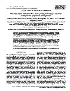
Fig. 1. Development of the apical region in the wild type and cuc1 cuc2 embryos. (A) Schematic diagram of the apical region of the wild-type embryo viewed from above. CP, cotyledon primordia region; PS, presumptive SAM region; BCM, boundary region of cotyledon margins. (B-D) Scanning electron micrograph (SEM) images of (B) wild-type seedling at 3 days postgermination viewed from above; (C) wild-type embryo at the heart stage; (D) cuc1 cuc2 embryo at the heart stage. Arrowheads indicate ectopic bulging of BCM. Scale bars, 100 µm (B) and 40 µm (C,D). c, cotyledon primordia; co, cotyledons; sa, SAM; bcm, boundaries of cotyledon margins.
Shoot meristem and cotyledon formation 1565 Scanning electron microscopy (SEM) Seedlings or dissected embryos were fixed in FAA overnight at 4°C. Embryos were attached to poly-L-lysine coated coverslips before the
subsequent steps. Samples were then subjected to dehydration and critical point drying. Samples were mounted on stubs and coated with gold in an ion splutter coater before observation.
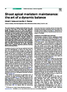
Fig. 2. Ectopic bulging of the presumptive SAM (PS) in cuc1 cuc2 embryos. Embryos were cleared and viewed with Nomarski optics. Lower arrowheads indicate the O′ line and upper arrowheads indicate the embryo apex. The distance between the arrowheads indicates the length of the PS in the apicalbasal direction. (A) Wild-type embryo at the globular stage. (B) Wild-type embryo at the heart stage. (C) cuc1 cuc2 embryo at the heart stage. (D-F) Higher magnifications of A, B and C respectively. (G) Mean lengths of PS in the apical-basal direction (n≥18). Error bars represent standard error. Scale bars, 20 µm (A-C) and 10 µm (D-F).
Fig. 3. CUC2 and STM mRNA expression in wild-type embryos. For definition of section planes, see Long and Barton (1998). (A-E) Early stage embryos probed with CUC2. Arrowheads indicate the protoderm cells in which CUC2 is not detected. (A) Frontal section of mid-globular embryo. (B) Sagittal section of late-globular embryo. (C) Frontal section of early-heart embryo. (D) Sagittal section of early-heart embryo. (E) Transverse section of torpedo embryo. (F-H) Early stage embryos probed with STM. Arrowheads indicate the protoderm cells in which STM is detected. (F) Frontal section of late-globular embryo. (G) Frontal section of early-heart embryo. (H) Transverse section of early heart embryo. (I,J) Bending cotyledon embryos probed with CUC2 in frontal (I) and transverse (J) sections. (K,L) Bending cotyledon embryos probed with STM in frontal (K) and transverse (L) sections. Diagrams in B,D,E,H,J,L represent frontal view of each embryo with red line indicating the section plane. Scale bars, 40 µm. c, cotyledon primordium.
1566 M. Aida, T. Ishida and M. Tasaka Clearing of embryos and seedlings For visualization of embryos or seedling vasculature, ovules or seedlings were cleared as previously described (Aida et al., 1997). Nomarski images (in Fig. 2) were processed in Adobe Photoshop so that cell walls could be clearly seen. In situ hybridization Digoxigenin-labeled RNA probes were synthesized with in vitro transcription using T3 or T7 RNA polymerase according to manufacturer’s instruction (Boehringer Mannheim). Templates for transcription of CUC2 antisense probes were derived from a PCRamplified 558 bp fragment corresponding to the 3rd exon (Aida et al., 1997) or a reverse transcriptase PCR-amplified 1140 bp fragment containing the whole coding region of the CUC2 cDNA (unpublished data). Both probes gave identical results. The 558 bp fragment does not contain the conserved NAC domain to prevent cross hybridization to other NAC-box containing genes. Templates for an STM antisense probe correspond to the region that spans amino acids 81-382 and includes the 3′UTR (Long et al., 1996). Control experiments were performed with or without the sense probes of CUC2 or STM made from the above templates, and no signal above background was detected. Tissues were fixed, dehydrated, and embedded as described by Lincoln et al. (1994). 8 µm sections were cut and attached to 3aminopropyltriethoxysilane-coated slides (MATSUNAMI). Section pretreatment and hybridization were performed according to the method of Lincoln et al. (1994), except that hybridization of CUC2 probes was performed at 45°C. Immunological detection was performed as described by Coen et al. (1990).
(Fig. 2A,D). Some of the hypodermal cells in this region divide transversely by the heart stage, resulting in two or three cells (Fig. 2B,E; Barton and Poethig, 1993). In spite of an increase in the number of cells, the length of the PS in the apical-basal direction did not changed markedly during the transition from the globular to heart stages (Fig. 2G). In cuc1 cuc2 heart stage embryos, however, the length was significantly greater compared to the wild type at the same stage (Fig. 2G; P