PATHOLOGY ONCOLOGY RESEARCH
Vol 8, No 1, 2002
Article is available online at http://www.webio.hu/por/2002/8/1/0068
METHODS The Image Pyramid System – an Unbiased, Inexpensive and Broadly Accessible Method of Telepathology Péter GOMBÁS,1 Jeremy N SKEPPER,2 László HEGYI3 1
Division of Pathology, MI Central Hospital, Városligeti Fasor 9-11, Budapest, H-1071, Hungary; 2Multi-Imaging Centre, University of Cambridge, Department of Anatomy, CB2 3DY and 3Department of Medicine, University of Cambridge, Addenbrooke’s Hospital, Level 6, ACCI, Hills Road, Cambridge, CB2 2QQ, UK
Although computerised information technology, location expressed as a percentage of the total inforincluding the Internet is broadly used and globally mation present in the histological sample. We refer accessible it is still not a significant form of profes- to this as the percentage of explicit versus implicit sional communications in diagnostic histopatholo- information. Another major source of bias may be gy. The high cost of interactive dynamic telepathol- found in the information transmitted in written or ogy systems makes their use limited outside the verbal discussion with a remote consultant. We richest economies. In contrast static telepathology have developed a system of static telepathology, the systems are relatively cheap but in practice their image pyramid, which attempts to minimise bias information content can be heavily biased by the by transferring all of the information in a section to choice of images sent by the consulting pathologist. the consultant. It is inexpensive and should prove The degree of this bias may be regarded simply as to be widely accessible. (Pathology Oncology the amount of information transferred to a remote Research Vol 8, No 1, 68–73, 2002) Keywords: education, image pyramid, remote consultation, telemedicine, telepathology
Introduction Telepathology is defined as the practice of interactive diagnostic pathology performed at a remote location by viewing images on a visual display unit instead of directly on a bright field light microscope.1 Telepathology systems can be divided into dynamic telepathology (DT) and static telepathology (ST). In the DT systems, images are seen interactively on line as they are captured from the microscope. In contrast, ST techniques use static images, which have been captured and archived in a digital format and made available for access from a remote location.2 Although computerised information transfer is a universally accepted method of global communication, the use of the Internet is still not a significant part of routine diagnostic work in pathology. One of the drawbacks of the inexpenReceived: Dec 17, 2001; accepted: Febr 8, 2002 Correspondence: Péter GOMBÁS, M.D., Division of Pathology, MI Central Hospital, Városligeti fasor 9–11, H-1071 Budapest, Hungary; Tel: (36)-1-3227-620, fax: (36)-1-3220-892; E-mail:
[email protected]
© 2002 Arányi Lajos Foundation
sive ST systems is that the proportion of histological information from the slide that is transferred to the remote location is often low. This can make the ST consultation biased to the selection of images and of dubious value. Our aim was to develop a new ST system, which minimises bias in telepathological consultation. The so-called pyramid system can be implemented at low cost and could easily be made readily accessible to pathologists in countries with less buoyant economies. The major advantage of this system lies in the system of image capture.3 We have maximised the information in our cases by systematically recording the whole histological section or smear in overlapping fields of view captured with a x40 objective lens. These images are then stitched together as montages with software developed by PG. This moderately high-resolution montage is reduced in size electronically to provide a low magnification overview of each sample. Higher magnification images that retain the original resolution of the individual images that make up the montage are viewed at different levels by selection portions of the entire montage. This has the disadvantage that the image files are large.3 The alternative is to capture larger original fields of view at
The Image Pyramid System for Telepathology
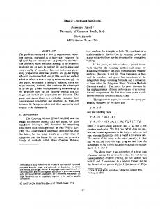
Figure 1. Comparison of “pyramidal zooming” with “traditional digital enlargement”. a-b. Histological image of gastric mucosa c. No pixellisation occurs in pyramidal image architecture. d. Pixellisation at traditional zooming.
lower magnification and inevitably lower resolution and to enlarge them electronically. In this situation image files are small but resolution is poor at higher magnification (Figure 1.). The advantage of the former is the ability to retain detail at higher magnification for all of the area of the original sample. The consulting pathologist therefore, has the advantage of a DT system at a fraction of its cost. The only disadvantage is the time required to capture the images and make the montages for presentation. Materials and methods More than one thousand images of smears and sections of surgical pathological specimens were digitised and archived. Approximately one hundred of them were sent to remote locations. Images were captured and digitised using an Olympus BH2 bright field light microscope, which was equipped with dry objective lenses of x4, x10, x20 and x40, with numerical apertures of 0.13, 0.3, 0.46 and 0.7, respectively. Images were captured systematically from overlapping fields of view until the whole area of the section or smear was collected, using charge coupled device (CCD) video camera Sony DXT-390 P attached to the microscope. The CCD camera was connected to a PC Pentium II Celeron 533 Mhz with 64 Megabyte RAM running Windows 98 and with a Matrox Meteor II/ Multichannel 4 frame grabber. The PC was connected to a local area network and the captured images were stored on the hard drive of a file server until they were joined together as montages. Vol 8, No 1, 2002
69
The complete information content of a section or smear was termed the implicit information.4 The information in one or more images captured with the bright field microscope and transferred to remote location was called the explicit information.4 The degree of bias in such a consultation can be expressed as a percentage: (explicit information ÷ implic it information) x 100. If the images are captured in a format that is at, or close to the theoretical resolution of the light microscope (approximately 0.25 µm) this will simply translate to: (the area of the images sent ÷ the total area of tissue on the slide) x 100. If the images are not captured at close to this resolution the consultation will be further biased by that disparity. This of course can be estimated. The correlation between image resolution and the numeric aperture of an objective lens is described by the formula: δ = 0.61 x (λ/n x sin ω) where (δ) is the distance between two discrete points, (0,61) is a correlation coefficient, (λ) is the wavelength of light, (n) is the refraction coefficient of the mounting medium, (ω is half the angle subtended by the illuminating light and (n x sinω) is the numerical aperture of the objective lens. In this case the dry, X40 lens used to capture images, had a resolution of approximately 0.4 µm. In theory it is therefore only possible to capture one half of the microscopical data available in the slide. In practice the effective loss is unlikely to be anywhere near this level as there is very little pathologically important information in a smear or in a histological section that even approaches the resolution of the light microscope. Information transfer As a first phase of this study an archive of original images and montages was placed in a directory on a file server connected to a LAN of 10 Megabyte bandwidth using transport control protocol/internet protocol (TCP/IP). In the second part of the study the efficacy with which images could be transferred using the LAN were examined. Images of histopathological cases were transferred for case consultation using five different combinations of the transmission of text and graphic files. The success and the professional effectiveness, for pathological consultation, of the five variations were compared. The combinations of transmissions were 1. electronic mail (e-mail) transfer of text files, 2. email transfer with graphic file attached, 3. graphic files loaded on a static World Wide Web (WWW) page and text files transferred by e-mail, 4. graphics and text files loaded on a static WWW page and 5. The image pyramid system implemented on the graphic surface of the Internet. The Image pyramid A digital montage, of entire histological sections or smears, captured with a x40 objective lens was used to create the so-called image pyramid. To achieve the best sig-
70
GOMBÁS et al
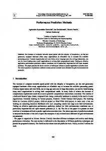
nal to noise ratio images were captured at high magnification and subsequently reduced in size electronically to give low magnification overviews of the samples. This method was chosen, as the use of higher magnification objective lenses in conjunction with montage software gave the best balance between final image resolution and the largest area of sample presented to the remote location. Images were initially stored in BMP file format. They were transformed to JPEG format with a resolution of 800 x 600 pixels. Each pixel had an effective size of 0.31 x 0.31 micrometres, which is well in excess of the resolution of the x40 objective lens used in this study. They were joined together in the form of a montage using image processing software SLICE© with each original image contributing 248 x 186 micrometres of area to the montage. After connecting adjacent images using SLICE© in coordinates of x and y, montages were produced. These begin at the first image level (IL0), which includes all of the slide‘s graphic information at the highest resolution of the system. This is a large image whose size will increase with increasing area of montage and is zoomed out or compressed to generate low magnification images of the entire sample. Similarly small areas of the original montages can be selected to produce relatively high resolution images of selected regions. The size of IL0 pictures were reduced in sequential steps IL1, IL2, etc. Each IL represent individual magnifications within the image pyramid. The size of a page and the graphic information presented in its various JPEG files varied according to the actual size of the montage collected. This is of course proportional to the area of the original section or smear. Image pyramids of various subjects were prepared with short case descriptions written in free text format (FTF) in conjunction with appropriate series of additional JPEG files. These may show the relevant results of additional examinations, which were carried out in connection with the case, including special stains, immunohistochemistry, ECG, computerised tomography, magnetic resonance imaging, etc. (Figure 2.) Image pyramids were then transferred by File Transfer Protocol (FTP) onto a web server in a system of hierarchically organised HTML files. Static WWW pages created by SLICE© on this way can be made available for consultation all over the world. Requests for electronic consultation were sent by e-mail, with details of the Unified Resource Locator (URL) of the page. Results Five of different modes of text and graphic file transmission were investigated. 1) We found that text files describing a surgical pathology report including clinical data, gross specimen information, tissue sampling procedures and histological findings were suitable as requests for brief expert advice.
Figure 2. Image Pyramid on the Word Wide Web. Pyramid may be completed with solitaire images representing special stainings, immunohistochemical reactions and other supplementary diagnostic images.
However, in the absence of an image of the histopathological section, which was the subject of consultation, text files alone proved the least informative method of pathological consultation. The opinion of the consultant pathologist is being dependent on the comprehensiveness of the report written by the consulting pathologist. This system proved to be strongly biased which could lead to serious misapprehensions. 2) Graphic files can be sent to remote locations as attachments to e-mail. However, set data quotas on local email servers often limit the amount of data that can be sent. In many cases numerous e-mails may need to be sent to transfer the information related to a single case. We also found that both the speed of transfer and storage capacities of e-mail-servers was diverse and the values for these were almost totally unpredictable to the sender. Opening and/or downloading large graphic files attached to the main body of e-mail can also be problematic. This can be due to the lack of accessibility to fast and practical mail-reader software in some regions. We concluded that using graphic file attachments is neither an optimal nor an effective method for static telepathology. 3-4) The use of WWW servers was found to be faster and easier than transferring case information by e-mail. Loading text, graphics and HTML files onto a WWW server’s sub-directory proved more advantageous, mainly for the remote consultant, than attaching image files to email. Uploading files on a WWW server is easy and they are readily accessible from the remote site. We found that previews of HTML files were very useful because they offered freedom to the consultant to examine the details he required independent of any prejudice from the consulting pathologist. The method of using WWW servers proved to be practical and inexpensive. Text and image files stored PATHOLOGY ONCOLOGY RESEARCH
The Image Pyramid System for Telepathology
on a WWW server could be linked to, and organised as, static HTML pages, using inexpensive commercially available software, by medical staff without special programming skills. Although using WWW servers seemed to be a simple way of ST, without further systematic improvements and arrangements of graphic files to an edited system the consultation could still be strongly biased. Simple JPEG files presented on an static HTML page showing a limited area of the histological section, selectively chosen by the pathologist on one side, often left out zones with histological characteristics of the disease, which otherwise would have been vital to a meaningful diagnosis. Once the web page has been loaded and accessed by the remote consultant they and the consulting pathologist can communicate as the images are viewed by opening a interactive notepad on that page. However, communicating by a combination of e-mail and using a WWW server in this way cannot eliminate the bias inherent in viewing only the images selected by the consulting pathologist. 5) Image pyramids built on the surface of the Internet make pathological consultation user-friendly, inexpensive, easily accessible and virtually unbiased. As the consultant has access to the entire area of each sample he can make his own assessment of that sample. In particular he has the ability to systematically examine the sample in his own preference at low and high magnification with no significant loss of biological detail at up to the equivalent of direct visualisation on a microscope using a x40 objective lens and x10 eyepiece lenses. Another obvious advantage of this version of ST is that once a library of cases are loaded onto a server they can be accessed by the consultant pathologist and examined and the case discussed with the consulting pathologist at any time. The DT system relies on access to the microscope facility and more stringent co-ordination between the two parties. Discussion Pathological information was first transferred electronically in 1968 when individual images were sent from the Logan Airport to Massachusetts General Hospital in Boston, USA.1 This system, which used transmitted images as the basis of consultation with specialists at a remote site, is known as static telepathology (ST). The first European ST presentations were performed in France5 and in Germany.6,7 In France six private laboratories in the area of Dijon were connected. In Germany static image consultation using commercially available modems and normal acoustic telephone lines also took place between the Departments of Pathology, Thoraxklinik, Heidelberg, Klinikum Heckeshorn, Berlin, Germany and Klinikum Baumgatrner Höhe, Vienna, Austria.7 Almost twenty years after the first ST transmission the first dynamic telepatholVol 8, No 1, 2002
71
ogy (DT) system was introduced in 1986. This initial demonstration of the DT system, utilising the SBS3Comsat satellite, was performed in the USA between Fort William Beaumont Army Medical Centre, El Paso, Texas, and the Communication Satellite Corporations, Washington DC, in collaborations with members of the Armed Forces Institute of Pathology and the US Surgeon General’s Office.1 In Europe the first DT system was installed a few years later, in 1990. This system connected two institutes in Norway, the University of Tromsoe and Kirkenes Hospital. An ISDN line was used with a transfer bandwidth of 2 Mbit per second.8 In the nineties, telepathology systems have been developed and spread all over the world.9 More countries of Central Europe have been involved.10 In the last years of the 20th century the new cyber age, reliant on the Internet has provided unprecedented developments both in ST and DT.9,10 The continuing improvements of global communication have resulted in the transformation of remote consultation in routine surgical pathology.2 The development of integrated telematic systems has greatly facilitated graphic data transfer from integrated digital databases and/or image archives to remote locations.10,11 One of the major experiences of the last decade was the duality of progression in telepathology epitomised by the improvements in DT and ST. At present relatively few institutes apply DT technology while the use of ST systems is increasing. The development of DT and ST systems and their apparent competition may reflect the novelty of different telepathology applications.9 Studies in telemedicine emphasise the three main factors, which, in general, seem to determine the choice and application of telemedicine systems, namely quality, cost and accessibility.12 The quality of ST and DT systems, in terms of diagnostic accuracy, appear different. Using a ST method in 144 cases Halliday et al found 88.2% coincidence between host and remote telepathological diagnoses13. In another study Viellefond et al found only 86% diagnostic precision14. However, in some hands DT systems have been shown to be more reliable. Callas at al consulted 294 cases using a DT system and they found no qualitative difference between direct examination and remote video diagnoses.22 Dunn et al, who found that the diagnostic precision of their DT system achieved 98,5%16, have published similar results. This variation between the two systems is most likely due to the relatively high degree of bias accompanied ST applications in the past where the consulting pathologist selects what information will be sent to the remote specialist. This bias was also experienced in the present study when only a proportion of the implicit information, chosen by the host was transferred to the remote sites using either e-mail or WWW servers. To reduce this bias to minimal levels we have developed a new ST system, the pyramid, which allows the remote consultant to
72
GOMBÁS et al
have unrestricted access to the total implicit information of the slide or smear. The other major difference between ST and DT systems, in favour of the former, is cost.17 Installation costs of one bilateral DT station may mount up to £100,000, which may be increased further by service costs including the expense of installing and maintaining a broad bandwidth communication channel.18 The installation cost of a ST station with a moderately high resolution, digital camera is much less, at around £15,000-25,000. Costs may vary depending on the sophistication of the systems.19 The cost of desktop computers, high-resolution cameras, microscope optics and software tend to be less in ST systems. At present, the cost differential between ST and DT systems is approximately one order of magnitude. If cost is the final deciding factor on what system to choose the image pyramid ST system can be made even cheaper by using a conventional film camera. The resolution of photographic film is greater than that of the highest resolution digital camera. Therefore the quality of the objective lens remains the limiting factor. If images are captured on film they can then be transferred to digital format using a moderately high-resolution flat bed scanner and fed into the montage software. One of the benefits of global cyber technology is that the Internet enables a standardised access to all remote servers throughout the world, from any terminal connected to the Internet. Increase of Internet use in the health service is rapid, inestimable in value and pretty well globally available.19. So, why are pathologists afraid of implementing cyber age tools in their daily routine? Main factors might include human performance in the practice of telepathology20 as well as human acceptance of teleconsultation services.21 Diagnostic errors in ST may occur due to wrong case interpretation, poor digital image quality and inappropriate field selection.9,22 The pyramid system described in the present study allows remote experts to access data from difficult cases independent of host selection and with full access to the implicit information of a sample thus minimising bias. The pyramid system on the WWW also enables unbiased panel discussions with experts6,7 and the opportunity to train junior colleagues and students.11,23,24 There may also be methods available to standardise quality control applications.3,25 Pyramidal image architecture is an easy way to create a “virtual slide ”3,26,27 using huge graphic files both on local network and on the WWW. In summary, the pyramid system is an inexpensive and broadly accessible system of static telepathology. Using this version of the ST system telepathological consultation becomes practically unbiased and independent of the location of the collaborators in each case. No special additional technological conditions are required to apply the pyramid system although time needs to be devoted to captur-
ing and archiving images. Thus the image pyramid is novel and useful development of ST for consultation, expert discussion and teaching, globally. Acknowledgements We are grateful to Alex Sossick of the Wellcome Trust CRC Institute of Cancer and Developmental Biology, University of Cambridge, Cambridge, UK for much valuable advice. We are also thankful to Dr Adrian Carpenter of Wolfson Brain Imaging Centre, University of Cambridge, UK for providing the server space during the time of the study, and Laszlo Csink of John von Neumann Faculty of Informatics, Kalman Kando Polytechnic, Budapest, Hungary, for providing the server space to demonstrate examples of telepathology and Szilveszter Juhos for expert computer assistance.
References: 1. Weinstein RS, Bloom KJ, Rozek LS: Telepathology and the networking of pathology diagnostic services. Arch Pathol Lab Med 111:646-652, 1987. 2. Kayser K, Szymas J, Weinstein R: Telepathology. Telecommunication, Electronic Education and Publication in Pathology. (1st ed). Springer Verlag: Berlin, 97-150, 1999. 3. Wells CA, Softer C: Telepathology: a diagnostic tool for the millennium? J Pathol 191:1-7, 2000. 4. Raggett J, Bains W: Artificial Intelligence from A to Z. Chapman and Hall, London. 1992. 5. Martin E, Dusserre P, Fages A, et al: Telepathology: a new tool of pathology? Presentation of a French national network. Zentralbl Pathol 138:419-423, 1992. 6. Kayser K, Oberholzer M, Weisse G, et al: Long distance image transfer: First results of its use in histopathological diagnosis. APMIS 99:808-814, 1991. 7. Kayser K, Drileck M, Rahn W: Aids of telepathology in intraoperative histomorphological tumor diagnosis and classification. In Vivo 7:395-398, 1993. 8. Eide TJ, Nordrum I: Current status of telepathology. APMIS 102:881-890, 1994. 9. Weinstein RS, Bhattacharyya AK, Graham AR, et al: Telepathology: a ten-year progress report. Hum Pathol 28:1-7, 1997. 10. Gombás P, Szende B, Stotz G: Future aspects and benefits of telematic networks used in pathology for countries of central Europe (CCE). Elec J Pathol Histol; 2:963-968, 1996. 11. Schubert E, Gross W, Siderits RH, et al: A pathologist-designed imaging system for anatomic pathology signout, teaching, and research. Semin Diagn Pathol 11:263-73, 1994. 12. Bashshur RL: Telemedicine effects: cost, quality, and access. J Med Syst 19: 81-91, 1995. 13. Halliday BE, Bhattacharyya AK, Graham AR, et al: Diagnostic accuracy of an international static-imaging telepathology consultation service. Hum Pathol 28:17-21. 1997. 14. Viellefond A, Staroz F, Fabre M, et al: Martin-Pop V, Martin E, Got C, Franc B. Reliability of the anatomopathological diagnosis by static image transfer. Arch Anat Cytol Pathol 43:246-50, 1995. 15. Callas PW, Leslie KO, Mattia AR, et al: Diagnostic accuracy of a rural live video telepathology system. Am J Surg Pathol 21:812-819, 1997 16. Dunn BE, Almagro UA, Choi H, et al: Dynamic-robotic telepathology. Department of Veterans Affairs feasibility study. Hum Pathol 28:8-12, 1997.
PATHOLOGY ONCOLOGY RESEARCH
The Image Pyramid System for Telepathology
17. Poremba C, Pickhardt N: Economic evaluation of telepathology. Pathologe 19: 18-24, 1998. 18. Sowter C, Wells CA: Telepathology: assessment of the implications and applications of telepathology for practical diagnostic pathology. [editorial ] J Clin Pathol 51: 14-715, 1998. 19. Petersen I, Wolf G, Roth K, Schluns K: Telepathology by the Internet. J Pathol 191:8-11, 2000. 20. Weinstein RS, Bloom KJ, Krupinski EA et al: Human performance studies of the video microscope component of a dynamic telepathology system. Zentralbl Pathol 138:399-403, 1992. 21. Mairinger T, Netzer TT, Schoner W et al:. Pathologist’ attitudes to implementing telepathology. J Telemed Telecare 4:41-46, 1998. 22. Weinberg DS, Allaert FA, Dussere P et al: Telepathology diagnosis by means of digital still images: an international validation study. Hum Pathol 27: 111-118, 1996. 23. Weinstein RA: Futurist meets the 21 century: love at first sight [editorial ] Hum Pathol 31: 1-2, 2000.
Vol 8, No 1, 2002
73
24. Katt EC, Dennis SE: Web-based pathology education. Arch Pathol Lab Med 122:745-749, 1998. 25. Kayser K, Kayser G. Recent development of telepathology in Europe with specific emphasis on quality assurance. Anal Quant Cytol Histol 21:319-21, 1999. 26. Gombas P, Skepper JN, Hegyi L: The Image Pyramid – digital slide in the surgical pathology. http://ultra.obuda.kando.hu/~medinfo/tp/ 2001. 27. Gombas P: Informational aspects of telepathology on routine surgical pathology Anal Cell Pathol 21:141-147, 2000. 28. Harris T, Leaven T, Heidger P, Kreiter C: Comparison of a virtual microscope laboratory to a regular microscope laboratory for teaching histology Anat Rec 265:10-4, 2001. 29. Leong FJ, J. McGee JO: Automated complete slide digitization: a medium for simultaneous viewing by multiple pathologists. J Pathol 195:508-514, 2001. 30. Saltz HJ: Digital Pathology – The big picture Hum Pathol; 2000; 31:779-780.