ID: 16013636
Biparametric MRI and expression of 11 genes in apparently benign tissue for the detection of prostate cancer: prospective registered clinical trial
Ivan Jambor, Ileana Montoya Perez, Pekka Taimen, Saeid Alinezhad, Riina-Minna Väänänen, Terhi Tallgrén, Esa Kähkönen, Otto Ettala, Kari Syvänen, Matthias Nees, Markku Kallajoki, Tapio Pahikkala, Peter J. Boström, Kim Pettersson, Hannu J. Aronen Department of Diagnostic Radiology, University of Turku, Turku, Finland Medical Imaging Centre of Southwest Finland, Turku University Hospital, Turku, Finland Department of Biotechnology, University of Turku, Turku, Finland Department of Surgery, Division of Urology, Turku University Hospital, Turku, Finland Department of Pathology, University of Turku and Turku University Hospital, Turku, Finland
ID: 16013636
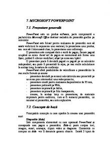
Improved Prostate Cancer Diagnosis - Combination of Magnetic Resonance Imaging and Biomarkers (IMPROD, NCT01864135) PROSTATE CANCER SUSPICION
MRI Surveillance
Biopsy
benign
Cancer
Active surveillance
surgery
radiation
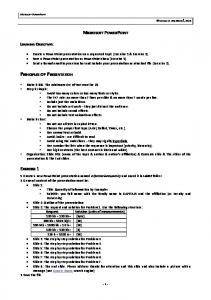
ID: 16013636 PCa suspicion Clinical suspicion, PSA 2.5-20
Exclusion criteria • Previous PCa Dg • Bx within 6 mo • Prostate surgery • Clinical infection • MRI contraindication
Information & consent
MRI (T2wi & DWI) LAB: serum & urine for marker research
TRUS Bx
MRI lesion present (Likert 3-5)
No MRI lesions (Likert score 1-2):
• MRI targeted x2 (only dominant) • Standard 12 cores • 2 core for biomarker research
• Standard 12 cores • 2 core for biomarker research
Fast and simple
ID: 16013636
PRO3 study (1) • • •
55 patients, ~60-70 min MRI (T2w, DWI, DCE-MRI, 1H-MRS) Two centers, April 2011 - March 2013 Prospective central data evaluation
IMPROD clinical trial, NCT01864135 • • •
150 patients, ~12-15 min MRI (T2w, DWI) One center, May 2013 – February 2015 Prospective data evaluation
Multi-IMPROD clinical trial, NCT02241122 • • •
600 patients, ~12-15 min MRI (T2w, DWI) Four centers, March 2015 Prospective central data evaluation
1 - Jambor et al. JMRI, 2014, doi: 10.1002/jmri.24682
Fast and simple
ID: 16013636
MRI protocol •
Only clinically available MR hardware, sequences, reconstruction
Biopsy protocol •
No image fusion of MRI with TRUS
•
2 targeted cores for the dominant lesion
•
12 systematic cores
•
2 cores for biomarkers
Fast and simple
ID: 16013636
MRI protocol • • •
3T Verio (Siemens, Erlagen) Surface array coils TSE • Transversal: 0.6×0.6×3.0mm3, TR/TE 8640/101ms • Sagittal: 0.7×0.6×3.0mm3, TR/TE 8640/101ms
•
•
bipolar gradient scheme, SE, epi read-out • 0, 100, 200, 300, 500 s/mm2, 2.0×2.0×3.0mm3, TR/TE 5543/80 ms • 0, 1500 s/mm2 s/mm2, 2.0×2.0×5.0mm3, TR/TE 5000/87 ms • 0, 2000 s/mm2 s/mm2, 2.0×2.0×5.0mm3 TR/TE 5000/87 ms Overall imaging time 14-17 minutes
•
Importable imaging protocol freely available –
[email protected]
Fast and simple
ID: 16013636
MRI reports Date of biopsy: Date of MRI: 08/05/2013 Clearly highly suspicious lesion (highly probably cancer) is present in the PZ, left apex, extending into mid-gland. DWI findings are suggestive for Gleason grade 4. No signs of extra-capsular extension and/or seminal vesicle invasion are present. Likert score: 5/5 The size of lesion: CCxRLxAP: 12x18x12 mm The size of prostate: CCxRLxAP: 29x40x29 mm In addition: - Representative MR images
mRNA biomarkers •
ID: 16013636
The RNA transcript levels of ACSM1, AMACR, CACNA1D, DLX1, PCA3, PLA2G7, RHOU, SPINK1, SPON2, TMPRSS2-ERG and TDRD1
•
measured with RT-PCR assays using a target-specific oligonucleotide probes
•
RT-PCR data were normalized to internal control
1 - http://dx.doi.org/10.1016/j.urolonc.2015.12.014
Methods
ID: 16013636
•
Regularized logistic regression classifier
•
Leave-pair-out cross validation, L1 and L2 norm
•
Clinically significant prostate cancer was defined as Gleason score 3+4 or higher based on the highest Gleason score of systematic and targeted biopsy
Results Features fPSA Likert fPSA and Likert Genes (11) Biomarkers (8) Biomarkers and Likert (9) All features (20)
ML Alg. Log. Reg. L2 Log. Reg. L1 Log. Reg. L2 Log. Reg. L1 Log. Reg. L2 Log. Reg. L1 Log. Reg. L2 Log. Reg. L1 Log. Reg. L2 Log. Reg. L1 Log. Reg. L2 Log. Reg. L1 Log. Reg. L2 Log. Reg. L1
ID: 16013636 LPOCV AUC 0.732 0.732 0.924 0.924 0.930 0.937 0.645 0.614 0.808 0.804 0.924 0.909 0.900 0.927
ID: 16013636
Conclusion The 11 studied genes provided limited added value to bpMRI.
bpMRI demonstrated high diagnostic accuracy for SPCa detection and addition of fPSA resulted in only minor improvement.
[email protected]