GENERAL SECTION
Nabees Man Singh Pradhan: MICRPP for supracondylar fracture
ORIGINAL ARTICLE
Minimally Invasive-Closed Reduction and Percutaneous Pinning for Supracondylar Fractures of the Humerus in Children Vol. 01 | No. 02 | Issue 02, Dec 2014
Nabees Man Singh Pradhan,1 Rojan Tamrakar,2 Bidur Gyawali,2 Toya Raj Bhatta,2 Balakrishnan M. Acharya,1 Suman Shrestha,1 Saroj Krishna Shrestha,3 Achyut Rajbhandari,4 Utsab Shrestha5 Associate Professor, 2Lecturer, 5Resident Department of Orthopedics, Patan Academy of health Sciences, Laliptur, Nepal 1
Professor, 4 Senior Consultant Om Hospital and Research Center, Chabhill, Kathmandu, Nepal 3
ISSN: 2091-2749 (Print) 2091-2757 (Online)
Correspondence Dr. Nabees Man Singh Pradhan Associate Professor, Department of Orthopedics Patan Academy of Health Sciences, Lalitpur, Nepal Email:
[email protected] Phone: +977-9851094037 Peer reviewed by Prof. Dr. Jay N Shah Patan Academy of Health Sciences Dr. Ashis Shrestha Patan Academy of Health Sciences
ABSTRACT Introductions: Although Closed Reduction and Percutaneous Pinning is the gold standard of treatment for Supracondylar fractures (SC) in children, reduction is not always easy. Minimally Invasive, Closed Reduction and Percutaneous Pinning (MI-CRPP) reduces the soft tissue trauma and provides easy reduction. We have reviewed the success rate of minimally invasive reduction technique and its outcome. Methods: We reviewed the charts of 155 children (97 male, 58 female) age ranging from 2 to 14 years with SC fractures of the humerus who were operated with minimally invasive closed reduction and precutaneous pinning from November 2008 to June 2014 at Patan Hospital and Om Hospital. They were followed up for a mean of eight (4 to 24) weeks. The K-wires were removed at 4 to 6 (average 4.28) weeks. Results: Male children were affected more than female with the ratio being 97 to 58. Right side was affected more than left (ratio 89 to 66). Post-operatively, there were six (3.87%) ulnar nerve injury and eight (5.16%) patients came with superficial pin tract infection. One hundred and thirteen (72.9%) had excellent, 35 (22.58%) good, five (3.23%) fair and two (1.3%) poor results at the eight week follow-up which was improved to 144 (92.9%) excellent, seven (4.5%) good, three (1.9%) fair and one (0.65%) poor results at the 14 week follow-up. Conclusions: Closed reduction of supracondylar fractures of the humerus in children with minimally invasive technique prior to K-wire fixation is a relatively simple, safe and effective method of achieving satisfactory reduction and good functional outcome. Keywords: cubitus varus deformity, K wire fixation,minimally invasive closed reduction and precutaneous pinning, supracondylar fractures Plain Language Summary The study was done to study the success rate of closed reduction with minimally invasive technique in supracondylar fractures in children and its outcome. The procedure was found to be highly successful for fracture reduction and facilitated the percutaneous fixation of the fractures. With the high success rate of reduction, the need for ORIF in such fractures can be eliminated or at least minimized, hence reducing the morbidity and associated complications.
Journal of Patan Academy of Health Sciences. 2014 Dec;1(2):15-18
15
Nabees Man Singh Pradhan: MICRPP for supracondylar fracture INTRODUCTIONS Supracondylar fractures (SC) of humerus is a common occurrence in children(male slightly more than female) with peak age of incidence occurs between 5-8 years.1-3 It is usually accompanied with marked swelling which presents a formidable challenge for reduction and immobilisation.4-6 Closed manipulation and Open Reduction and Internal Fixation (ORIF) can result in nerve injury, soft tissue injury etc.7,8 Closed Reduction and Percutaneous Pinning (CRPP) has now been considered the gold standard in treatment of SC fractures. However, reduction is not always easy, mainly because of the swelling or when they present late as in the context of third world countries. To avoid all these pitfalls of ORIF, minimally invasive closed reduction and percutaneous pinning (MI-CRPP) can address the issue of difficult reduction of SC fractures. But the efficacy of this new modality of treatment particularly in our community is still unexplored. This study has been conducted to assess the success rate of reduction in SC fractures of the humerus and also study its outcome. METHODS This is a cross sectional study conducted at Patan Academy of Health Sciences and Om Hospital and Research Centre. Chart review of children undergoing MI-CRPP for SC fracture of humerus from November 2008 to June 2014 was done. Children from 2 to 14 years with Gartland type fracture 3A and 3B were included. Children aged below two years were excluded and so were children with bilateral fractures, with previous contralateral fractures around the elbow, GustilloAndersson Grade II and III fractures, and those with associated ipsilateral distal forearm fractures were also excluded. The data on follow up were also retrieved from the record. The follow-ups were arranged as follows: the first follow- up on the seventh day to inspect the wound; the second follow-up on the second week for wound inspection or suture removal and to see the pin configuration; the third follow-up on the fourth week for the removal of plaster slab and pins if permitted by radiological findings and to start physiotherapy; the fourth follow-up on the eighth week post-operatively to see the range of movement (ROM) and carrying angle of the elbow; and the final follow-up on the twelfth week post-operatively.
16
Results for functional outcome using Flynn criteria,9 for reduction using post-operative radiographs, and or other complications like compartment syndrome, nerve injury, infection rate, elbow stiffness and myositis ossificans were recorded. Ethical clearance for the was taken from institutional review committee. RESULTS The charts of 155 children (97 male, 58 female) age ranging from 2 to 14 years (mean six years) were reviewed. Right sided injury were 89 and left sided were 66. The time of operation ranged from the day of injury to the 15th day of injury, with the mean time of operation being 5.5 days. Preoperatively, three cases were open fractures, six cases had nerve injuries (median nerve three, ulnar nerve two and radial nerve one) and there were no cases of vascular injuries. Postoperatively, there were six (3.87%) ulnar nerve injury and eight (5.16%) patients came with superficial pin tract infection, which subsided with antibiotics and/or pin removal. Callus formation was seen in all patients at the fourth week post-operatively before removing the K-wires. The fracture united in all cases before the wire removal. In our study, 113 (72.9%) had excellent, 35 (22.58%) good, five (3.23%) fair and two (1.3%) poor results at the eight week follow-up which was improved to 144 (92.9%) excellent, seven (4.5%) good, three (1.9%) fair and one (0.65%) poor results at the 14 week follow-up. During this study, complications like vascular injury, compartment syndrome, myositis ossifications, significant mal-union and non-union were not encountered. In the present series, eight (5.16%) patients developed superficial pin-tract infections. No deep infection or septic arthritis was found. Distal pin migration was seen in four (2.58%) patients, which were not significant and so required no re-pinning. We did not have any loss of reduction in our series. Radiological assessments were performed immediately the day after surgery, at four weeks(some at six weeks). Clinical assessment was performed immediately after the procedure to looks for neurovascular compromise, and at four or six weeks, eight weeks, 12 weeks and some after six months. Fracture healing was defined as callus bridging of three out of four cortices, seen on both lateral and antero-posterior X-ray. One hundred and fifty five patients meeting the inclusion criteria were included in the study. Twelve patients were lost to follow up who never turned up after K-wire removal and were discarded from the study.
Journal of Patan Academy of Health Sciences. 2014 Dec;1(2):15-18
Nabees Man Singh Pradhan: MICRPP for supracondylar fracture reduction with minimum soft-tissue trauma is required to achieve the best possible functional outcome.10-14 Treatment modalities vary, but the primary aim is to try and achieve anatomical reduction by the simplest way possible before the fracture is stabilized.
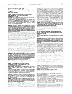
Figure 1. Immediate post operative x-ray after CRPP of fractures humerus in children
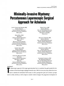
Figure 2. X- ray after 4 weeks after CRPP of fractures humerus in children
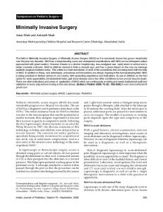
Figure 3. X-ray after 8 weeks after CRPP of fractures humerus in children
DISCUSSIONS Supracondylar fractures of the humerus (SC fracture) is the most common pediatric fracture around the elbow. The associated swelling and late presentation make closed reduction difficult.4-6 Remodeling capability of a child’s bone used to be the basis of closed manipulation and acceptance of inadequate reduction. But most authors now agree that accurate Journal of Patan Academy of Health Sciences. 2014 Dec;1(2):15-18
Malunion of SC fractures commonly results in cubitus varus deformity which is a result of coronal rotation, or medial tilt of the distal fragment.10,15 Smith et.al suggested that residual medial tilt after reduction is the most important factor in varus angulation, with isolated rotational deformities being corrected by compensatory rotation at the shoulder.16 This concept has become popular in understanding the sequel of alteration in carrying angle.17 Although open reduction can be used to achieve anatomical reduction before internal fixation, it invariably results in the scarring of extensor mechanism, thus resulting in elbow stiffness. Closed reduction is not always easy, either because of swelling of the elbow or due to late presentations. The use of an artery forcep to aid reduction has a high success rate and also preserves the extensor mechanism with only a minimal trauma to the soft tissue. In this study, the results at eighth post-operative week showed excellent results in around seventy percent of patients, which increased to more than ninety percent at 14th week post operatively. We believe that this increase of range of motion of the elbow was because of the physiotherapy. Those patients who had good or fair results were having severe soft tissue injuries or repeated closed reduction performed in another center. Khan et.al obtained 88% excellent, four percent good and four percent poor results in his study.18 Tiwari et.al observed 88 percent satisfactory results, among which 42% were excellent, in his series of late-presenting supracondylar fractures of humerus in children.19 These two studies are comparable to our study. In this series, ten patients (6.45%) had nerve injury preoperatively, out of which five had median, three ulnar and two radial. Six cases (3.87%) reported to have ulnar nerve injury after the procedure. Fourteen of the nerve injuries recovered within 12 weeks post-operatively and two were lost to follow-up after pin removal. The incidence of post-operative ulnar injuries has been estimated to range from 5 to 19%.20 In the present study, 15 patients presented within six hours of injury, 47 patients between 6 to 24 hours, 38 patients between 24 to 48 hours, 29 patients between 48 to 72 hours and 26 patients after 72 hours. The mean time of surgery from the time of hospital admission ranged from 12 hours to 84 hours, which was chiefly because of the unavailability of operation theatre. The maximum delay in performing the MI-CRPP was 15 days
17
Nabees Man Singh Pradhan: MICRPP for supracondylar fracture after injury. Satisfactory alignment could be achieved in all the fractures reduced with minimally invasive reduction technique. The configuration of the pins depended chiefly upon the fracture configuration and the comfort level of the operating surgeon. In the present series, eight (5.16%) patients developed superficial pin-tract infections, which subsided after of oral antibiotics and/or pin removal. No deep infection or septic arthritis was found. Pirone et.al found superficial pin-tract infection in two percent of cases with no deep infection and septic arthritis.21 The larger number of pin tract infection in our study was probably because of poor hygienic and living conditions of the patients. Gordon et.al observed pin-tract migration in six percent of cases and Lee noticed the loss of reduction in seven percent of cases.22,23 In the present series, the distal pin migration was seen in four (2.58%) patients, which were not significant and so required no re-pinning. We did not have any loss of reduction in our series. CONCLUSIONS Closed reduction of supracondylar fractures of the humerus in children with minimally invasive technique prior to K-wire fixation is a relatively simple, safe and effective method of achieving satisfactory reduction and good functional outcome. REFERENCES 1. Cheng JC, Ng BK, Ying SY, Lam PK. A 10-year study of the changes in the pattern and treatment of 6,493 fractures. J Pediatr Orthop. 1999 May-Jun;19(3):344-50. 2. Dimeglio A. Growth in pediatric orthopaedics. In: Morrissy RT, Weinsten SL, editors. Lovell and Winters’s Pediatric Orthopaedics. 6th ed. Philadelphia: Lippincott Williams and Wilkins, 2006: p. 35-65.
8. Kallio PE, Foster BK, Paterson DC. Difficult supracondylar elbow fractures in children: analysis of percutaneous pinning technique. J Pediatr Orthop. 1992;12:11-5. 9. Flynn JC, Matthews JC, Benoit RJ. Blind pinning of displaced supracondylar fracture of humerus in children. Sixteens’ years experience with long term follow up. J Bone Joint Surg Am. 1974;56:263-72. 10. Labelle H, Bunnell WP, Duhaime M. Cubitus Varus Deformity following supracondylar fracture of humerus in children. J Pediatr Orthop. 1982;2:539-46. 11. D’ambrosia RD. Supracondylar fractures of the humerus - prevention of cubitus varus. J Bone Joint Surg Am. 1972;54:60-6. 12. Attenborough CG. Remodeling of the humerus after supracondylar fractures in childhood. J Bone Joint Surg Br. 1953;35:386-95. 13. BoydDW, Aronson DD. Supracondylar fractures of the humerus: a prospective study of percutaneous pinning. J Pediatr Orthop. 1992;12:789-94. 14. Cheng JC, Lam TP, Shen WY. Closed reduction and percutaneous pinning for type III displaced supracondylar fractures of the humerus in children. J Orthop Trauma. 1995;9:511-5. 15. Kallio PE, Foster BK, Paterson DC. Difficult supracondylar elbow fractures in children: analysis of percutaneous pinning technique. J Pediatr Orthop. 1992;12:11-5. 16. Smith L. Supracondylar fractures of the humerus treated by direct observation. Clin Orthop Relat Res. 1967;50:37-42. 17. Mann TS. Prognosis in supracondylar fractures. J Bone Joint Surg Br. 1963;45:516-22. 18. Khan AQ, Goel S, Abbas M, Sherwani MK. Percutaneous K-wiring for Gartland type III supracondylar humerus fractures in children. Saudi Med J. 2007; 28(4):603-6. 19. Tiwari A, Kanojia RK, Kapoor SK. Surgical management for late presentation of supracondylar humeral fracture in children. J Orthop Surg (HongKong). 2007;15(2):177-82.
3. Otsuka NY, Kasser JR. Supracondylar fractures of the humerus in children. J Am Acad Orthop Surg. 1997; 5:19-26.
20. McGraw JJ, Akbarnia BA, Hanel DP. Neurological complications resulting from supracondylar fractures of the humerus in children. J Pediat Orthop. 1986;6:647-50.
4. Takahara M, Sasaki I, Kimura T, Kato H, Minami A, Ogino T. Second fracture of distal humerus after cubital varus malunion of a supracondylar fracture in children. J Bone Joint Surg Br. 1998;80(5):791-7.
21. Pirone AM, Graham HK, Krajbich JI. Management of displaced extension-type supracondylar fractures of the humerus in children. J Bone Joint Surg Am. 1988;70:641-50.
5. Gartland JJ. Management of supracondylar fracture of humerus in children. Surg Gynecol Obstet. 1959;109:145–54.
22. Gordon JE, Patton CM, Luhmann SJ, Bassett GS, Schoenecker PL. Fracture stability after pinning of displaced supracondylar distal humerus fractures in children. J Pediatr Orthop. 2001;21:313-8.
6. Mitchell WJ, Adams JP. Supracondylar fracture of humerus in Children; a ten year review. J Am Med Assn. 1961;175:573-7. 7. Herzenberg JE, Koreska J, Carroll NC, Rang M. Biomechanical testing of pin fixation techniques for pediatric supracondylar elbow fractures. Orthop Trans. 1988;12:678-9.
18
23. Lee SS, Mahar AT, Miesen D, Newton PO. Displaced pediatric supracondylar humerus fractures: biomechanical analysis of percutaneous pinning techniques. J Pediatr Orthop. 2002;22:440-3.
Journal of Patan Academy of Health Sciences. 2014 Dec;1(2):15-18